Risks of heart procedures Dr Kiran Lakkireddy Cardiologists Hyderabad, Kondapur

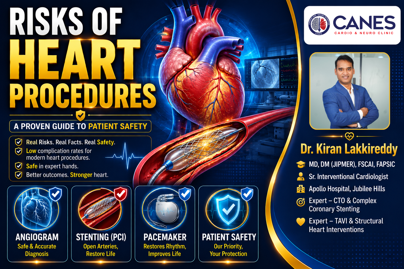
Risks of Heart Procedures: A Proven Guide to Patient Safety

●   Lifestyle & Prevention

Risks of Heart Procedures: A Proven Guide to Patient Safety

Table of Contents

Book a Consultation

Prevention is better than cure. Talk to our specialists today.
Risks of heart procedures Dr Kiran Lakkireddy Cardiologists Hyderabad, Kondapur

If your cardiologist has recommended an intervention, it is entirely natural to feel anxious about the risks of heart procedures. When you hear medical terms like “complications” or “surgery,” the fear of the unknown can quickly become overwhelming. However, when we look closely at the actual medical data, the numbers are incredibly reassuring. Most modern cardiac interventions are minimally invasive, highly routine, and designed with your long-term safety as the absolute top priority.

By understanding exactly what these procedures involve and looking at the real chances of complications based on published medical research, you can replace fear with confident, informed decision-making. At CANES Cardio & Neuro Clinic, guided by Dr. Kiran Lakkireddy, we believe in absolute transparency. This comprehensive guide will clearly explain the anatomical purpose of each treatment, the published statistics, and the specific safety protocols your care team uses to protect you.

Quick Answer

While all medical interventions carry some level of risk, the risks of heart procedures are generally very low for planned, routine cases. On average, diagnostic tests and standard stenting have a major complication rate of less than 1%. Modern cardiology relies heavily on minimally invasive catheter techniques, which significantly reduce the chances of severe bleeding, infection, and prolonged recovery compared to traditional open-heart surgery.

Should I Be More Worried About the Procedure or the Heart Disease Itself?

For most patients, untreated heart disease carries a significantly greater risk of permanent disability or death than the actual procedure designed to treat it.

Key Takeaway: The risks of heart procedures are closely managed and monitored by a medical team, whereas the progression of an untreated blocked artery or a failing valve is highly unpredictable and often fatal.

It is completely normal to focus on the immediate anxiety of entering a hospital. However, it is vital to weigh that short-term anxiety against the long-term reality of your condition. If a cardiologist recommends a stent or a valve repair, it is because your heart is currently in a dangerous, unstable state.

Declining a necessary intervention means leaving a severe blockage or electrical fault untreated. This dramatically increases your daily risk of suffering a sudden, massive heart attack, developing irreversible heart failure, or experiencing a fatal cardiac arrest. Medical interventions are recommended only when the benefit of fixing the heart overwhelmingly outweighs the statistical risk of the procedure.

What Are the Real Risks of Heart Procedures?

The real risks of heart procedures depend entirely on your specific heart condition, your age, and whether the procedure is planned or an emergency. However, on average, the risk of a major complication for a routine intervention is exceptionally low, often sitting below 1%.

Key Takeaway: The complication numbers you see online are population averages; your personal risk may be much lower, which is why your care team takes specific steps to keep those numbers as low as possible for you.

When reading through this guide, remember that numbers can be reassuring once you see them in proper medical context. A 1% risk means that out of 100 people, 99 experience absolutely no severe issues. Furthermore, your cardiology team spends extensive time planning your specific procedure to mitigate any personal health challenges, such as existing kidney issues or a history of bleeding.

Visual Scale: Procedure Risk Levels

To help put things into perspective quickly, here is how cardiologists generally classify the risk levels of common interventions.

Procedure

Average Risk Level

Diagnostic Angiogram

Very Low

Routine PCI (Stenting)

Low

Permanent Pacemaker

Low

TAVI (Valve Replacement)

Moderate

High-Risk PCI

Higher

What Is a Diagnostic Cardiac Catheterisation?

A diagnostic cardiac catheterisation (angiogram) is a test where a thin tube is guided through your wrist or groin, and contrast dye is injected to make your arteries show up clearly on an X-ray. It lets your doctor see exactly where any blockages are.

Key Takeaway: This is the most basic heart procedure and is often the very first step before anything else is done, carrying a mortality risk of just 0.05–0.1% in planned cases.

This is a very safe procedure. The risk of mortality is around 0.05–0.1% for planned (elective) cases, rising to about 1% only in emergency situations. A heart attack or stroke during the test is similarly rare at 0.05–0.1%.

The more common issues are minor. Bruising or a small lump at the access site occurs in 1–3% of people having the test through the groin, and less than 0.5% through the wrist (which is why many doctors prefer using the wrist). Some people have a mild allergic reaction to the contrast dye—about 1–3%—though severe reactions are extremely rare. The dye can also occasionally affect your kidneys temporarily, more so if you already have kidney problems.

What Are the Risks of Angioplasty and Stenting (PCI)?

Percutaneous Coronary Intervention (PCI) is a procedure where a small balloon is inflated to open a blocked artery, and a stent (a tiny mesh tube) is left in place to keep the artery open. It restores blood flow without open-heart surgery.

Key Takeaway: For planned PCI in stable patients, the risk of death is exceptionally low, around 0.1–0.5%. It remains the safest and most effective way to clear a blocked artery.

If the catheterisation shows a blocked artery, PCI is often done at the same time. If you are having PCI during an active heart attack, the risk is naturally higher—around 3–7%—because you are already unwell when the procedure starts. A small number of patients, about 1–3%, experience a minor heart attack signal during the procedure itself, often without any physical symptoms.

The stent can occasionally clot off in the days afterward (stent thrombosis), affecting around 0.5–1.5% of patients. This is exactly why it is so important to keep taking your blood-thinning medication as prescribed; stopping early is the main cause of this complication. Over the longer term, the artery can re-narrow (restenosis) in 5–10% of people, even with modern drug-coated stents.

Why Are High-Risk PCI Procedures Different?

High-risk PCI is performed on patients with severe underlying problems, such as a very weak heart muscle, heavily calcified arteries, or cardiogenic shock. These cases require extra support equipment and are reviewed by a full team of specialists.

Key Takeaway: The elevated risks of heart procedures in this category reflect how sick the patient’s heart already was, rather than the procedure itself causing the danger.

Because the underlying heart condition is much more serious, the risks are understandably higher. In-hospital death ranges from 3–10% in stable high-risk patients. However, it can be as high as 20–50% if someone arrives already in cardiogenic shock.

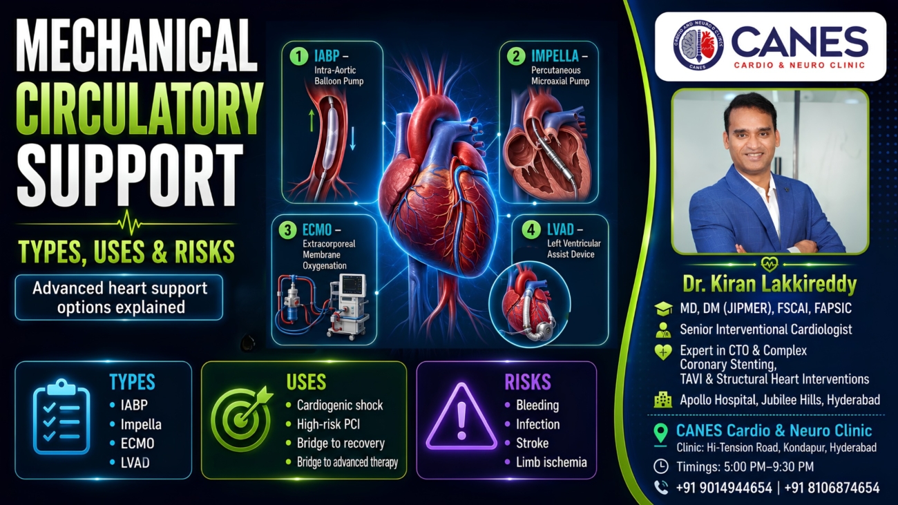
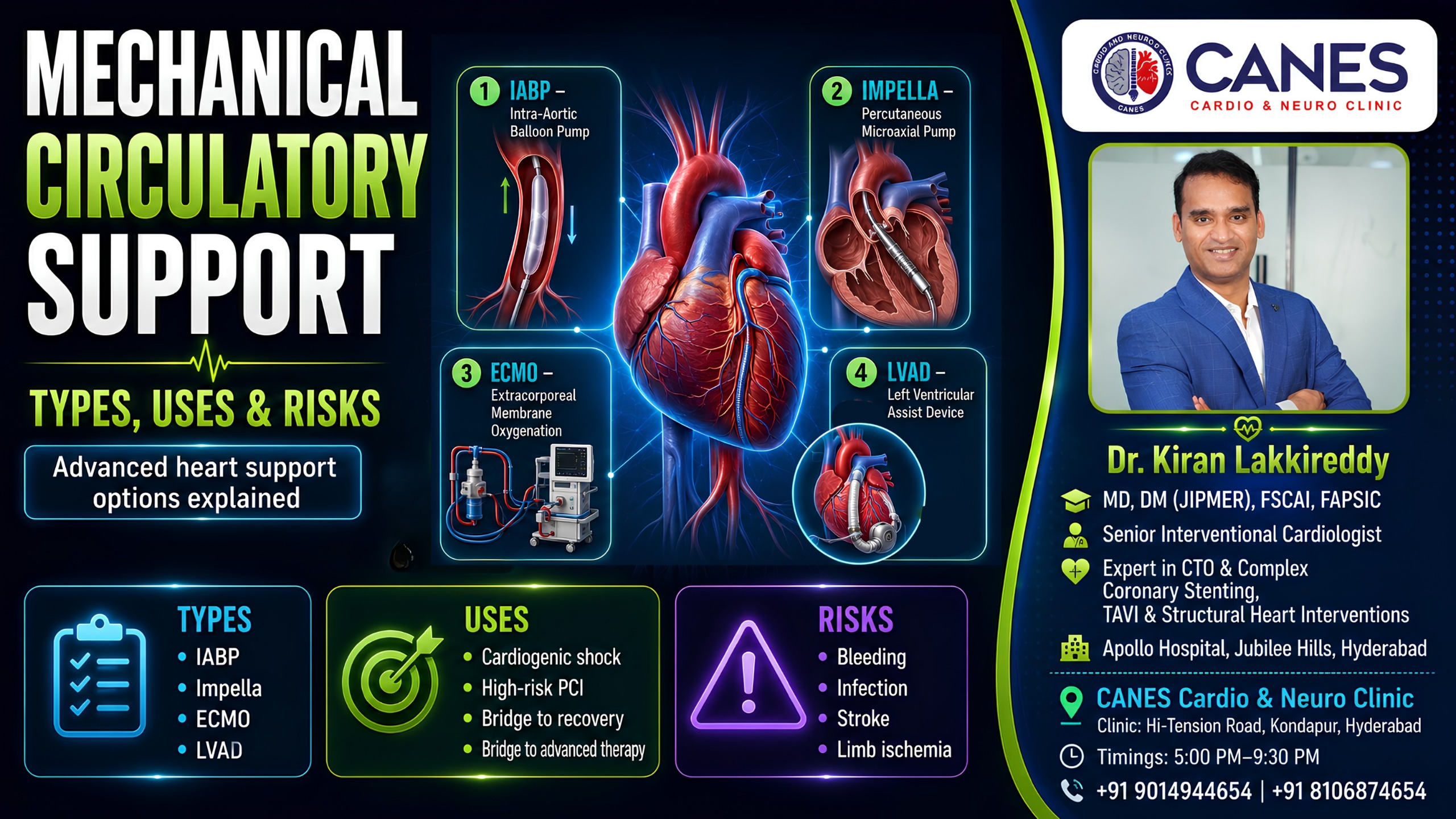
Roughly 2–5% of patients have a significant heart attack during the procedure, and about 3–8% may develop or worsen shock. Mechanical heart support devices, when used, carry their own risks (around 5–10%), such as reduced blood flow to the leg where the device is inserted.

What Are the Risks of a Permanent Pacemaker?

A permanent pacemaker is a small device placed under the skin near your collarbone, with wires running into your heart to keep your heart rate steady when it is beating too slowly.

Key Takeaway: This is a well-established, low-risk procedure with a death rate under 0.1%, offering a highly reliable solution for slow heart rhythms.

The most common complications are a small blood collection at the implant site (2–5%) and minor infection of the wound (1–2%). Both are usually manageable without major treatment.

More serious infection of the device or leads happens in about 0.5–1% of cases and would need the device to be removed and replaced. Occasionally, a wire can shift out of position (1–3%) and need repositioning. A collapsed lung from the vein puncture during insertion happens in 0.5–2% of cases. The device itself typically lasts 10–15 years before the battery needs replacing.

How Do ICD and CRT Devices Compare to Pacemakers?

An ICD can deliver a shock to correct a dangerous rapid heart rhythm, while a CRT adds an extra wire to help both sides of your heart beat in better coordination for heart failure.

Key Takeaway: These devices are more complex than standard pacemakers, meaning the procedural risks are slightly higher (death around 0.1–0.5%).

The trickiest part of CRT implantation is placing the third wire on the left side of the heart. This anatomical challenge fails in about 5–10% of attempts, and even when successful, the wire can shift position afterward in up to 10% of cases.

One of the more emotionally difficult complications is an inappropriate shock—where the device fires when it did not need to. This happens in roughly 5–10% of patients per year, though settings are usually adjusted afterward to reduce this. About 20–30% of CRT patients do not notice the symptom improvement they were hoping for; this is called non-response and is a limitation of the therapy rather than a complication.

At a Glance: Comparing the Risks of Heart Procedures

Procedure Type

Primary Purpose

Average Mortality Risk

Most Common Minor Issue

Diagnostic Angiogram

Locate blockages

0.05–0.1% (Planned)

Bruising at access site (1-3%)

Routine PCI (Stenting)

Open blocked artery

0.1–0.5% (Stable)

Artery re-narrowing over time

Permanent Pacemaker

Fix slow heartbeats

< 0.1%

Small blood collection (2-5%)

TAVI Valve Replacement

Replace aortic valve

1–2% (Low risk)

Needing a new pacemaker (10-25%)

What Is TAVI and What Are Its Safety Rates?

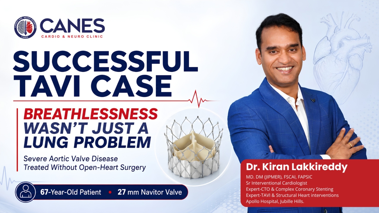
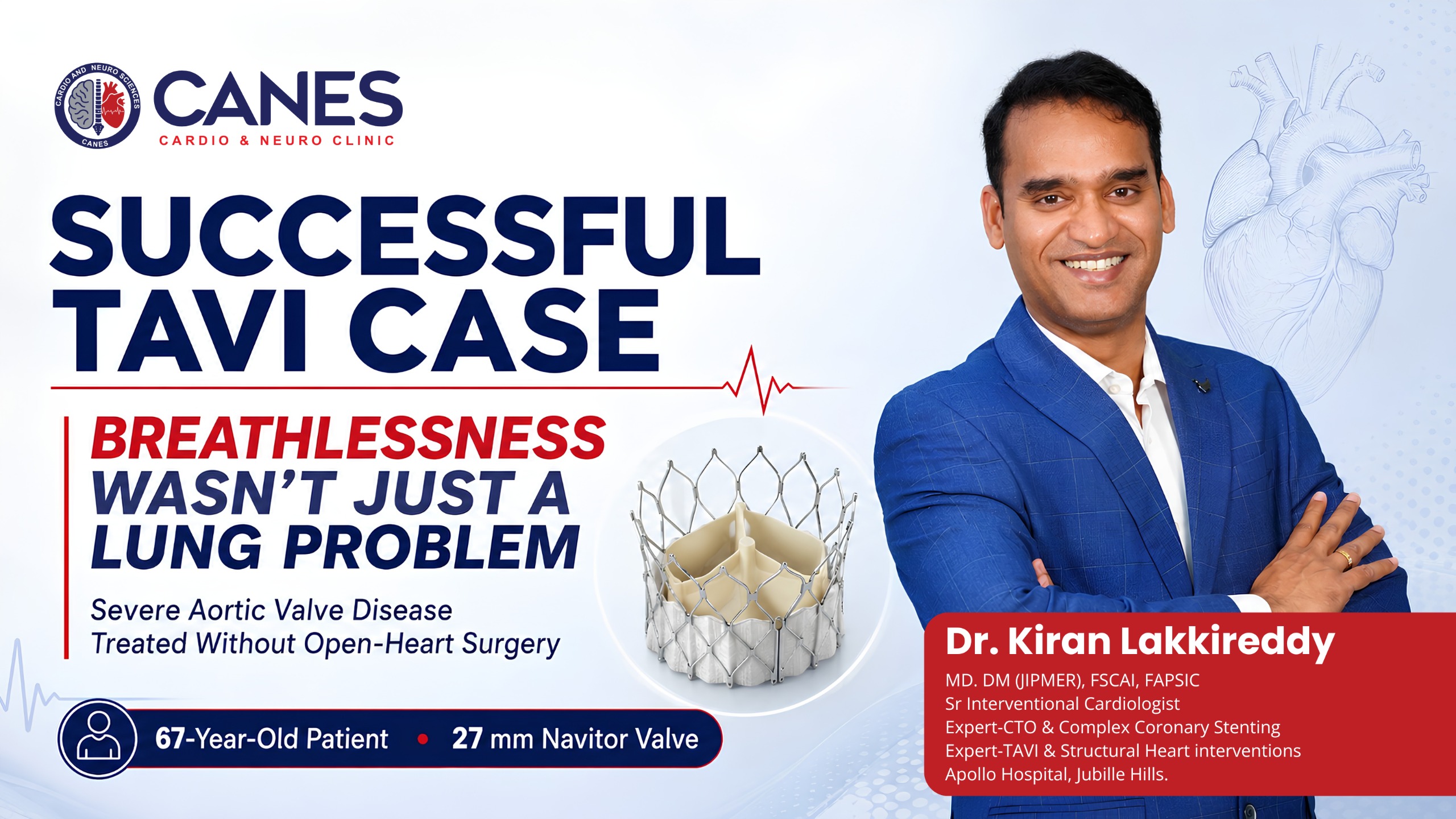
TAVI replaces a worn-out aortic valve through a catheter, usually via the groin, without open-heart surgery. It is typically offered to people whose surgical risk is too high for traditional operations, though it is increasingly used for lower-risk patients too.

Key Takeaway: TAVI provides a life-saving alternative to open surgery with death rates within 30 days ranging from 1–2% in lower-risk patients to 2–5% in higher-risk patients.

Stroke occurs in 2–5% of patients within the first month. One of the more common after-effects is needing a permanent pacemaker afterward because the new expanding valve can disrupt your heart’s electrical pathways; this happens in 10–25% of cases depending on the device used.

Some leakage around the new valve (paravalvular leak) is seen in 3–10% of patients, and major complications at the artery access site occur in 3–7%. Serious complications like the valve frame tearing the aortic annulus are rare, under 1%, but are treated as emergencies when they occur.

What Is MitraClip Valve Repair?

MitraClip treats a leaking mitral valve by clipping the two leaflets together so they close more effectively, done through a catheter from the groin under general anaesthesia.

Key Takeaway: Mortality within 30 days is around 1–3% for degenerative valve disease and 3–7% for heart failure patients, offering symptom relief without open-chest trauma.

The clip occasionally only grips one of the two leaflets properly (single leaflet attachment) in about 3–8% of cases, which can mean the leak is not reduced as much as hoped. In 10–15% of patients, some degree of leakage remains after the procedure. Serious complications from the catheter crossing into the left side of the heart, such as a fluid buildup around the heart, occur in 0.5–2% of cases.

What Are the Risks of the Watchman Device (LAAO)?

The Watchman device seals off a small pouch in the heart where blood clots tend to form in people with atrial fibrillation, offering a permanent alternative to taking long-term blood thinners.

Key Takeaway: This is one of the lower-risk structural procedures available, with an average mortality rate well under 1%.

Fluid around the heart requiring drainage occurs in 1–3% of cases. A small clot can form on the surface of the device itself in 3–7% of patients, which is exactly why you will usually stay on some blood-thinning medication for a period after the procedure, even though the goal is to eventually stop it. Stroke around the time of the procedure occurs in just 1–2% of cases.

Myth vs. Fact: Common Myths About Heart Procedures

Understanding the true risks of heart procedures means separating internet myths from proven medical facts.

Common Myth

Medical Fact

All heart procedures are highly dangerous.

Most planned, routine procedures have major complication rates well below 1%.

Stents cure heart disease permanently.

Stents fix blockages, but lifestyle changes and medication are required to prevent new ones.

Pacemakers stop the heart from failing.

Pacemakers regulate the electrical rhythm, not the physical pumping strength of the heart muscle.

Open-heart surgery is the only way to fix valves.

Advanced structural techniques like TAVI can now replace valves through a tiny catheter.

The Patient Safety Checklist: Preparing for Your Procedure

Minimizing the risks of heart procedures requires teamwork between you and your doctor. Use this actionable checklist to prepare safely:

  • Review Medications: Provide your doctor with a complete list of all prescriptions, especially blood thinners and diabetes medications.

  • Check Kidney Function: Ensure you have recent blood work done so your doctor knows exactly how well your kidneys will handle the contrast dye.

  • Report Allergies: Clearly state any allergies you have to iodine, shellfish, or previously used anesthetics.

  • Plan Your Discharge: Arrange for a trusted family member or friend to drive you home and monitor you for the first 24 hours.

What Questions Should You Ask Your Cardiologist?

Every number above is a population average. Your own personal risk depends entirely on your specific heart condition, your age, your other health conditions, and how the procedure is performed. If you are due to have any of these procedures, it is worth asking your doctor:

  1. What is my specific, personal risk percentage based on my current health profile?
  2. What would realistically happen to my heart if I chose not to have this procedure?
  3. What specific steps does the surgical team take to reduce these risks for someone in my situation?

A good conversation with your cardiologist before the day of the procedure is one of the best ways to walk in feeling informed rather than just nervous.

Expert Cardiac Care at CANES Clinic

Navigating a cardiac diagnosis requires a medical team that combines technical precision with open, empathetic communication. At CANES Cardio & Neuro Clinic, we prioritize thorough patient education and evidence-based procedural care. We understand that discussing surgical risks can cause anxiety, and we are dedicated to providing clear, authoritative answers.

Guided by Dr. Kiran Lakkireddy, an experienced interventional cardiologist specializing in complex structural heart procedures, our facility ensures that every diagnostic angle is fully explored. Whether you require a routine angiogram, complex high-risk stenting, or advanced pacemaker management in Hyderabad, our team utilizes the latest protocols to keep your personal risk as low as absolutely possible.

(Disclaimer: This article is for general educational purposes and is not a substitute for personalised medical advice. Always discuss your individual risks and options with your treating cardiologist.)

Key Points to Remember

  • Context Matters: Published complication rates are averages; your specific risk profile depends heavily on your age and the severity of your heart disease.

  • Routine vs. Emergency: Planned, elective procedures like standard angiograms carry extremely low complication risks, while emergency interventions inherently carry higher risks.

  • Medication is Crucial: Taking your prescribed antiplatelet medication after a stent is placed is the most important step you can take to prevent dangerous blood clots.

  • Structural Alternatives: Advanced techniques like TAVI and MitraClip offer patients life-saving valve repairs through a simple catheter, completely avoiding the severe trauma of open-heart surgery.

Frequently Asked Questions (FAQs)

Is it normal to feel chest pain during a stent procedure?

Yes, it is very common to feel a brief sensation of chest pressure or mild pain exactly at the moment the cardiologist inflates the balloon inside your artery. This pressure typically resolves as soon as the balloon is deflated.

How long does it take to recover from a cardiac catheterisation?

If the procedure was performed through your wrist, recovery is very fast; you can typically walk immediately and go home the same day. If accessed through the groin, you will need to lie completely flat for a few hours.

Can a pacemaker be affected by household appliances like microwaves?

Modern pacemakers have excellent internal shielding. Standard household appliances, including microwaves and televisions, are completely safe to use without interfering with your device.

Why do I need blood thinners after getting a stent if the blockage is fixed?

A new metal stent is seen as a “foreign object” by your immune system. Blood thinners prevent blood platelets from sticking to the bare metal and forming a dangerous clot before your body naturally heals over it.

What is the difference between a pacemaker and an ICD?

A pacemaker is designed only to speed up a heart that is beating too slowly. An ICD does this as well, but it also has the ability to deliver a strong internal shock to stop a chaotic, dangerously fast heart rhythm.

Are the risks of TAVI lower than traditional open-heart valve surgery?

For many patients, especially the elderly or those with multiple health conditions, TAVI is significantly safer than open-heart surgery because it avoids the need for a heart-lung bypass machine and large chest incisions.

Difference Between a Heart Attack and Heart Block

Difference Between a Heart Attack and Heart Block

●   Lifestyle & Prevention

What Is the Difference Between a Heart Attack and Heart Block?

Table of Contents

Book a Consultation

Prevention is better than cure. Talk to our specialists today.

Difference Between a Heart Attack and Heart Block

The short answer: A heart attack is a “plumbing” problem where a blocked artery stops blood flow to the heart muscle, while a heart block is an “electrical” problem where the nerve signals that tell the heart to beat are delayed or stopped.

When you sit down in a doctor’s office and hear the word “block” in relation to your heart, it is entirely natural to feel a wave of anxiety. In everyday conversation, medical terminology often gets mixed up. Because both a “heart attack” and a “heart block” involve something being obstructed, many patients assume they are dealing with the exact same illness.

However, they are two completely distinct cardiac conditions. They have different root causes, produce different physical symptoms, and require entirely different life-saving treatments. Understanding the difference between a heart attack and heart block is not just about learning medical vocabulary; it is about recognizing the right warning signs so you can seek the correct emergency care without delay.

At CANES Cardio & Neuro Clinic, recognized as the best cardiologist clinic in Hyderabad, we believe in speaking to our patients in plain, regular English. Guided by Dr. Kiran Lakkireddy, a highly trusted heart specialist, our team is dedicated to replacing fear with clear, actionable knowledge. In this comprehensive master guide, we will break down the exact differences between these two conditions, how they are diagnosed, and the modern treatments we use to fix them.

1. The Short Answer: The Plumbing vs. Electrical Analogy

To easily understand the difference, imagine your heart is a house: a heart attack is a clogged water pipe, while a heart block is a cut electrical wire.

The easiest way to grasp how your heart functions is to think of it like a well-built house. To keep a house running smoothly, you need two completely separate systems. First, you need a plumbing system (pipes) to bring fresh water into the rooms. Second, you need an electrical system (wires) to carry current to the light switches so the house can function. Your heart works the exact same way.

When we talk about a heart attack, we are talking about a catastrophic failure in the plumbing system. The blood vessels (coronary arteries) that deliver oxygen-rich blood to the heart muscle become clogged with cholesterol. On the other hand, when we talk about a heart block, we are looking at a failure in the electrical system. The internal wiring (conduction system) that sends the spark telling your heart muscle to squeeze experiences a delay or a complete break.

2. What Exactly is a Heart Attack (Myocardial Infarction)?

A heart attack is a life-threatening emergency where a ruptured cholesterol plaque forms a blood clot, completely blocking oxygenated blood from reaching a section of the heart muscle.

A heart attack is formally known in the medical world as a myocardial infarction (MI). Your heart is a working muscle, constantly pumping 24 hours a day. Like your biceps or your calf muscles, it needs a constant supply of fresh, oxygen-rich blood to survive. This blood is delivered by a network of vessels wrapped around the outside of the heart, known as the coronary arteries.

The problem begins slowly over decades. A waxy, fatty substance called cholesterol plaque builds up along the smooth inner walls of these coronary arteries—a condition we call atherosclerosis. A heart attack usually happens when one of these unstable plaques suddenly cracks or ruptures. Your body thinks this crack is an injury, so it rushes blood cells (platelets) to the area to form a clot. Unfortunately, inside a tiny artery, this clot acts like a cork in a bottle, completely blocking blood flow. The heart muscle below the blockage begins to suffocate and die, leading to permanent damage.

Recognizing the Symptoms of a Heart Attack

Heart attack symptoms are usually sudden and painful, heavily featuring crushing chest pressure, shortness of breath, and pain radiating down the arm or jaw.

Because a heart attack involves muscle tissue actively losing oxygen, the symptoms are usually related to severe physical distress. This is an absolute medical emergency. If you experience these warning signs, you must act immediately:

  • An intense, crushing chest pain or a feeling of heavy, squeezing pressure right in the center of your chest.

  • Pain or deep discomfort that radiates outward to your left arm, jaw, neck, or upper back.

  • Sudden, severe shortness of breath, making you feel like you cannot catch your breath even while sitting perfectly still.

  • Profuse, unexplainable cold sweating.

  • Severe nausea, vomiting, or symptoms that feel exactly like severe acid reflux or indigestion.

  • A sudden wave of lightheadedness or a deep, lingering feeling of impending doom.

3. What Exactly is a Heart Block (Atrioventricular Block)?

A heart block is a rhythm disorder where the electrical signals that tell the bottom chambers of the heart to beat are delayed or completely stopped, resulting in a dangerously slow pulse.

A heart block, officially called an atrioventricular (AV) block, is a condition that directly affects the electrical conduction system of the heart. For your heart to pump blood smoothly, it relies on a highly coordinated electrical spark. This spark starts in the upper right chamber at the sinoatrial (SA) node, which acts as your natural pacemaker. The signal then travels down to the atrioventricular (AV) node—a gateway—before spreading to the lower chambers (ventricles), telling them to squeeze and push blood out to your body.

A heart block happens when these vital electrical signals are abnormally delayed or entirely blocked at that AV node gateway. Because the message to “squeeze” isn’t getting through properly, the bottom chambers beat too slowly or out of sync. This can be caused by natural aging and scarring of the heart tissues, congenital defects you are born with, side effects from strong blood pressure medications, or even severe electrolyte imbalances.

Understanding the Three Degrees of Heart Block

Heart blocks are categorized into three levels: first-degree (mild delay), second-degree (skipped beats), and third-degree (complete electrical failure requiring immediate intervention).

Cardiologists classify this electrical issue into three specific degrees based on how severely the signal is disrupted:

  1. First-Degree Heart Block: The electrical signal slows down as it passes through the AV node, but every single signal eventually makes it to the bottom chambers. This is very common, often harmless, and rarely causes any physical symptoms.
  2. Second-Degree Heart Block: Some electrical signals are completely dropped at the gateway. Your heart will actually skip a beat, leading to a slower, noticeably irregular pulse that can make you feel fatigued.
  3. Third-Degree (Complete) Heart Block: No electrical signals reach the ventricles from the top chambers. To survive, the bottom chambers rely on a backup pacemaker system, which is incredibly slow (often 30-40 beats per minute). This is highly dangerous and inadequate for keeping you conscious during physical activity.
Recognizing the Symptoms of a Heart Block

Heart block symptoms are characterized by a lack of oxygen to the brain, causing sudden dizziness, chronic fatigue, fainting spells, and an abnormally slow pulse.

Unlike a heart attack, a heart block doesn’t typically cause crushing pain because the muscle isn’t suffocating. Instead, because your heart is beating too slowly, your brain and body are not getting enough blood flow. Symptoms of a blocked electrical pathway include:

  • Frequent episodes of dizziness, lightheadedness, or feeling like the room is spinning.

  • Chronic, unexplained fatigue where you have absolutely no energy to complete normal daily tasks.

  • Syncope (fainting) or near-fainting spells, which often require evaluation from both a cardiologist and a neurologist to rule out brain issues.

  • Shortness of breath that only happens when you try to walk upstairs or do mild physical activity.

4. Side-by-Side Comparison: Myocardial Infarction vs. AV Block

While both involve blockages, heart attacks are urgent plumbing emergencies causing chest pain, whereas heart blocks are electrical issues causing a slow heart rate and fainting.

To make the difference between a heart attack and heart block perfectly clear, we use this simple, highly factual clinical comparison table.

FeatureHeart Attack (Myocardial Infarction)Heart Block (Atrioventricular Block)
The Primary IssuePlumbing (A blocked artery stops blood flow).Electrical (Nerve signals are delayed or stopped).
The Root CauseRuptured cholesterol plaque and a blood clot.Tissue aging, scarring, or medication side effects.
Effect on Heart RateOften rapid (tachycardia) due to stress and adrenaline.Abnormally slow (bradycardia) or skipped beats.
Primary SymptomsCrushing chest pain, arm/jaw pain, cold sweats.Dizziness, frequent fainting, extreme daily fatigue.
Medical UrgencyAn absolute emergency requiring immediate hospital care.Urgency depends on the degree; 3rd degree is critical.

5. Can a Heart Attack Actually Cause a Heart Block?

Yes, if a heart attack blocks the specific artery that feeds blood to the heart’s electrical wiring, the lack of oxygen can damage the wiring and cause a secondary heart block.

It is very important to understand that while these are two different conditions, they can absolutely happen at the same time. In fact, a plumbing issue can directly cause an electrical issue.

Here is how it happens: The electrical nodes in your heart (like the AV node) are living tissues that need their own blood supply to function. For most people, the AV node gets its blood from a branch of the right coronary artery. If you suffer a heart attack because a massive clot forms in that right coronary artery, the AV node is suddenly starved of oxygen. When that delicate electrical tissue is damaged by the lack of blood (ischemia), it stops conducting signals properly. In this specific scenario, you experience a dangerous heart block as a direct, secondary complication of the initial heart attack.

6. How Do We Treat a Heart Attack? (Emergency Management)

Heart attacks are treated urgently by reopening the blocked artery using blood-thinning medications, balloon angioplasty, and placing a stent to restore healthy blood flow.

Managing a heart attack is a race against the clock. The primary goal is to minimize the amount of heart muscle that permanently dies. In cardiology, we frequently say “time is muscle.” As soon as you arrive at the emergency room, the medical team goes to work:

  • Acute Medications: You are immediately given aspirin or strong antiplatelet drugs to stop the blood clot from growing. Nitroglycerin is given to help dilate your blood vessels and ease the intense chest pain.

  • Reperfusion Therapy (Opening the Pipe): This is the ultimate fix. Dr. Kiran Lakkireddy specializes in Percutaneous Coronary Intervention (PCI). He threads a tiny catheter through your wrist or groin up to the blocked artery, inflates a microscopic balloon to crush the plaque, and leaves a metal mesh tube (a stent) behind to keep the artery permanently propped open.

  • Long-Term Rehabilitation: After the stent is placed, recovery begins. You will take daily medications like statins to aggressively lower cholesterol and beta-blockers to let the heart rest. You will also participate in cardiac rehab to slowly and safely rebuild your cardiovascular fitness.

7. How Do We Treat a Heart Block? (Rhythm Management)

Heart blocks are managed by adjusting or stopping heart-slowing medications, or in severe cases, by implanting a permanent pacemaker to keep the heart beating at a normal rate.

Treating an electrical issue requires a completely different approach. The treatment depends entirely on how severe the block is and how badly the symptoms are affecting your daily life.

  • Observation and Medication Adjustments: For a mild, first-degree heart block, we simply monitor your ECG yearly. If you have a second-degree block caused by taking too high a dose of a blood pressure medication (like a beta-blocker), we will simply adjust or stop that medication to let your heart rate speed back up naturally.

  • Permanent Pacemaker Implantation: For severe, third-degree heart blocks, medication cannot fix the broken wiring. The permanent solution is a pacemaker. This is a small, highly sophisticated device we implant just under the skin near your collarbone. It has thin wires that drop down into your heart chambers. The pacemaker acts as an intelligent backup system. It watches your heart, and if it notices that your natural electrical signal has dropped, it instantly sends a painless micro-shock to the bottom chamber, forcing it to squeeze right on time.

8. Prevention Strategies for Overall Heart Health

You can prevent both plumbing and electrical heart issues by adopting a plant-heavy diet, exercising daily, managing chronic stress, and prioritizing high-quality sleep.

While you cannot change your genetics or completely stop the aging process of your electrical pathways, you have massive control over your vascular health. Keeping your arteries clean is the best way to ensure your heart muscle and its electrical nodes stay healthy.

  • Dietary Choices: Focus on a Mediterranean-style diet. Drastically reduce highly processed foods and trans fats, which directly build the cholesterol plaques that cause heart attacks.

  • Consistent Movement: Aim for 30 minutes of brisk walking or moderate exercise every day. This keeps your blood vessels flexible and helps lower your resting blood pressure.

  • Neurological and Sleep Health: Chronic stress and poor sleep directly damage the heart. Untreated sleep apnea puts enormous electrical strain on the heart at night. If you snore heavily or wake up exhausted, consulting with a neurology or sleep specialist is vital for preventing future cardiac arrhythmias.

Why Choose CANES Clinic for Your Heart Health?

CANES Clinic offers comprehensive care from the best cardiologist in Hyderabad, combining advanced interventions with personalized, conversational care to heal your heart and brain.

Whether you are dealing with the frightening aftermath of a blocked artery or navigating the confusing symptoms of a slow, electrical rhythm disorder, expert medical guidance is non-negotiable. Symptoms like chest tightness, unexplainable fatigue, or sudden fainting should never be ignored or brushed off as just “getting older.”

At CANES Cardio & Neuro Clinic, recognized as a leading heart and brain clinic in Hyderabad, we treat the whole patient. Dr. Kiran Lakkireddy provides state-of-the-art diagnostics and minimally invasive treatments for complex cardiac issues. Furthermore, by working closely with Dr. Soumya Reddy, a premier neurologist, we ensure that complex symptoms involving both the heart and the brain (like fainting spells) are diagnosed accurately under one roof. We take pride in explaining your health to you in regular, easy-to-understand English so you can make confident decisions about your care.

📍 Location: 3rd Floor, Tapasavi Commercial Complex, Hi-Tension Rd, Kondapur, Hyderabad

📞 Call Us: +91 9014944654

🌐 Book an Appointment: Visit the CANES Clinic Website

Frequently Asked Questions (FAQs)

Q: Can a routine ECG scan detect both a heart attack and a heart block?
A: Yes. An Electrocardiogram (ECG) easily shows the specific wave patterns that indicate a heart muscle is currently lacking oxygen, and it clearly visualizes the delayed or missing electrical signals that define a heart block.
 
Q: Is a complete heart block considered a fatal medical condition?
A: While a mild first-degree block is harmless, a complete third-degree heart block is highly dangerous. Without prompt medical intervention (like a pacemaker), it drastically reduces blood flow to the brain and can lead to fatal cardiac arrest.
 
Q: Will I need traditional open-heart surgery if I have a blocked coronary artery?
A: Not usually. The vast majority of heart attacks today are treated using a minimally invasive procedure called an angioplasty, where a small stent is placed through your wrist or groin to open the artery without cracking the chest.
 
Q: Can extreme stress or daily anxiety actually cause a heart block?
A: While severe anxiety can flood your body with adrenaline and cause a rapid, pounding heartbeat, it does not cause the physical tissue scarring or electrical delays that create a true heart block.
 
Q: How long does a permanent pacemaker battery last after it is implanted?

A: Modern pacemakers are highly reliable, with batteries typically lasting between 7 to 12 years depending on usage. When the battery runs low, the generator under your collarbone is simply swapped out during a minor outpatient procedure.

Q: If I am diagnosed with a heart block, do I need to change my diet?

A: While diet doesn’t directly fix electrical wiring, adopting a heart-healthy diet prevents cholesterol plaque from building up, ensuring your heart’s electrical nodes continue receiving the rich blood supply they need to function.

Sudden Cardiac Arrest in Young Athletes- Dr Kiran Lakkireddy Cardiologist

Why Sudden Cardiac Arrest Happens in Young Athletes: 6 Hidden Causes

●   Lifestyle & Prevention

Why Sudden Cardiac Arrest Happens in Young Athletes: 6 Hidden Causes

Table of Contents

Book a Consultation

Prevention is better than cure. Talk to our specialists today.

Sudden Cardiac Arrest in Young Athletes- Dr Kiran Lakkireddy Cardiologist

There are few medical events as concerning as a young, seemingly well-conditioned athlete collapsing on the playing field. To the general public, athletes represent the peak of physical health and cardiovascular endurance. Therefore, when sudden cardiac arrest in young athletes occurs, it challenges common assumptions about health, leaving families, coaches, and communities seeking clear medical answers.

At CANES Cardio & Neuro Clinic, recognized as a leading heart and brain clinic in Hyderabad, we believe that demystifying these events is the first step toward effective prevention. Guided by Dr. Kiran Lakkireddy, a top cardiologist in Hyderabad with over 15 years of expertise in complex cardiac care, our clinic advocates for advanced sports cardiology and proactive screenings.

In this comprehensive medical guide, we will break down the physiological reasons why sudden cardiac arrest in young athletes occurs, the underlying genetic conditions that often evade standard physicals, the warning signs that warrant attention, and the screening protocols designed to protect athletes.

1. The Critical Distinction: Heart Attack vs. Cardiac Arrest

To understand why athletes collapse, it is necessary to correct a widespread public misconception: a cardiac arrest is distinct from a heart attack.

heart attack is primarily a circulatory problem. It occurs when a coronary artery becomes blocked by cholesterol plaque, cutting off blood flow to a section of the heart muscle. This is typically a condition associated with older age, poor diet, and long-term lifestyle factors.

cardiac arrest, however, is an electrical problem. It occurs when the heart’s internal electrical system malfunctions, causing the heart to beat chaotically (ventricular fibrillation) or stop beating entirely. The heart immediately ceases pumping blood to the brain and vital organs, causing the individual to lose consciousness. Without immediate CPR and defibrillation, it progresses rapidly.

When a young, fit athlete collapses, it is almost exclusively an electrical failure triggered by a hidden structural or genetic abnormality.

2. The 6 Hidden Causes of Sudden Cardiac Arrest in Young Athletes

Why does the electrical system fail in a young, healthy heart? The intense physical exertion, adrenaline spikes, and dehydration associated with competitive sports can act as a catalyst, triggering pre-existing but undetected conditions. Here are the six primary causes:

1. Hypertrophic Cardiomyopathy (HCM)

Hypertrophic Cardiomyopathy is the leading medical cause of sudden cardiac arrest in young athletes. HCM is an inherited genetic condition characterized by the abnormal thickening and stiffening of the muscular walls of the heart’s left ventricle.

While the heart may appear larger on an x-ray, this thickened muscle is disorganized at a cellular level. As the muscle thickens inward, it severely limits the volume of blood the heart chamber can hold and physically obstructs blood flow out of the heart during intense exercise.

Furthermore, this abnormal, disorganized muscle tissue is highly susceptible to electrical misfires. During peak physical exertion, these misfires can trigger dangerous arrhythmias.

2. Congenital Coronary Artery Anomalies

Some individuals are born with coronary arteries that are routed abnormally. Instead of resting securely on the surface of the heart, an anomalous artery might be situated between two main chambers or large blood vessels.

During intense physical exertion, the heart expands and requires more oxygen. This expansion can physically compress the misplaced artery, suddenly reducing blood supply to the heart muscle. This acute lack of oxygen can trigger an electrical arrest.

3. Long QT Syndrome (LQTS)

Long QT Syndrome is an inherited electrical disorder. The heart’s electrical system requires a precise, measured amount of time to “recharge” between beats. In patients with LQTS, this recharging phase is abnormally prolonged.

The danger arises when the heart rate increases rapidly. When an athlete with LQTS experiences a sudden surge of adrenaline such as sprinting to the finish line, reacting to a starting gun, or diving into cold water the heart’s rhythm can destabilize.

This destabilization can trigger a severe arrhythmia, such as ventricular fibrillation, leading to a sudden loss of consciousness.

4. Arrhythmogenic Right Ventricular Cardiomyopathy (ARVC)

ARVC is a rare genetic condition where the healthy muscle tissue of the heart’s right ventricle gradually breaks down and is replaced by fatty, fibrous scar tissue.

This encroaching scar tissue directly disrupts the heart’s normal electrical pathways, creating a high risk for arrhythmias.

Research indicates that intense, endurance-based sports (like marathon running, rowing, or competitive cycling) place significant mechanical strain on the right ventricle. This strain can accelerate the progression of ARVC, making athletic participation a high-risk activity for those with the gene.

5. Myocarditis (Post-Viral Heart Inflammation)

Unlike genetic conditions, myocarditis is an acquired condition. It involves the inflammation of the heart muscle, typically caused by a recent viral infection (such as influenza, COVID-19, or Coxsackievirus).

If an athlete resumes intense training before the viral inflammation has fully resolved, the swollen and irritated heart cells are placed under immense stress. This stress can disrupt the electrical stability of the heart, leading to a sudden cardiac event.

6. Commotio Cordis (Blunt Force Trauma)

Commotio cordis is a physical phenomenon rather than a pre-existing disease. It occurs when an athlete sustains a blunt, forceful impact directly to the chest such as being struck by a baseball, a hockey puck, or a lacrosse ball.

If this specific impact occurs at a precise 20-millisecond window during the heart’s electrical cycle (the repolarization phase), the mechanical force can instantly send the heart into ventricular fibrillation.

3. The Importance of Advanced Sports Cardiology Screenings

The standard physical examination required by most athletic programs generally involves listening to the heart with a stethoscope. While helpful, this basic exam is often inadequate for detecting silent electrical and structural heart diseases.

To properly assess the risk of sudden cardiac arrest in young athletes, advanced screening is recommended. At the leading heart and brain clinic in Hyderabad, our sports cardiology protocol includes:

  1. A 12-Lead Electrocardiogram (ECG): This non-invasive test maps the heart’s electrical pathways, helping to detect disorders like Long QT Syndrome, ARVC, or pre-excitation syndromes.
  2. A 2D Echocardiogram: An ultrasound of the heart that provides detailed visual data on muscle thickness (ruling out HCM) and confirms proper valve function and structural integrity.

4. The Chain of Survival: Emergency Action on the Field

If a collapse occurs, the response time is critical. Survival rates drop significantly for every minute that passes without intervention. The standard emergency protocol includes:

  1. Recognize: If an athlete drops suddenly and is unresponsive, assume a cardiac event.
  2. Call: Alert emergency medical services immediately.
  3. Perform CPR: Initiate hands-only CPR to manually pump blood to the brain and vital organs.
  4. Use an AED: An Automated External Defibrillator is the definitive tool to restore a normal heart rhythm. Athletic facilities should have accessible AEDs, and staff should be trained in their use.

5. Comprehensive Preventive Care at CANES Clinic

If your child is engaged in competitive sports, ensuring their cardiovascular system is structurally and electrically sound provides vital reassurance and safety.

At CANES Cardio & Neuro Clinic, Dr. Kiran Lakkireddy utilizes advanced diagnostic imaging to evaluate young athletes. Our goal is to identify risks early, provide evidence-based guidance, and ensure athletes can participate safely in the sports they love.

📍 Location: 3rd Floor, Tapasavi Commercial Complex, Hi-Tension Rd, Kondapur, Hyderabad
📞 Call Us: +91 9014944654
🌐 Book an Appointment: https://canesclinic.com/

Frequently Asked Questions (FAQs)

Q: Can an athlete return to sports after surviving a cardiac arrest?diologist in Hyderabad?
A: Historically, athletes were disqualified from competitive sports following a cardiac arrest. Today, depending on the specific diagnosis, some athletes can safely return to competition with the implantation of an ICD (Implantable Cardioverter Defibrillator) and strict, ongoing medical supervision from a specialized cardiologist.
 
Q: Are energy drinks linked to cardiac arrest in youth?
A: Consuming high amounts of heavily caffeinated energy drinks can cause electrolyte imbalances and overstimulate the nervous system. In a young athlete with an underlying, undiagnosed heart condition, this extreme stimulation can act as a trigger for a severe arrhythmia.
 
Q: Is an echocardiogram or an ECG better for screening athletes?
A: They serve different, complementary purposes. An ECG records the electrical activity and is best for finding rhythm disorders like Long QT. An echocardiogram uses ultrasound to look at the physical structure of the heart, making it the best tool for identifying thickened muscles (HCM) or anomalous arteries. A comprehensive screening uses both.
 
Q: What is the difference between a sports clearance physical and a cardiac screening?
A: A standard sports physical checks general health, joint mobility, and listens to the heart with a stethoscope to clear a student for play. A cardiac screening specifically targets the heart using medical technology (like an ECG) to look for hidden genetic or structural defects that a stethoscope cannot hear.
 
Q: Is an ECG screening required for all high school athletes?

A: While not legally mandated in all regions, global cardiology associations and preventive health organizations strongly recommend that competitive athletes undergo a baseline ECG screening at least once during their teenage years to rule out undetected genetic conditions.


Q: How is Commotio Cordis prevented in sports like baseball or cricket?

A: Prevention relies on the use of certified, impact-absorbing chest protectors for players in high-risk positions (such as catchers or batters). Additionally, coaching players on proper mechanics to turn their bodies and avoid direct, frontal chest impacts is a necessary safety strategy.


Heart Palpitations - Dr Kiran Lakkireddy - CANES Clinic

Heart Palpitations: 7 Signs When It’s Normal and When You Should Worry

●   Lifestyle & Prevention

Heart Palpitations: 7 Signs When It’s Normal and When You Should Worry

Table of Contents

Book a Consultation

Prevention is better than cure. Talk to our specialists today.

Heart Palpitations - Dr Kiran Lakkireddy - CANES Clinic

Heart palpitations are one of the most common reasons people seek a cardiac consultation. Patients often sit in our office and describe terrifying sensations a “racing heart,” “skipped beats,” a sudden “fluttering,” or a feeling that their heart is “pounding” right out of their chest. While these physical sensations can trigger intense anxiety, it is important to know that not all of them indicate underlying heart disease.

Understanding the critical difference between physiological (normal) and pathological (abnormal) heart rhythms is the key to knowing when simple reassurance is enough and when immediate medical evaluation is necessary.

At CANES Cardio & Neuro Clinic, recognized as a leading cardiac clinic in Hyderabad, we believe in empowering our patients with knowledge. Guided by Dr. Kiran Lakkireddy, a top heart specialist in Hyderabad, we help patients decode their cardiac symptoms. In this master guide, we will explore exactly what heart palpitations are, why they happen, and the critical warning signs that mean you should seek immediate care.

1. What Are Heart Palpitations?

In the simplest terms, heart palpitations are an acute awareness of your own heartbeat. Under normal circumstances, your heart beats steadily in the background without you ever noticing it. However, when the rhythm becomes irregular, significantly faster, or much more forceful, your nervous system alerts you, and you begin to actually feel the mechanical pumping of your heart.

2. Physiological (Normal) Palpitations: The Harmless Flutters

Physiological palpitations are completely benign and temporary. They are usually triggered by external or reversible lifestyle factors and do not indicate underlying heart disease in the vast majority of cases.

Physiologically, these palpitations are caused by an adrenaline surge (increased sympathetic nervous system activity), which temporarily increases your heart rate and the force of contraction. The heart itself remains structurally normal.

Common Harmless Triggers Include:

  • 1. Heavy physical exertion or intense exercise.

  • 2. Emotional stress, sudden anxiety, or panic attacks.

  • 3. High caffeine intake (coffee, strong tea, energy drinks).

  • 4. Nicotine consumption from smoking or vaping.

  • 5. Excessive alcohol intake.

  • 6. Temporary bodily stress like fever, severe dehydration, or lack of sleep.

Typical Features of Normal Palpitations:

  • 1. They are short-lasting episodes, resolving in seconds to minutes.

  • 2. They occur during or immediately after a specific trigger.

  • 3. They settle quickly with physical rest or mental relaxation.

  • 4. They occur without any associated pain or dizziness.

3. Pathological (Abnormal) Palpitations: Hidden Health Issues

On the other hand, pathological heart palpitations may indicate an underlying heart rhythm disorder (an arrhythmia) or a systemic disease. These should never be ignored.

Cardiac Causes:

  • 1. Premature beats (PACs or PVCs), where the heart fires an electrical signal too early.

  • 2. Supraventricular Tachycardia (SVT), a rapid heartbeat originating above the ventricles.

  • 3. Atrial Fibrillation or Atrial Flutter, where the upper chambers beat out of coordination.

  • 4. Ventricular Tachycardia, a rare but highly dangerous rapid rhythm in the lower chambers.

Non-Cardiac Systemic Causes:

  • 1. Severe anemia (low red blood cell count).

  • 2. Thyroid disorders, particularly an overactive thyroid (hyperthyroidism).

  • 3. Critical electrolyte imbalances involving potassium or magnesium.

  • 4. Hormonal shifts during menopause or pregnancy.

4. 7 Warning Signs: When You Should Worry

You must not ignore heart palpitations if they are accompanied by “red flag” symptoms. If you experience any of the following, your heart may be struggling to pump blood effectively:

  1. Dizziness or Lightheadedness: Feeling like the room is spinning.
  2. Syncope (Fainting): Passing out or experiencing “near-fainting” spells.
  3. Chest Discomfort: A heavy, aching, or tight pain in the center of the chest.
  4. Shortness of Breath: Gasping for air even while resting.
  5. Sudden Rapid Onset: A resting heart rate that suddenly spikes above 150 beats per minute.
  6. Persistent Irregularity: A heartbeat that feels chaotic and does not settle down after a few minutes.
  7. Unprovoked Episodes: Palpitations that happen while you are completely at rest with no obvious trigger.

Note: A family history of sudden cardiac death drastically increases the urgency of these symptoms.

5. Diagnosis: When to Consult a Top Heart Specialist in Hyderabad

If you experience palpitations with fainting, chest pain, or a sustained rapid heart rate, seek emergency immediate consultation.

For recurrent, unexplained flutters that reduce your exercise tolerance or cause chronic fatigue, schedule a routine consultation at the leading cardiac clinic in Hyderabad. Dr. Kiran Lakkireddy will likely recommend specific diagnostic testing:

  • 1. Electrocardiogram (ECG): To capture your baseline electrical rhythm.

  • 2. Holter Monitoring: A wearable device used for 24–48 hours to capture intermittent, random events.

  • 3. Echocardiography (2D Echo): An ultrasound to visually assess the structural integrity of your heart muscles and valves.

6. Simple Measures to Reduce Benign Palpitations

If your doctor has ruled out serious disease, you can manage benign palpitations by modifying your daily habits. Reduce your intake of caffeine and alcohol, ensure you are drinking plenty of water, and establish a strict, healthy sleep schedule. Practicing stress reduction techniques like yoga or mindful meditation can also drastically calm an overactive nervous system.

Reclaim Your Peace of Mind at CANES Clinic

Most heart palpitations are entirely harmless, especially in young, healthy individuals. However, the presence of warning signs should never be dismissed. Early evaluation can detect potentially serious conditions and provide massive reassurance when the cause is benign.

📍 Location: 3rd Floor, Tapasavi Commercial Complex, Hi-Tension Rd, Kondapur, Hyderabad
📞 Call Us: +91 9014944654
🌐 Book an Appointment: https://canesclinic.com/

Frequently Asked Questions (FAQs)

Q: Can stomach gas or acidity cause heart palpitations?

A: Yes, severe acid reflux or trapped gas can irritate the vagus nerve, which runs close to the digestive tract and the heart, occasionally triggering harmless, temporary skipped beats.

Q: Should I go to the ER if my heart is racing?

A: If your resting heart rate suddenly exceeds 130-150 beats per minute and does not slow down with deep breathing, or if you feel dizzy and short of breath, you should go to the ER immediately.

Q: Does high blood pressure cause the heart to flutter?

A: While high blood pressure itself does not directly cause fluttering, the chronic strain it puts on the heart muscle can eventually lead to arrhythmias like Atrial Fibrillation, which do cause palpitations.

Q: Can anxiety cause heart palpitations all day long?

A: Severe, chronic anxiety keeps your body in a constant “fight or flight” state, continuously flooding your system with adrenaline, which can cause a rapid, pounding heartbeat that lasts for hours.

Heart Attacks in the Early Morning | Dr Kiran Lakkireddy | Best Cardiologist in Hyderabad

Why Do Most Heart Attacks Occur in the Early Morning? 5 Common Reasons

●   Lifestyle & Prevention

Why Do Most Heart Attacks Occur in the Early Morning? 5 Common Reasons

Table of Contents

Book a Consultation

Prevention is better than cure. Talk to our specialists today.

Heart Attacks in the Early Morning | Dr Kiran Lakkireddy | Best Cardiologist in Hyderabad

Cardiovascular statistics reveal a frightening reality: the risk of suffering a cardiac event is not distributed evenly throughout the day. In fact, medical data shows that you are significantly more likely to experience a cardiovascular emergency between the hours of 6:00 AM and noon. This phenomenon, widely known in the medical community as the “morning surge,” makes understanding why heart attacks in the early morning occur a critical component of cardiovascular health.

At CANES Cardio & Neuro Clinic, recognized as a leading heart and brain clinic in Hyderabad, we believe that proactive education saves lives. Your body undergoes massive physiological shifts as it transitions from deep sleep to wakefulness. For a healthy individual, this transition is seamless. However, for someone with underlying coronary artery disease, this “reboot” sequence can trigger a life-threatening crisis.

In this comprehensive guide, Dr. Kiran Lakkireddy, a top cardiologist in Hyderabad, breaks down the five most common biological reasons for this morning danger zone and what you can do to protect your heart.

1. The Sudden Surge of Stress Hormones

The human body operates on a natural 24-hour internal clock known as the circadian rhythm. To help you wake up and prepare for the physical demands of the day, your body begins releasing a surge of stress hormones—primarily cortisol, adrenaline, and noradrenaline—in the early hours of the morning.

While these hormones give you the necessary energy to get out of bed, they also put an immediate strain on your cardiovascular system. Adrenaline causes the heart to beat faster and with more force. For someone with weakened heart muscles or existing arterial plaque, this sudden demand for increased oxygen and energy can trigger heart attacks in the early morning.

2. Dangerous Spikes in Blood Pressure

As you move from a horizontal, resting position to standing upright, your autonomic nervous system must rapidly adjust to keep blood flowing to your brain against the force of gravity. This results in a sharp, sudden spike in blood pressure.

This morning blood pressure surge puts immense mechanical stress on the inner walls of your arteries. If you have vulnerable, cholesterol-filled plaques built up inside your coronary arteries, this sudden pressure wave can cause a plaque to rupture. When a plaque ruptures, the body attempts to heal it by forming a blood clot, which can completely block blood flow to the heart muscle.

3. Increased Blood Platelet "Stickiness"

Blood chemistry changes significantly depending on the time of day. Research indicates that blood platelets—the microscopic cell fragments responsible for clotting—are at their highest level of “stickiness” during the early morning hours.

Evolutionarily, this thick, sticky blood may have protected our ancestors from excessive bleeding if they were injured while hunting at dawn. In the modern world, however, sticky platelets are a severe liability. If a plaque ruptures during the morning surge, these highly reactive platelets will quickly form a massive, aggressive clot, making heart attacks in the early morning far more likely.

4. Sluggish Clot-Busting Defenses

Your body has a natural defense mechanism called the fibrinolytic system, which is responsible for breaking down dangerous blood clots before they can cause a blockage. Unfortunately, the efficiency of this system also fluctuates with your circadian rhythm.

Studies show that your body’s ability to naturally dissolve clots is at its absolute lowest point during the early morning. Therefore, if a clot forms due to a ruptured plaque and sticky platelets, your body is biologically least equipped to break it down between the hours of 6:00 AM and 10:00 AM.

5. Undiagnosed Obstructive Sleep Apnea (OSA)

Sleep apnea is a common but highly dangerous condition where a person repeatedly stops breathing for short periods throughout the night. These pauses in breathing cause oxygen levels in the blood to plummet, sending the body into a state of panic.

To compensate for the lack of oxygen, the brain triggers a massive release of adrenaline, drastically raising blood pressure while you are still asleep. By the time morning arrives, the heart has already endured hours of intense, oxygen-deprived stress. This nocturnal strain is a massive catalyst for early morning cardiac events.

6. How to Protect Yourself from the Morning Surge

While you cannot stop your biological clock, you can minimize the impact of the morning surge. A top cardiologist in Hyderabad will often recommend the following protective measures:

  • 1. Don’t Rush Out of Bed: Set your alarm 10 minutes early. Sit on the edge of your bed, stretch gently, and allow your blood pressure to stabilize before standing up.

  • 2. Hydrate Immediately: Drink a full glass of water as soon as you wake up to help thin your blood and reduce platelet stickiness.

  • 3. Optimize Medication Timing: Discuss taking your blood pressure medications at bedtime with your doctor. Chronotherapy (timing medication to your body clock) can help blunt the morning surge.

  • 4. Treat Sleep Apnea: If you snore loudly or wake up fatigued, undergo a sleep study. Using a CPAP machine can drastically lower your risk of morning cardiac events.

World-Class Preventive Care at CANES Clinic

Do not wait for an emergency to prioritize your cardiovascular health. At CANES Cardio & Neuro Clinic, a leading heart and brain clinic in Hyderabad, we specialize in identifying hidden risks before they result in a crisis. Through advanced screenings like Ambulatory Blood Pressure Monitoring and 2D Echo imaging, we can map your exact cardiovascular health.

Take the first step toward a safer, healthier heart today.
📍 Location: 3rd Floor, Tapasavi Commercial Complex, Hi-Tension Rd, Kondapur, Hyderabad
📞 Call Us: +91 9014944654
🌐 Book an Appointment: https://canesclinic.com/

Frequently Asked Questions (FAQs)

Q: Why are heart attacks generally more severe in the morning?

A: Because the body’s natural clot-busting system is sluggish in the early hours, the blood clots that form are often larger and more stubborn, leading to more extensive damage to the heart muscle.

Q: Can a morning workout cause a heart attack?

A: For individuals at high risk or those unaccustomed to heavy exercise, intense morning workouts can exacerbate the natural adrenaline and blood pressure surge. High-risk patients are often advised to exercise in the late afternoon.

Q: Does taking aspirin at night help prevent morning heart attacks?

A: Low-dose daily aspirin can reduce platelet stickiness, and taking it before bed may offer enhanced protection during the morning surge. However, you must consult your cardiologist before starting daily aspirin therapy.

Q: How do I know if my morning chest pain is a heart attack?

A: If morning chest pain feels like heavy pressure, squeezing, or fullness, and is accompanied by shortness of breath, cold sweats, or pain radiating to your jaw or left arm, seek emergency medical help immediately.

MitraClip Procedure | Dr Kiran Lakkireddy | Best Cardiologist In Hyderabad

What Is the MitraClip Procedure? 7 Benefits, Risks & Recovery Explained

●   Lifestyle & Prevention

What Is the MitraClip Procedure? 7 Benefits, Risks & Recovery Explained

Table of Contents

Book a Consultation

Prevention is better than cure. Talk to our specialists today.

MitraClip Procedure | Dr Kiran Lakkireddy | Best Cardiologist In Hyderabad

For individuals suffering from a leaky heart valve, the physical toll can be exhausting. Simple activities like climbing stairs or walking to the mailbox can leave you breathless and severely fatigued. In the past, the only way to fix this issue was through highly invasive open-heart surgery. Today, however, cardiovascular medicine offers a groundbreaking, minimally invasive alternative.

If you have been diagnosed with mitral valve regurgitation, you are likely asking: what exactly is the MitraClip Procedure?

At CANES Cardio & Neuro Clinic, recognized as a leading heart and brain clinic in Hyderabad, we are dedicated to bringing the latest cardiac innovations to our patients. Guided by Dr. Kiran Lakkireddy, a top cardiologist in Hyderabad, we help high-risk patients repair their hearts without the trauma of traditional surgery. In this comprehensive guide, we will explain how this procedure works, its incredible benefits, potential risks, and what to expect during recovery.

1. Understanding the Problem: What is Mitral Valve Regurgitation?

To understand the MitraClip Procedure, you first need to understand the mitral valve. This valve is located between the heart’s two left chambers (the left atrium and left ventricle). Its job is to act like a one-way door, opening to let oxygen-rich blood flow in, and tightly closing to prevent it from flowing backward.

When someone has mitral valve regurgitation, the two flaps (leaflets) of this valve do not close completely. As a result, blood leaks backward into the lungs every time the heart pumps. This forces the heart to work twice as hard to push blood to the rest of the body, eventually leading to severe heart failure if left untreated.

2. How Does the MitraClip Procedure Work?

The MitraClip Procedure is a transcatheter edge-to-edge repair (TEER). This means the cardiologist repairs the valve without ever opening the patient’s chest.

During the treatment, the doctor makes a tiny puncture in a large vein in your groin (the femoral vein). A long, flexible tube called a catheter is carefully guided up to your heart using advanced ultrasound and X-ray imaging. The MitraClip a tiny device about the size of a dime is passed through this tube.

The device functions just like a miniature clothespin. The cardiologist uses it to grasp the two floppy flaps of the leaking mitral valve and clip them firmly together in the center. This creates a secure, bow-tie-shaped opening that allows blood to flow forward while stopping the dangerous backward leak.

3. 7 Major Benefits of the MitraClip Procedure

Choosing the MitraClip Procedure offers a multitude of advantages, particularly for elderly patients or those too fragile for open-heart surgery.

  1. Minimally Invasive: Because it relies on a catheter, there is no need to saw through the breastbone or stop the heart.

2. Rapid Symptom Relief: Patients often notice an immediate improvement in their breathing and energy levels as soon as they wake up.

3. Shorter Hospital Stay: The average hospital stay is only 1 to 3 days, compared to the week or more required for open-heart surgery.

4. Accelerated Recovery: Most patients return to their normal, light daily activities within a week.

5. Lower Surgical Risk: It is a life-saving option for high-risk patients who were previously considered “inoperable.”

6. Reduced Hospitalizations: By effectively treating the leak, it drastically reduces future emergency room visits for heart failure complications.

7. Improved Quality of Life: You can finally enjoy a more active, independent lifestyle without the constant burden of severe fatigue.

4. Potential Risks and Complications

While the MitraClip Procedure is remarkably safe, transparency is vital in medical care. Every cardiac intervention carries a degree of risk, which a top cardiologist in Hyderabad will discuss with you.

Potential risks include:

  • 1. Bleeding or Bruising: Minor bleeding at the catheter insertion site in the groin is the most common issue.

  • 2. Device Dislodgement: In extremely rare cases, the clip can detach from one side of the valve leaflet.

  • 3. Infection: As with any hospital procedure, there is a small risk of infection, which is strictly managed with antibiotics.

  • 4. Arrhythmias: Some patients may experience temporary irregular heartbeats as the heart adjusts to its new blood flow dynamics.

5. Recovery: What to Expect After Your MitraClip Procedure

The recovery process for this intervention is refreshingly straightforward.

  • 1. The First 24 Hours: You will be closely monitored in the cardiac intensive care unit. You must keep your leg straight for a few hours to allow the groin puncture to seal.

  • 2. Going Home: Once discharged, you will be given specific instructions. You may experience mild soreness in your groin or a slight sore throat from the ultrasound probe used during the procedure.

  • 3. The First 30 Days: You should avoid heavy lifting, strenuous exercise, and intense straining for about four weeks. However, daily walking is highly encouraged to keep your circulation healthy.

  • 4. Follow-Up: You will return to the clinic for a follow-up echocardiogram so Dr. Kiran Lakkireddy can visually confirm the clip is holding perfectly.

World-Class Cardiac Care at CANES Clinic

Do not let a leaking heart valve rob you of your vitality. At CANES Cardio & Neuro Clinic, a leading heart and brain clinic in Hyderabad, we are fully equipped with the diagnostic tools and clinical expertise required to safely manage complex structural heart disease.

Take the first step toward breathing easier and living better.
📍 Location: 3rd Floor, Tapasavi Commercial Complex, Hi-Tension Rd, Kondapur, Hyderabad
📞 Call Us: +91 9014944654
🌐 Book an Appointment: https://canesclinic.com/

Frequently Asked Questions (FAQs)

Q: How long does the MitraClip device last inside the heart?

A: The MitraClip is designed to be a permanent implant that stays in your heart for the rest of your life. Long-term clinical studies show it remains highly effective and durable without needing to be replaced.

Q: Will I be awake during the MitraClip procedure?

A: No, the procedure is performed under general anesthesia, so you will be completely asleep and pain-free. A specialized echocardiogram is used down your throat to guide the doctor, which is why you must be asleep.

Q: Can I undergo an MRI scan if I have a MitraClip?

A: Yes, the MitraClip is made of a specialized metal alloy (cobalt-chromium) that is MRI-conditional and safe. However, you should always inform your imaging technician about your heart implant before any scan.

Q: Is the MitraClip the exact same thing as a valve replacement?

A: No, this procedure repairs your own natural valve by clipping it together, rather than cutting it out and replacing it with an artificial one. It is a repair technique specifically designed for high-risk regurgitation patients.

TAVI Procedure | | Dr. Kiran Lakkireddy | Best Cardiologist | Canes clinic

7 Life-Saving Facts About the TAVI Procedure: Benefits & Recovery

●   Lifestyle & Prevention

7 Life-Saving Facts About the TAVI Procedure: Benefits & Recovery

Table of Contents

Book a Consultation

Prevention is better than cure. Talk to our specialists today.

TAVI Procedure | | Dr. Kiran Lakkireddy | Best Cardiologist | Canes clinic

For many years, patients suffering from severe aortic valve stenosis had only one major option: traditional open-heart surgery. However, modern cardiovascular medicine has been completely transformed by the TAVI Procedure (Transcatheter Aortic Valve Implantation). This revolutionary, minimally invasive treatment allows doctors to replace a diseased heart valve without the need for a large chest incision or a grueling recovery period.

When facing complex heart conditions, finding the right medical expertise is crucial. Dr. Kiran Lakkireddy, widely regarded as the best cardiologist in Hyderabad, utilizes this advanced technology to help high-risk patients regain their energy, mobility, and longevity. At CANES Cardio & Neuro Clinic recognized as the best heart and brain clinic in Hyderabad we are dedicated to providing world-class, minimally invasive solutions.

In this master guide, we explain everything you need to know about the TAVI Procedure, from how the treatment works to your final stages of recovery.

1. What Exactly is the TAVI Procedure?

The TAVI Procedure is an advanced catheter-based technique used to treat aortic valve stenosis. This dangerous condition occurs when the heart’s aortic valve becomes rigid and narrows, preventing oxygen-rich blood from flowing properly to the rest of the body.

Instead of opening the chest cavity, an interventional cardiologist inserts a thin, flexible tube (catheter) through an artery, typically in the groin, and guides it directly to the heart. A replacement valve is then wedged into the old, damaged valve’s place. Once the new valve is expanded, it takes over the job of regulating blood flow immediately. Because it is vastly less invasive, the TAVI Procedure has become a preferred choice for older adults and those with multiple health complications.

2. Major Benefits of the TAVI Procedure

Choosing the TAVI Procedure over traditional open-heart surgery offers several life-changing benefits for patients:

  • 1. Minimally Invasive: There is no need to crack the breastbone (sternotomy), which significantly reduces physical trauma to the body.

  • 2. Faster Recovery Times: Most patients are back on their feet within days rather than the months required for open-heart surgery.

  • 3. Shorter Hospital Stays: Many patients are safely discharged to go home within 24 to 48 hours after the treatment.

  • 4. Immediate Symptom Relief: Patients frequently report instant relief from debilitating symptoms like chest pain, chronic fatigue, and shortness of breath.

  • 5. Lower Surgical Risk: For those who cannot survive the severe stress of a major operation, this procedure offers a safe, highly effective alternative.

3. Understanding the TAVI Procedure Treatment Process

The journey through a TAVI Procedure is streamlined and highly technical. Here is exactly what you can expect during the treatment phase:

Pre-Procedure Screening

Before the treatment, you will undergo a comprehensive series of tests, including a 3D CT scan and an echocardiogram. These precise images help Dr. Kiran Lakkireddy determine the exact size of the replacement valve needed and the safest entry point for the catheter.

The Intervention

The TAVI Procedure is usually performed under general anesthesia or conscious sedation. The cardiologist makes a small puncture in the femoral artery. Using advanced real-time X-ray imaging, the new biological valve is guided up into the heart. Once positioned precisely inside the diseased valve, a balloon is inflated to lock the new valve into place securely.

Immediate Cardiac Results

As soon as the delivery catheter is removed, the new valve begins functioning. Your heart no longer has to struggle to push blood through a calcified, narrowed opening, which instantly reduces cardiac strain and improves blood pressure.

4. Potential Risks of the TAVI Procedure

While the TAVI Procedure is fundamentally safer than open surgery for high-risk individuals, every medical intervention carries some level of risk. It is important to discuss these openly with your specialist:

  • 1. Vascular Complications: Minor damage or bleeding at the artery where the catheter was initially inserted.

  • 2. Rhythm Disturbances: Some patients may require a permanent pacemaker if the new valve presses against the heart’s electrical pathways.

  • 3. Stroke: There is a minimal risk of calcium debris or blood clots traveling to the brain during the procedure.

At CANES Clinic, we use the latest generation of protective devices and imaging technology to minimize these risks to the lowest possible clinical levels.

5. TAVI Procedure Recovery: What to Expect

The accelerated recovery timeline for the TAVI Procedure is one of its most attractive features. Unlike the grueling 6-to-12-week recovery associated with open-heart surgery, this minimally invasive approach allows for a rapid return to normalcy.

  • 1. The First 24 Hours: You will be closely monitored in an intensive recovery room. Most patients are encouraged to sit up and even walk a few steps by the evening.

  • 2. Returning Home: Most patients are discharged within two days. You will be given specific, easy-to-follow instructions on caring for the small puncture site in your groin.

  • 3. The First Week: You must avoid heavy lifting, but light, consistent walking is highly encouraged to promote healthy circulation.

  • 4. Follow-up Care: You will visit the clinic for a follow-up echocardiogram to ensure the new valve is seated perfectly and functioning at optimal capacity.

6. Why Choose the Best Heart and Brain Clinic in Hyderabad?

When it comes to the structural integrity of your heart, medical expertise matters above all else. Finding the best cardiologist in Hyderabad ensures that your procedure is handled with precision and care.

At CANES Cardio & Neuro Clinic, we pride ourselves on being the best heart and brain clinic in Hyderabad. We provide a holistic environment where top-tier cardiology and neurology experts work collaboratively to ensure your overall vascular and nervous system health is protected. From initial diagnostic imaging to structured post-procedure cardiac rehabilitation, we provide end-to-end support.

Don’t let a failing heart valve hold you back from living a full life.
📍 Location: 3rd Floor, Tapasavi Commercial Complex, Hi-Tension Rd, Kondapur, Hyderabad
📞 Call Us: +91 9014944654
🌐 Book an Appointment: https://canesclinic.com/

Frequently Asked Questions (FAQs)

Q: How long does a replacement valve from a TAVI procedure last?

A: Most modern TAVI valves are engineered to last between 7 and 10 years, depending on the patient’s age and daily activity level. Ongoing clinical studies continue to demonstrate excellent long-term durability for these high-tech replacement valves.


Q: Is the TAVI procedure performed under full general anesthesia?
A: While many procedures are done under general anesthesia, an increasing number can be performed with local anesthesia and conscious sedation. This “minimalist” approach allows for even faster recovery times and less postoperative grogginess.


Q: Can I drive after having a TAVI procedure?

A: Most patients are strictly advised to wait at least one week before returning to driving to ensure the vascular entry site has fully healed. Your cardiologist will provide specific medical clearance based on your individual healing progress during your follow-up.


Q: Who is the best candidate for a TAVI procedure today?

A: Individuals with severe symptomatic aortic stenosis who are at intermediate or high risk for traditional open-heart surgery are the ideal candidates. However, due to its excellent success rates, it is increasingly being offered to younger, low-risk patients as well.


Heart Failure in Young Indians | Dr. Kiran Lakkireddy | Canes clinic

Why Heart Failure in Young Indians is Rising: 5 Crucial Insights

●   Lifestyle & Prevention

Why Heart Failure in Young Indians is Rising: 5 Crucial Insights

Table of Contents

Book a Consultation

Prevention is better than cure. Talk to our specialists today.

Heart Failure in Young Indians | Dr. Kiran Lakkireddy | Canes clinic

Over the past few years, a startling trend has emerged within the walls of my cardiology practice. I am increasingly seeing patients with severely weakened heart function who are still in the prime of their lives many in their early 30s and 40s. While heart disease was once considered a condition of the elderly, Heart Failure in Young Indians is becoming a modern health crisis that we can no longer afford to ignore.

At CANES Cardio & Neuro Clinic in Kondapur, Hyderabad, we are committed to moving beyond just treating symptoms. We believe in understanding the “why” behind the numbers. In this master guide, Dr. Kiran Lakkireddy, Senior Interventional Cardiologist, explores the unique factors driving this shift and what you can do to protect your future.

1. Understanding Heart Failure in Simple Terms

A common misconception is that heart failure means the heart has stopped working entirely. In medical terms, it means the heart is no longer pumping efficiently enough to meet the body’s metabolic needs.

For most young patients, this inefficiency stems from structural damage. Whether the heart muscle has become too stiff (diastolic failure) or too weak to contract (systolic failure), the result is the same: organs receive less oxygen, and fluid begins to back up into the lungs and extremities.

2. What Makes the Indian Population Different?

Statistics show that the “Indian Phenotype” differs significantly from Western populations. In India, cardiovascular diseases tend to occur:

  • 1.Nearly 10 years earlier than in Europe or North America.

  • 2.With more aggressive progression, often leading to complications at a younger age.

  • 3. In individuals who appear “fit,” but harbor hidden metabolic risks.

This premature onset is the primary reason why Heart Failure in Young Indians is a growing concern for healthcare providers across the country.

5 Crucial Reasons for the Rise in Early Heart Failure

1. Early-Onset Coronary Artery Disease (CAD)

The most common cause of a weakened heart is a prior heart attack. We are seeing a massive spike in young patients with significant arterial blockages. Even without obvious external risk factors, genetic predispositions and high levels of Lipoprotein(a) can lead to early plaque buildup. A single heart attack in your 30s can leave behind permanent scar tissue, significantly reducing the heart’s pumping capacity.

2. The Rapid Rise of "Young" Diabetes and Hypertension

India is often called the diabetes capital of the world. High blood sugar and uncontrolled blood pressure are no longer “old age” problems; they are increasingly diagnosed in people in their 20s. Chronic hypertension forces the heart to pump against higher resistance, causing the muscle to thicken and eventually fail.

3. Lifestyle Factors and "Hustle Culture"

The modern Indian workspace often demands long hours, leading to:

1.Sedentary Behavior: Minimal physical movement for 10+ hours a day.

2. High-Stress Environments: Chronic cortisol spikes that damage the vascular lining.

3.Poor Nutrition: Dependence on high-sodium, processed “convenience” foods.

4.Sleep Deprivation: The heart’s primary time for repair is during deep sleep.

4. Post-Infection and Inflammatory Effects

In the wake of recent global viral outbreaks, we have observed a higher incidence of myocarditis (inflammation of the heart muscle). This inflammation can weaken the heart rapidly, sometimes leading to acute heart failure in individuals who had no prior history of cardiac issues.

5. The Danger of Late Diagnosis

Perhaps the biggest challenge is the “it won’t happen to me” mindset. Young adults often dismiss fatigue or mild breathlessness as “just stress” or “being out of shape.” By the time they seek medical help, the heart function may have already dropped to dangerous levels.

Symptoms You Should Never Ignore

SymptomWhat It Might Indicate
BreathlessnessFluid buildup in the lungs (pulmonary edema).
Swollen Ankles/FeetThe heart is struggling to pump blood back from the lower body.
Persistent FatigueLow cardiac output means muscles aren’t getting enough oxygen.
Paroxysmal Nocturnal DyspneaWaking up gasping for air shortly after falling asleep.

Prevention: Taking Control of Your Heart Health

“Prevention is not just better than a cure; in the case of heart failure, it is the only way to ensure a full, active lifespan.” — Dr. Kiran Lakkireddy

To prevent Heart Failure in Young Indians, we recommend the following protocol:

1. Know Your Numbers: Get a baseline screening for BP, Sugar, and Cholesterol by age 25.

2. Screen Early: If you have a family history of heart disease, consider a 2D Echo or TMT (Treadmill Test) early.

3. Prioritize Movement: 30 minutes of aerobic exercise daily can reverse early arterial stiffening.

4.Manage Stress: Practice mindfulness or yoga to lower your resting heart rate.

Expert Care at CANES Cardio & Neuro Clinic

At CANES Clinic, we utilize state-of-the-art diagnostics and personalized treatment plans to identify risks before they become life-altering events. Whether you need a routine check-up or advanced management for established heart conditions, our team is here to guide you.

Don’t ignore the warning signs. Your heart deserves a chance to heal.

📍 Location: 3rd Floor, Tapasavi Commercial Complex, Hi-Tension Rd, Kondapur, Hyderabad
📞 Call Us: +91 9014944654
🌐 Book Online: https://canesclinic.com/

Frequently Asked Questions (FAQs)

Q: Can a young person recover from heart failure?
A:

With early diagnosis and the right combination of medications (like Beta-blockers and ACE inhibitors), the heart can undergo “reverse remodeling” and significantly improve its pumping function.

 
Q: Is heart failure in young adults always hereditary?
A: While genetics play a major role in India, lifestyle factors like smoking, high-stress jobs, and poor diet are increasingly becoming the primary drivers of early heart dysfunction.
alkaline water-heart health | Dr kiran Lakkireddy

7 Shocking Truths About Alkaline Water for Heart Health You Must Know

●   Lifestyle & Prevention

7 Shocking Truths About Alkaline Water for Heart Health You Must Know

Table of Contents

Book a Consultation

Prevention is better than cure. Talk to our specialists today.

alkaline water-heart health | Dr kiran Lakkireddy

Alkaline water for heart health has become one of the most debated topics in the wellness industry. From celebrity endorsements to aggressive marketing campaigns, we are told that switching our water can “alkalize” our bodies and shield us from chronic diseases. But as a patient seeking the best cardiologist in Hyderabad, you deserve to know what the clinical science actually says.

At CANES Cardio & Neuro Clinic, we prioritize evidence-based medicine over marketing trends. While staying hydrated is essential for cardiovascular function, the claims surrounding high-pH water often blur the line between fact and fiction. In this guide, we break down the reality of alkaline water and what truly keeps your heart beating strong.

1. Understanding the Basics of Alkaline Water

To evaluate the impact of alkaline water for heart health, we must first understand what it is. Standard drinking water typically has a neutral pH of 7. Alkaline water has a higher pH level, usually between 8 and 9.5. This alkalinity is achieved either naturally through minerals picked up from rocks or artificially through a process called electrolysis.

Manufacturers often add minerals such as:

Magnesium: Vital for electrical signaling in the heart.
Calcium: Necessary for muscle contraction.
Potassium: Helps regulate heart rhythm.

2. The Myth of "Alkalizing" Your Blood

The most common claim is that drinking high-pH water can change your blood’s pH level to prevent disease. However, the human body is a masterpiece of self-regulation known as homeostasis. Your stomach is naturally highly acidic to digest food, and your kidneys and lungs constantly work to keep your blood pH strictly between 7.35 and 7.45.

Regardless of how much alkaline water for heart health you consume, your blood pH will not change. If it did, it would actually be a medical emergency. Therefore, the idea that you can “balance” your body’s acidity through water is scientifically inaccurate.

3. Does It Actually Prevent Heart Disease?

When patients visit the best cardiologist in Hyderabad, they often ask if alkaline water can lower blood pressure or clear arteries. Currently, there is no robust peer-reviewed evidence suggesting that alkaline water prevents heart attacks, reduces cholesterol, or improves circulation.

Heart health is managed through:

  • Strict blood pressure control.
  • Managing LDL (bad) cholesterol levels.
  • Maintaining healthy glucose levels for diabetics.
  • Consistent physical activity.

Water type is a lifestyle preference, but it is not a clinical treatment for heart disease.

4. Alkaline Water for Heart Health and Electrolyte Balance

The minerals found in alkaline water—potassium and magnesium—are indeed “heart-friendly.” Magnesium, in particular, helps prevent arrhythmias (irregular heartbeats). However, the concentration of these minerals in bottled alkaline water is usually too low to make a therapeutic difference.

If you have a genuine mineral deficiency, a bottle of expensive water will not fix it. You would require medical-grade supplements or a diet rich in leafy greens, nuts, and seeds prescribed by a healthcare professional.

5. Potential Benefits for Acid Reflux (GERD)

Interestingly, while the alkaline water for heart health connection is weak, there is some evidence regarding the digestive system. A study published in Annals of Otology, Rhinology & Laryngology suggested that water with a pH of 8.8 can denature pepsin, the enzyme responsible for acid reflux.

Since chest pain from acid reflux is often mistaken for heart pain, some patients may “feel” better heart-wise simply because their indigestion has eased. However, this is a gastric benefit, not a cardiovascular one.

6. Safety Concerns and Kidney Health

For most healthy people, drinking alkaline water in moderation is safe. However, at CANES Clinic, we urge caution for specific groups. Those with chronic kidney disease may struggle to filter out the excess minerals found in some alkaline brands. Always consult with your specialist before making significant changes to your mineral intake.

7. What Actually Protects Your Heart?

Instead of spending a fortune on premium water, the best cardiologist in Hyderabad will tell you to focus on the “Essential Eight” of heart health.

  • Hydration: Drink plenty of clean, filtered water (pH doesn’t matter as much as volume).

  • Salt Intake: Reducing sodium is far more effective at lowering blood pressure than drinking alkaline water.

  • Prescription Adherence: Take your heart medications as directed.

  • Sleep: Aim for 7-9 hours of quality rest to allow your heart to recover.

Conclusion: Fact vs. Marketing

The final takeaway is simple: Alkaline water for heart health is a lifestyle choice, not a medical necessity. It is not harmful, but it is certainly not a miracle cure for cardiovascular issues. Clean, plain water is more than enough to keep your heart hydrated and functional.

If you are concerned about your heart rhythm, blood pressure, or overall cardiovascular health, it is time to stop guessing with “health waters” and start talking to an expert.

Book Your Consultation at CANES Cardio & Neuro Clinic Dr. Kiran Lakkireddy and our expert team are here to provide the advanced care you need.
📍 Location: 3rd Floor, Tapasavi Commercial Complex, Kondapur, Hyderabad.
📞 Call Us: +91 9014944654
🌐 Website: www.canesclinics.com

Frequently Asked Questions (FAQs)

Q: Can alkaline water lower my high blood pressure?
A: There is no clinical evidence that alkaline water reduces hypertension; instead, you should focus on low-sodium diets and medications prescribed by a specialist. Regular monitoring and lifestyle changes remain the gold standard for managing blood pressure effectively.
 
Q: Is it safe to drink alkaline water every day?
A: For most healthy individuals, daily consumption is safe, but those with kidney issues should avoid it due to the high mineral content. Always choose clean, filtered water as your primary source of hydration for consistent heart and kidney health.
 
Q: Does alkaline water help with heart palpitations?
A: While minerals like magnesium in the water are good for heart rhythm, the levels are usually too low to treat active palpitations or arrhythmias. If you experience frequent heart flutters, consult the best cardiologist in Hyderabad for a professional ECG evaluation.
 
Q: Can alkaline water replace heart medications?
A: No, alkaline water is a beverage and cannot replace life-saving medications like statins, beta-blockers, or blood thinners. Never stop your prescribed medical treatment in favor of dietary trends without consulting your neurologist or cardiologist.
 
 
How Caffeine Affects Your Heart | Dr Kiran Lakkiredy

How Caffeine Affects Your Heart: Warning Signs & Risks

●   Lifestyle & Prevention

How Caffeine Affects Your Heart: Warning Signs & Risks

Table of Contents

Book a Consultation

Prevention is better than cure. Talk to our specialists today.

How Caffeine Affects Your Heart | Dr Kiran Lakkiredy

If you are like most people, your morning doesn’t officially start until you’ve had your first cup of coffee. Whether it is a strong coffee, a comforting cup of green tea, or a midday energy drink, caffeine is the fuel that keeps our busy lives moving. But have you ever paused to wonder exactly how caffeine affects your heart?

While moderate caffeine consumption is generally considered safe for most healthy adults, crossing that fine line into excessive intake can put unnecessary stress on your cardiovascular system. At CANES Cardio & Neuro Clinic in Kondapur, Hyderabad, we frequently see patients experiencing unsettling symptoms simply because of their daily caffeine habits.

In this post, Dr. Kiran Lakkireddy, Senior Interventional Cardiologist, breaks down the science behind caffeine, the warning signs you shouldn’t ignore, and how to enjoy your favorite brew safely.

1. The Science: How Caffeine Affects Your Heart

The Science: How Caffeine Affects Your Heart

When you consume caffeine, it is rapidly absorbed into your bloodstream. It works primarily by blocking adenosine, a chemical in your brain that promotes relaxation and sleepiness. In response, your body triggers a mild “fight-or-flight” response, prompting the release of adrenaline.

This sudden adrenaline surge has a direct and measurable impact on your cardiovascular system:

  • Increased Heart Rate: You might notice your pulse quickening, a condition clinically known as tachycardia.
  • Elevated Blood Pressure: Caffeine causes your blood vessels to constrict temporarily, which can lead to a short-term spike in blood pressure.
  • Stronger Contractions: Your heart may begin to beat with a bit more force than usual.

For a healthy individual, these effects are usually harmless, peaking within 30 to 60 minutes and fading within a few hours. However, if you consume massive doses or have an underlying heart condition, the impact can be much more severe.

2. Coffee vs. Energy Drinks: Understanding the Hidden Risks

Coffee vs. Energy Drinks: Understanding the Hidden Risks

It is crucial to distinguish between a standard cup of coffee and commercial energy drinks when discussing heart health.

A standard cup of brewed coffee contains about 80–100 mg of caffeine. General cardiology guidelines suggest that up to 400 mg of caffeine per day is perfectly safe for the average healthy adult.

Energy drinks, on the other hand, are a different story. They often pack 150–300 mg of caffeine into a single can, along with massive amounts of sugar and additional stimulants like taurine and guarana. This cocktail amplifies the stimulation of your nervous system, leading to a much higher risk of sudden blood pressure spikes and dangerous heart rhythm disturbances (arrhythmias).

3. Warning Signs: When Is Caffeine Too Much?

Warning Signs: When Is Caffeine Too Much?

Listening to your body is the best way to protect your heart health. If you are consuming too much caffeine, your body will send you warning signals. Watch out for these common symptoms:

  • Heart Palpitations: A sensation that your heart is fluttering, pounding, or skipping a beat (often caused by Premature Ventricular Contractions, or PVCs).
  • Racing Heartbeat: A resting heart rate that stays unusually high long after you’ve finished your drink.
  • Dizziness or Lightheadedness: Often a result of sudden blood pressure fluctuations or dehydration, as caffeine acts as a mild diuretic.
  • Chest Discomfort: A heavy, tight, or aching feeling in your chest.
  • Severe Anxiety and Jitters: Feeling wired, nervous, or physically unable to sit still.

Note: If you ever experience severe chest pain, extreme shortness of breath, or fainting spells after consuming stimulants, seek emergency medical care immediately.

4. Who Should Avoid High Caffeine Intake?

Who Should Avoid High Caffeine Intake?

While a morning latte is fine for most, certain individuals should heavily limit or entirely avoid highly caffeinated beverages:

  • Patients with Arrhythmias: If you have a history of irregular heartbeats, such as Atrial Fibrillation, stimulants can easily trigger episodes.
  • Individuals with High Blood Pressure: Chronic overconsumption can make hypertension much harder to manage and control.
  • People with Structural Heart Disease: Extra strain on a weakened heart muscle can be dangerous.
  • Pregnant Women: Medical guidelines suggest limiting intake to under 200 mg per day.

5. Tips for Heart-Healthy Caffeine Consumption

Tips for Heart-Healthy Caffeine Consumption

You don’t necessarily have to give up your favorite beverages to keep your heart healthy. Follow these simple tips:

  1. Know Your Limits: Try to stick to the recommended 400 mg daily limit.
  2. Stay Hydrated: Drink plenty of water throughout the day to counteract caffeine’s diuretic effect.
  3. Avoid Energy Drinks: Opt for natural sources of caffeine like black coffee or tea instead of sugary, stimulant-heavy energy drinks.
  4. Monitor Your Body: If you feel jittery or notice palpitations, cut back on your intake the next day.

Consult the Experts at CANES Cardio & Neuro Clinic

If you frequently experience heart flutters, a racing pulse, or unexplained dizziness even on days when you haven’t had a cup of coffee, it’s time to get a professional evaluation. Ignoring these warning signs can lead to more severe cardiovascular complications down the road.

Dr. Kiran Lakkireddy and the expert team at CANES Cardio & Neuro Clinic offer comprehensive cardiac evaluations, including ECGs, Holter monitoring, and advanced structural heart care.

Take charge of your heart health today.

📍 Location: 3rd Floor, Tapasavi Commercial Complex, Hi-Tension Rd, Kondapur, Hyderabad

📞 Call Us: +91 9014944654

🌐 Book an Appointment: Visit our website or call our clinic directly to schedule your consultation.

Frequently Asked Questions (FAQs)

Q: Can caffeine cause a heart attack?
A: Moderate caffeine won’t cause a heart attack in healthy people, but extreme doses can dangerously spike blood pressure. Those with underlying heart conditions should strictly monitor their intake to avoid severe complications.
Q: How long does caffeine affect your heart rate?
A: Caffeine’s effects usually peak within 30 to 60 minutes, keeping your heart rate slightly elevated for up to 5 hours. However, it can take up to 10 hours for the stimulant to completely leave your bloodstream.
Q: Is a skipped heartbeat after drinking coffee normal?
A: A mild flutter or palpitation happens occasionally due to adrenaline and is usually harmless for healthy adults. However, if this happens frequently, you should schedule an evaluation with Dr. Kiran Lakkireddy to be safe.
Q: Does decaf coffee still affect your heart?
A: Decaf coffee contains only trace amounts of caffeine, making it a much safer cardiovascular choice. It won’t cause the rapid adrenaline spikes that regular coffee does, making it ideal for patients with high blood pressure.
Q: How do you calm your heart down from too much caffeine?
A: Drinking plenty of water helps flush the caffeine through your system and eases its dehydrating effects. Doing some light movement, like a slow walk, can also help burn off the excess adrenaline safely.