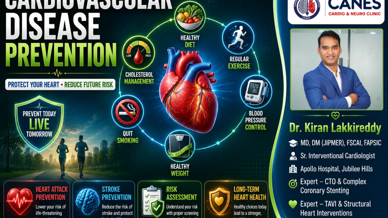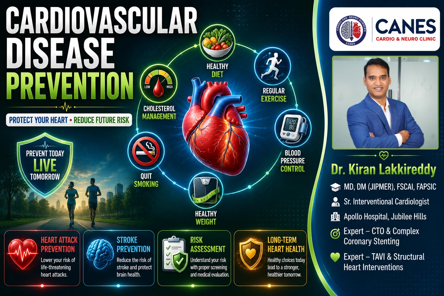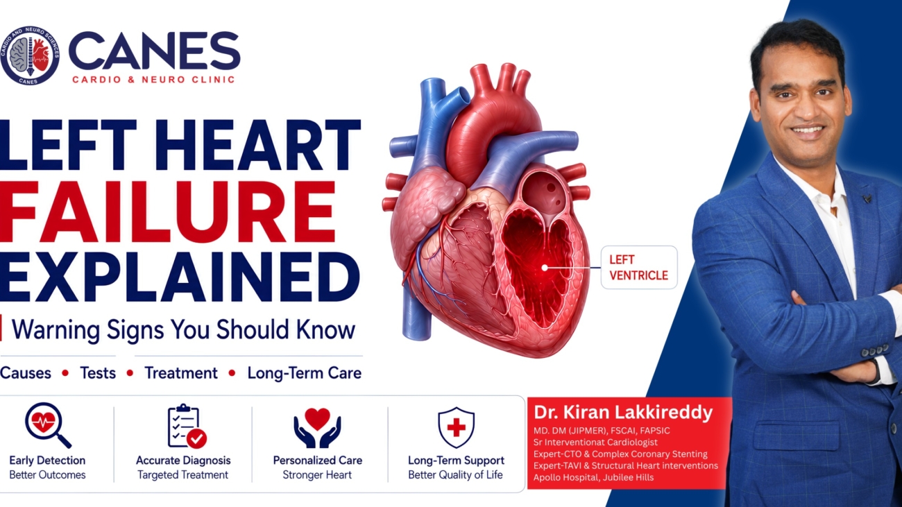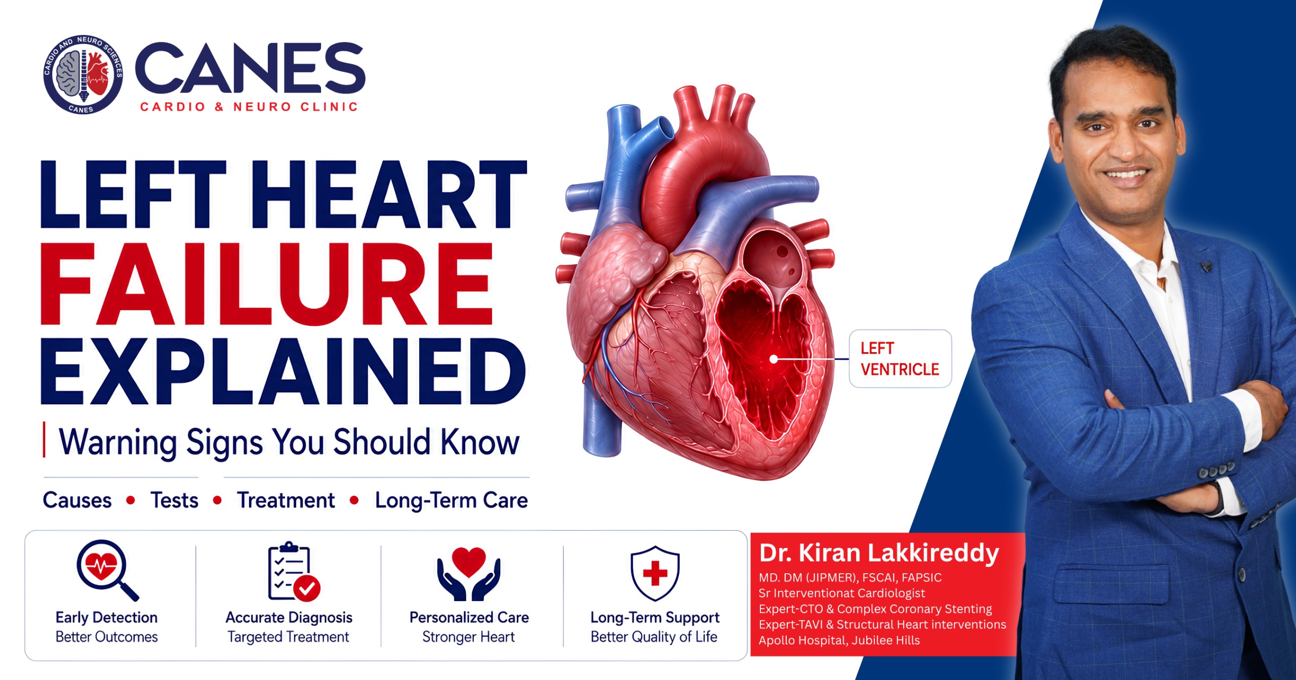
High Blood Pressure and Heart Health: Causes, Risks & Control

High Blood Pressure and Heart Health: Causes, Risks & Control

●   Lifestyle & Prevention

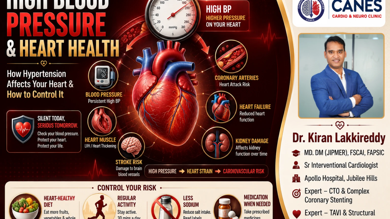
High Blood Pressure and Heart Health: How Hypertension Affects Your Heart and How to Control It

Table of Contents

Book a Consultation

Prevention is better than cure. Talk to our specialists today.
High Blood Pressure and Heart Health: Causes, Risks & Control

Regular physical activity is one of the most important lifestyle measures for maintaining your long-term well-being. The heart is a muscle, and just like other muscles in the body, it adapts and strengthens in response to regular physical activity. Over time, appropriate exercise can improve cardiovascular fitness and help your heart and circulation work much more efficiently.

The connection between exercise and cardiovascular health extends far beyond the heart itself. Regular physical activity can help improve blood pressure, lower cholesterol, boost insulin sensitivity, manage body weight, and heavily support emotional well-being. It is definitively associated with a lower risk of heart disease, stroke, and type 2 diabetes. However, exercise is not identical for every person. Someone who is generally healthy may require a very different program from someone recovering from a heart attack or living with heart failure.

At CANES Cardio & Neuro Clinic in Hyderabad, guided by Dr. Kiran Lakkireddy, an experienced interventional cardiologist, our approach emphasizes careful cardiovascular assessment and patient education. This comprehensive guide will explain exactly how physical activity protects your heart and how to begin exercising safely.

Quick Answer

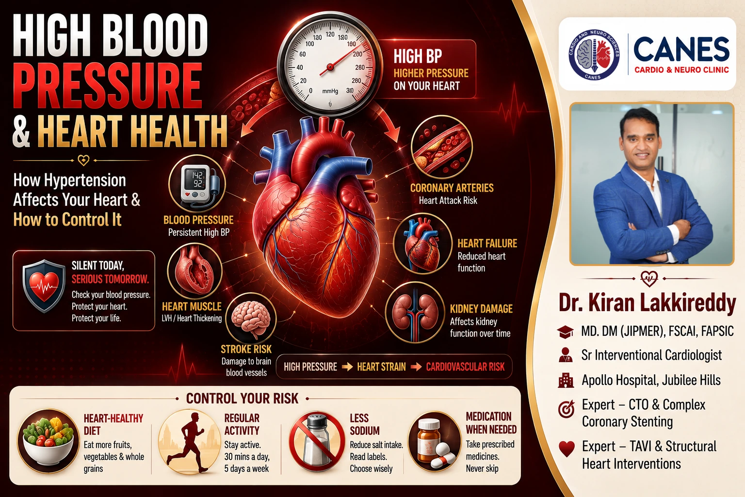
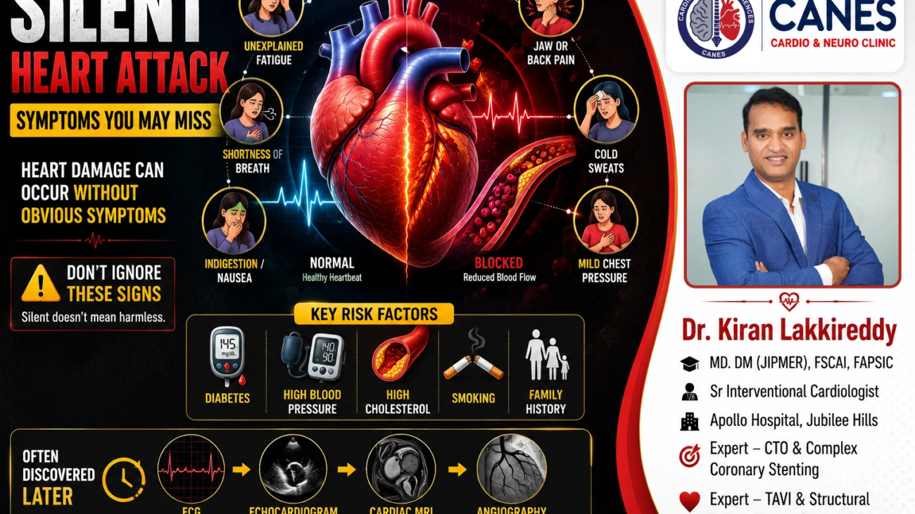
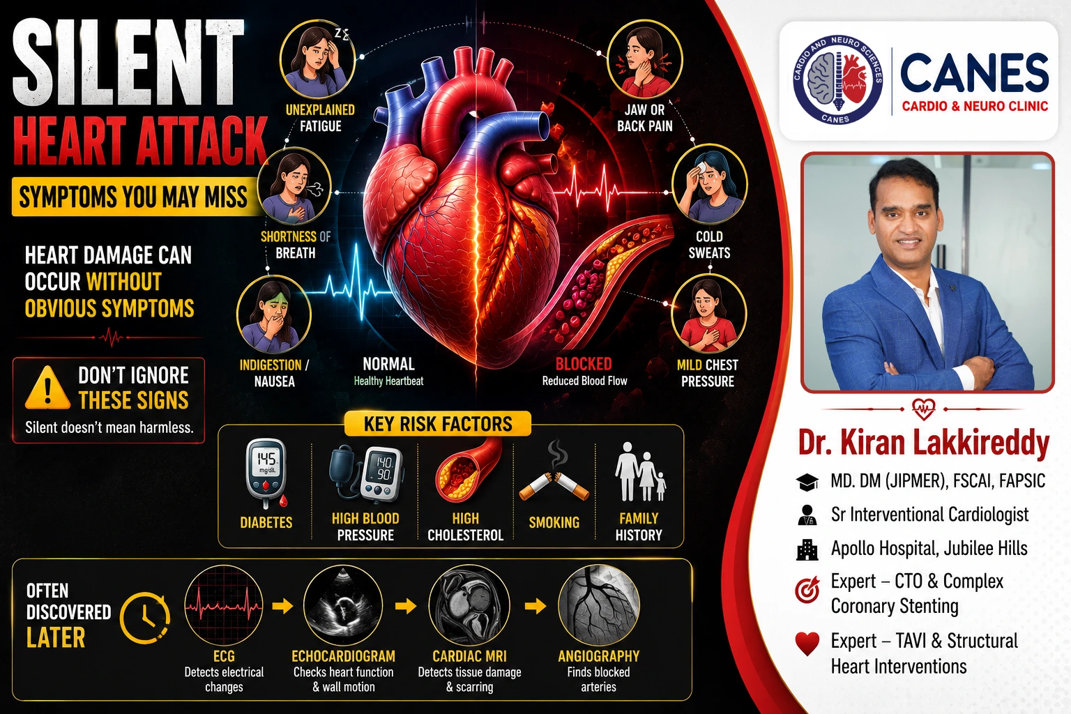
High blood pressure means that the pressure of blood against the artery walls remains higher than it should over time. Persistent hypertension makes the heart work harder and can contribute to changes in the heart muscle, artery disease, coronary artery disease, heart attack, heart failure, stroke, kidney disease and certain forms of aortic disease.

Most people with hypertension do not experience clear symptoms. That is why regular blood-pressure measurement is more reliable than waiting for symptoms. Treatment depends on the individual’s blood-pressure pattern, cardiovascular risk, medical history, kidney function, diabetes status, age, pregnancy status where relevant, and response to lifestyle measures.

How Does High Blood Pressure Affect the Heart?

Think of the heart as a pump and the arteries as the network of pipes through which blood travels. When pressure within that network stays elevated, the heart must generate more force to move blood forward. A single temporarily high reading does not mean that the heart has been damaged, but persistent hypertension over months and years can gradually change both the heart and blood vessels.

One of the most important changes is left ventricular hypertrophy (LVH), in which the muscular wall of the heart’s main pumping chamber becomes thicker. At first, this may be an adaptation to the increased workload. Over time, however, a thickened heart muscle can become stiffer and less efficient at relaxing. This can contribute to problems with how the heart fills and, in some people, eventually contribute to heart failure.

High blood pressure also interacts with other cardiovascular risk factors. A person with hypertension who also has high cholesterol, diabetes, smoking exposure, obesity, kidney disease or a strong family history may have a substantially different overall cardiovascular risk from someone with hypertension alone. That is why effective blood-pressure management should be viewed as one part of a broader strategy for protecting the heart.

High Blood Pressure and Heart Health at a Glance

Effect of Hypertension What Can Happen Why It Matters
Increased workload The heart pumps against greater resistance May contribute to heart-muscle thickening
Heart-muscle changes The left ventricle may become thicker or stiffer Can affect relaxation and filling
Artery damage Blood vessels may become less healthy over time Contributes to cardiovascular disease
Coronary artery disease Atherosclerotic plaque may develop in coronary arteries Can cause angina or heart attack
Heart failure The heart may eventually struggle to pump or fill effectively Can cause breathlessness, fatigue and fluid retention
Stroke Brain blood vessels may become blocked or rupture Can cause permanent disability or death
Kidney damage Small kidney blood vessels may be injured Can contribute to chronic kidney disease
Aortic disease Increased pressure can add stress to the aortic wall May contribute to certain aneurysms or dissections

What Is Blood Pressure?

Blood pressure describes the force exerted by circulating blood against the walls of the arteries. It is recorded using two numbers, expressed in millimetres of mercury (mm Hg).

What Is Systolic Blood Pressure?

The systolic blood pressure is the upper number. It represents the pressure in the arteries when the heart contracts and pumps blood into the circulation.

What Is Diastolic Blood Pressure?

The diastolic blood pressure is the lower number. It represents the pressure in the arteries while the heart relaxes between beats.

For example, a reading of 120/80 mm Hg has a systolic pressure of 120 and a diastolic pressure of 80.

Blood pressure naturally changes throughout the day. Physical activity, stress, pain, caffeine, smoking, sleep and even measurement technique can influence an individual reading. Therefore, one isolated high reading does not necessarily establish a diagnosis of hypertension.

Repeated measurements, home monitoring or ambulatory blood-pressure monitoring may be recommended depending on the circumstances.

Why Is High Blood Pressure Called a "Silent" Condition?

One of the most challenging things about hypertension is that many people feel completely normal while their blood pressure is elevated.

You may exercise regularly, go to work, sleep normally and have no chest pain or headache and still have high blood pressure. This is why symptoms are not a dependable way to determine whether your blood pressure is healthy.

Headaches and dizziness can occur for many reasons and should not be used as a personal blood-pressure test. The practical way to detect hypertension is to measure blood pressure correctly and consistently.

Routine blood-pressure checks are particularly important if you have risk factors such as increasing age, family history, excess body weight, diabetes, kidney disease, physical inactivity, smoking or obstructive sleep apnoea.

How Does High Blood Pressure Damage the Heart?

1. Hypertension Makes the Heart Work Harder

The heart must generate enough pressure to push blood throughout the body’s circulation. When arterial pressure remains elevated, the heart faces greater resistance every time it pumps.

Over time, the left ventricle may respond by becoming thicker. This is called left ventricular hypertrophy, or LVH.

A thicker heart muscle is not necessarily a healthier or stronger heart. As the muscle becomes thicker, it may become less flexible and less efficient at relaxing between contractions. Some people have no symptoms initially, while others may eventually develop reduced exercise tolerance, breathlessness, fatigue, chest discomfort or palpitations.

Controlling blood pressure is therefore important not simply because of the number displayed on a monitor, but because sustained pressure affects the structure and function of the cardiovascular system.

2. Can High Blood Pressure Cause Coronary Artery Disease?

Yes, hypertension is an important risk factor for coronary artery disease.

The coronary arteries supply oxygen-rich blood to the heart muscle. Persistent high blood pressure can contribute to vascular injury and commonly occurs alongside other risk factors such as high cholesterol, diabetes, smoking and obesity.

Over time, atherosclerotic plaque can develop within coronary arteries. If a coronary artery becomes significantly narrowed, a person may experience chest discomfort or angina, particularly during physical activity. If a plaque ruptures and a blood clot suddenly blocks the artery, a heart attack can occur.

Does controlling blood pressure guarantee that a heart attack will never happen? No. Heart attacks have multiple contributing factors, including cholesterol, diabetes, smoking, age, family history, obesity, physical inactivity and established coronary artery disease.

Blood-pressure control is therefore one important part of comprehensive cardiovascular prevention.

3. Can High Blood Pressure Cause Heart Failure?

Long-standing hypertension is an important risk factor for heart failure.

Heart failure does not mean that the heart has stopped. It means that the heart cannot pump or fill efficiently enough to meet the body’s needs.

Hypertension can contribute to heart failure through several pathways. The heart may initially become thicker in response to increased workload. Over time, that muscle can become stiff, making it more difficult for the ventricle to fill normally. In other people, prolonged cardiovascular stress or associated coronary artery disease can eventually weaken the heart’s pumping function.

Possible symptoms of heart failure include:

  1. Shortness of breath during activity
  2. Breathlessness when lying down
  3. Unusual tiredness
  4. Reduced exercise capacity
  5. Swelling of the feet, ankles or legs
  6. Rapid changes in body weight caused by fluid retention
  7. Persistent cough in some people
  8. Difficulty performing normal daily activities

These symptoms can have many causes. New or worsening breathlessness, swelling or reduced exercise tolerance should be medically evaluated rather than self-diagnosed.

4. How Does Hypertension Increase Stroke Risk?

The cardiovascular system includes the blood vessels supplying the brain. Persistent hypertension can damage these vessels and is one of the most important modifiable risk factors for stroke.

A stroke may occur when:

  1. An artery supplying the brain becomes blocked, or
  2. A blood vessel in the brain ruptures and bleeds.

Possible warning signs include sudden:

  1. Facial drooping
  2. Weakness or numbness, particularly on one side
  3. Difficulty speaking or understanding speech
  4. Vision changes
  5. Loss of balance or coordination
  6. Severe or sudden neurological symptoms

Suspected stroke is an emergency. Do not wait for symptoms to disappear or attempt to treat them at home.

5. Can High Blood Pressure Affect the Aorta?

The aorta is the body’s largest artery and carries blood from the heart to the rest of the circulation.

Persistent hypertension increases mechanical stress on arterial walls and is an important risk factor for certain forms of aortic disease. Other factors can also influence risk, including age, smoking, atherosclerosis, family history, genetics and certain connective-tissue disorders.

What Is an Aortic Aneurysm?

An aortic aneurysm is an abnormal enlargement or bulging of part of the aorta. Many aneurysms produce no symptoms and may be discovered during imaging performed for another reason.

What Is an Aortic Dissection?

An aortic dissection occurs when a tear develops in the inner layer of the aortic wall, allowing blood to enter between the layers of the vessel.

This is a life-threatening emergency.

Sudden severe chest or upper-back pain, fainting, marked breathlessness or other sudden cardiovascular symptoms require immediate emergency assessment.

Importantly, having hypertension does not mean that someone will automatically develop an aneurysm or dissection.

6. Can High Blood Pressure Damage the Kidneys?

The relationship between hypertension and kidney disease works in both directions.

The kidneys help regulate fluid balance and blood pressure. At the same time, persistently elevated blood pressure can damage the small blood vessels inside the kidneys and gradually reduce their filtering ability.

This means hypertension can contribute to chronic kidney disease, while kidney disease can make blood-pressure control more difficult.

Depending on your medical history, your healthcare professional may therefore recommend kidney-function testing as part of hypertension assessment.

7. Why Should Blood Pressure Be Considered With Other Risk Factors?

Blood pressure is only one part of cardiovascular risk.

Other important factors include:

  1. Cholesterol
  2. Diabetes
  3. Smoking
  4. Body weight
  5. Physical activity
  6. Kidney function
  7. Family history
  8. Age
  9. Existing cardiovascular disease
  10. Diet
  11. Sleep
  12. Obstructive sleep apnoea

A person with moderately elevated blood pressure and multiple additional risk factors may require a different approach from someone with similar readings but otherwise low cardiovascular risk.

The goal is therefore not simply to lower one number. The goal is to reduce overall cardiovascular risk while maintaining safe, sustainable blood-pressure control.

What Causes High Blood Pressure?

For many people, there is no single identifiable cause. This is commonly called primary hypertension.

Factors associated with a greater likelihood of developing hypertension include:

  1. Increasing age
  2. Family history
  3. Excess body weight
  4. Physical inactivity
  5. High dietary sodium intake
  6. Smoking or tobacco exposure
  7. Excessive alcohol consumption
  8. Diabetes
  9. Kidney disease
  10. Obstructive sleep apnoea
  11. Certain medicines
  12. Certain medical conditions

A smaller proportion of people have secondary hypertension, meaning their elevated blood pressure results from an identifiable underlying condition or factor.

Additional investigation may be considered when hypertension begins at an unusual age, is particularly severe, is difficult to control, or has other features that raise clinical suspicion.

What Are the Symptoms of High Blood Pressure?

Most people with hypertension have no obvious symptoms.

Very severe blood-pressure elevations can sometimes occur with symptoms such as:

  1. Severe headache
  2. Chest pain
  3. Shortness of breath
  4. Confusion
  5. Vision changes
  6. Neurological symptoms
  7. Nausea or vomiting
  8. Dizziness

Symptoms alone cannot tell you whether your blood pressure is high.

If symptoms are severe, sudden or associated with a severely elevated reading, seek appropriate medical care rather than trying to manage the situation yourself.

When Is Very High Blood Pressure an Emergency?

A reading above 180/120 mm Hg is severely elevated.

If you unexpectedly obtain a reading this high, sit quietly and repeat the measurement after at least one minute using correct technique.

If the repeat reading remains above 180/120 mm Hg and you have symptoms such as chest pain, shortness of breath, back pain, weakness, numbness, vision changes or difficulty speaking, seek emergency medical care immediately.

If the reading remains severely elevated without those symptoms, contact a healthcare professional promptly for advice. Do not independently increase, stop or change your medication based on a single reading.

Pregnancy-related hypertension requires separate medical guidance because thresholds and emergency considerations differ.

How Is High Blood Pressure Diagnosed?

Blood pressure changes naturally throughout the day.

An elevated reading can be influenced by:

  1. Recent physical activity
  2. Anxiety
  3. Pain
  4. Caffeine
  5. Smoking
  6. Certain medicines
  7. Incorrect cuff size
  8. Incorrect measurement technique
  9. Other temporary factors

For this reason, healthcare professionals may use repeated clinic measurements, home blood-pressure monitoring or ambulatory blood-pressure monitoring to establish a clearer picture.

Depending on your circumstances, assessment may also include:

  1. Blood tests
  2. Urine tests
  3. Kidney-function testing
  4. Cholesterol testing
  5. Blood-sugar testing
  6. ECG
  7. Echocardiography or other cardiac testing when appropriate
  8. Evaluation for secondary causes when clinically indicated

The exact assessment should be individualized.

Why Is Home Blood-Pressure Monitoring Useful?

Home monitoring can provide information that a single clinic reading cannot.

A consistent record can help your healthcare professional understand your usual blood-pressure pattern and determine whether treatment or lifestyle changes are working.

For more reliable readings:

  1. Use a validated blood-pressure monitor.
  2. Make sure the cuff is the correct size.
  3. Sit quietly before measuring.
  4. Keep your back supported.
  5. Keep both feet flat on the floor.
  6. Support your arm approximately at heart level.
  7. Avoid talking during the measurement.
  8. Follow the device and healthcare professional’s instructions.
  9. Record readings consistently.

Do not measure excessively or change medication based on individual readings unless your healthcare professional has given you specific instructions.

How Can High Blood Pressure Be Controlled?

Successful management usually combines lifestyle measures, treatment of other cardiovascular risk factors and medication when clinically indicated.

1. Follow a Heart-Healthy Eating Pattern

The DASH diet, which stands for Dietary Approaches to Stop Hypertension, is one well-studied eating pattern for blood-pressure management.

It emphasizes:

  1. Vegetables
  2. Fruits
  3. Whole grains
  4. Beans and legumes
  5. Nuts
  6. Fish and poultry
  7. Appropriate low-fat or fat-free dairy
  8. Foods lower in saturated fat
  9. Reduced sodium
  10. Limited added sugars

The goal is not to follow a restrictive “hypertension diet” for a few weeks and then return to old habits. The most useful eating pattern is one that can be maintained long term.

People with kidney disease, diabetes or other medical conditions may need individualized dietary advice.


2. Reduce Excess Sodium

Sodium can contribute to elevated blood pressure, particularly in people who are salt-sensitive.

Large amounts of sodium may come from:

  1. Packaged foods
  2. Processed meats
  3. Pickles
  4. Sauces
  5. Restaurant meals
  6. Fast foods
  7. Salty snacks
  8. Ready-to-eat foods

WHO recommends less than 2,000 mg of sodium per day, equivalent to less than 5 g of salt per day, for adults. Individual dietary needs may differ in certain medical conditions.

Reducing sodium works best as part of an overall heart-healthy eating pattern rather than simply removing table salt.


3. Stay Physically Active

Regular physical activity can support blood-pressure control, cardiovascular fitness, weight management and overall heart health.

Depending on your health and physical abilities, activities may include:

  1. Brisk walking
  2. Cycling
  3. Swimming
  4. Aerobic exercise
  5. Appropriate resistance training

People with known cardiovascular disease, severe uncontrolled hypertension, significant symptoms or other medical conditions should seek professional guidance before beginning strenuous exercise.


4. Work Toward a Healthy Body Weight

Excess body weight can contribute to hypertension and other cardiovascular risk factors.

For people who are overweight, gradual and sustainable weight reduction may improve blood pressure and metabolic health.

Extreme diets and rapid weight-loss programs are usually unnecessary. A more sustainable approach combines nutritious food, regular physical activity, adequate sleep and appropriate medical support.


5. Stop Smoking and Avoid Tobacco

Smoking damages blood vessels and substantially increases cardiovascular risk.

If you have hypertension, quitting smoking becomes even more important, particularly if you also have high cholesterol, diabetes or established cardiovascular disease.

Blood-pressure treatment does not replace smoking cessation.

Both matter.


6. Limit Alcohol

Excessive alcohol intake can contribute to high blood pressure and may interact with certain medicines or medical conditions.

If you drink alcohol, discuss an appropriate limit with your healthcare professional. People should not begin drinking alcohol for cardiovascular benefits.


7. Manage Stress and Sleep

Stress does not explain every case of hypertension, but chronic stress can make healthy routines more difficult and may contribute to behaviors associated with higher cardiovascular risk.

Helpful approaches may include:

  1. Regular physical activity
  2. Relaxation techniques
  3. Adequate sleep
  4. Breathing exercises
  5. Better time management
  6. Counselling when appropriate
  7. Addressing ongoing sources of psychological stress

Sleep also deserves attention. Loud snoring, witnessed pauses in breathing and significant daytime sleepiness can be signs of obstructive sleep apnoea, which is associated with hypertension.

Are Medicines Needed for High Blood Pressure?

Not everyone with elevated blood pressure needs the same treatment.

For some people, lifestyle measures may be an important initial component. For others, medication is appropriate because of:

  1. Persistently elevated blood pressure
  2. Existing cardiovascular disease
  3. Diabetes
  4. Chronic kidney disease
  5. High overall cardiovascular risk
  6. Previous heart attack or stroke
  7. Inadequate control despite lifestyle measures
  8. Other individual clinical factors

Common medication groups include:

  1. ACE inhibitors
  2. Angiotensin receptor blockers (ARBs)
  3. Calcium-channel blockers
  4. Thiazide-type diuretics
  5. Other medicines selected according to the individual’s condition

The choice of medication should be individualized.

Should You Stop Blood-Pressure Medicine When Your Reading Becomes Normal?

No. Do not stop, reduce or change blood-pressure medication simply because your readings have improved.

A normal reading may mean that your treatment is working.

Stopping medication without medical guidance can allow blood pressure to rise again and may increase cardiovascular risk.

If you believe your medication dose should change because of side effects, low readings or other concerns, discuss it with your treating healthcare professional.

What Blood-Pressure Target Should You Aim For?

There is no single target that every patient should copy from another person’s treatment plan.

Current guidelines differ in some aspects. The 2025 American Heart Association/American College of Cardiology guideline recommends an overall treatment goal of below 130/80 mm Hg for adults, while the 2024 European Society of Cardiology guideline recommends a systolic target of 120–129 mm Hg for most adults receiving blood-pressure-lowering treatment when tolerated.

The appropriate goal may depend on:

  1. Age
  2. Cardiovascular disease
  3. Diabetes
  4. Kidney disease
  5. Pregnancy
  6. Frailty
  7. Medication tolerance
  8. Other medical conditions

Can High Blood Pressure Be Lowered or Controlled?

Yes.

Lifestyle changes can substantially improve blood pressure in many people. Treating an identifiable secondary cause may also improve blood pressure in selected patients.

However, many people with primary hypertension require long-term management.

The goal is not necessarily to become “cured” or stop every medication. The more important objective is to achieve safe, sustainable blood-pressure control and reduce long-term cardiovascular risk.

Can You Have High Blood Pressure Even If You Feel Healthy?

Yes.

This is one of the most important facts about hypertension.

A person may:

  1. Exercise regularly
  2. Have no chest pain
  3. Have no headache
  4. Feel energetic
  5. Perform normal daily activities

and still have high blood pressure.

Symptoms are therefore not an adequate screening method.

Measurement is essential.

Does High Blood Pressure Always Cause Heart Disease?

No.

Having hypertension does not mean that a person will definitely develop heart disease, heart failure or stroke.

It means that cardiovascular risk is increased, particularly when blood pressure remains uncontrolled or occurs together with other risk factors.

Risk can be influenced by:

  1. How high the blood pressure is
  2. How long it has been elevated
  3. Whether it is adequately treated
  4. Smoking
  5. Cholesterol
  6. Diabetes
  7. Kidney function
  8. Weight
  9. Physical activity
  10. Family history
  11. Age
  12. Existing cardiovascular disease

Early detection and consistent management can therefore make an important difference.

How Do Some Guidelines Classify Blood Pressure?

Different organizations use different terminology, so classification should always be interpreted in context.

For example, the 2025 AHA/ACC framework classifies adult blood pressure as follows:

CategorySystolicDiastolic
Normal<120 mm Hg<80 mm Hg
Elevated120–129 mm Hg<80 mm Hg
Stage 1 Hypertension130–139 mm Hg80–89 mm Hg
Stage 2 Hypertension≥140 mm Hg≥90 mm Hg

This table is one guideline framework, not a universal classification used identically in every country or medical system.

A diagnosis should not be made from a single reading alone.

Myth vs Fact: High Blood Pressure and Heart Health

Common MythMedical Fact
I feel healthy, so my blood pressure must be normal.Hypertension often causes no noticeable symptoms. Measurement is needed to detect it.
High blood pressure only affects older people.Risk increases with age, but hypertension can also occur in younger adults.
If my blood pressure becomes normal, I can stop my tablets.Normal readings may indicate that treatment is working. Medication should not be changed without medical guidance.
Hypertension only causes headaches.Most people have no symptoms, while uncontrolled hypertension can affect the heart, brain, kidneys and blood vessels.
Only the upper number matters.Both systolic and diastolic readings provide important information.
Medicines are enough, so lifestyle does not matter.Medication and lifestyle measures often work together to reduce cardiovascular risk.
Lifestyle changes mean medication is never necessary.Some people require medication despite making appropriate lifestyle changes.
One high reading means I definitely have hypertension.Diagnosis generally requires appropriate assessment and often repeated measurements.
Heart disease is inevitable if I have hypertension.Hypertension increases risk, but appropriate control can reduce that risk.
Once blood pressure is controlled, follow-up is unnecessary.Ongoing monitoring helps confirm that treatment remains appropriate and effective.

What Should You Ask Your Cardiologist About High Blood Pressure?

If you have hypertension or repeatedly elevated readings, consider asking:

  1. Are my blood-pressure readings consistently high?
  2. What blood-pressure target is appropriate for me?
  3. Should I monitor my blood pressure at home?
  4. Which home blood-pressure monitor should I use?
  5. How often should I measure it?
  6. Do I have other cardiovascular risk factors?
  7. Should my cholesterol or blood sugar be checked?
  8. Do I need tests to assess my heart or kidneys?
  9. Could another medical condition be causing my hypertension?
  10. What dietary changes would be most useful for me?
  11. How much physical activity is appropriate?
  12. Should I reduce sodium?
  13. Do I need blood-pressure medication?
  14. What side effects should I watch for?
  15. What should I do if I miss a dose?
  16. Which blood-pressure readings should concern me?
  17. Which symptoms require emergency medical attention?
  18. When should I return for follow-up?

These questions can turn a routine appointment into a more productive conversation about your long-term cardiovascular health.

Hypertension and Cardiovascular Guidance at CANES Clinic

High blood-pressure management should be individualized.

Two people with similar blood-pressure readings may require different approaches depending on their:

  1. Age
  2. Symptoms
  3. Cardiovascular history
  4. Diabetes status
  5. Kidney function
  6. Cholesterol
  7. Medicines
  8. Family history
  9. Overall cardiovascular risk

At CANES Cardio & Neuro Clinic, patients can receive cardiovascular evaluation and understandable guidance regarding hypertension, cardiovascular risk factors and appropriate follow-up.

The clinic is guided by Dr. Kiran Lakkireddy, an experienced interventional cardiologist specializing in complex cardiology. The approach emphasizes careful assessment, patient education, shared decision-making and coordination of cardiovascular care when appropriate.

Location: 3rd Floor, Tapasavi Commercial Complex, Hi-Tension Road, Kondapur, Hyderabad

Contact: +91 9014944654

Routine clinic appointments are not a substitute for emergency care.

If you experience severe or persistent chest pain, sudden marked breathlessness, fainting, sudden weakness or numbness, difficulty speaking, severe neurological symptoms, loss of consciousness or another suspected cardiovascular emergency, seek immediate emergency medical attention.

Key Points to Remember

  1. High blood pressure often has no symptoms: Regular measurement is important for detection.
  2. The heart has to work harder: Persistent hypertension increases the resistance against which the heart pumps.
  3. The heart muscle may thicken: Long-standing hypertension can contribute to left ventricular hypertrophy.
  4. Coronary risk increases: Hypertension is an important cardiovascular risk factor for coronary artery disease and heart attack.
  5. Heart failure can develop: Long-term pressure overload can eventually impair normal heart function.
  6. The brain can also be affected: High blood pressure is a major modifiable risk factor for stroke.
  7. Kidneys and blood pressure are closely connected: Hypertension can contribute to kidney damage, while kidney disease can make blood pressure harder to control.
  8. Aortic health matters: Hypertension is an important risk factor for certain forms of aortic disease.
  9. Lifestyle changes are valuable: Healthy nutrition, physical activity, appropriate weight management, reduced sodium, smoking cessation and healthy sleep habits can support blood-pressure control.
  10. Medication may still be necessary: Lifestyle measures do not replace prescribed medication when medication is clinically indicated.
  11. Do not stop medication because readings improve: Changes should be made only with professional guidance.
  12. Treatment targets are individual: Ask your healthcare professional what blood-pressure range is appropriate for you.
  13. Long-term monitoring matters: Hypertension usually requires ongoing attention rather than one-time treatment.

Frequently Asked Questions (FAQs)

What is high blood pressure?

High blood pressure, or hypertension, occurs when pressure within the arteries remains elevated over time. Persistent hypertension increases the workload on the cardiovascular system and can increase the risk of heart, brain and kidney disease.

Can hypertension damage the heart?

Yes. Long-term uncontrolled hypertension can make the heart work harder and may cause structural changes such as left ventricular hypertrophy. It is also associated with coronary artery disease and heart failure.

Can high blood pressure cause a heart attack?

High blood pressure is an important risk factor for coronary artery disease and heart attack. It does not mean every person with hypertension will experience a heart attack, but controlling blood pressure is an important part of reducing cardiovascular risk.

Can high blood pressure cause heart failure?

Yes. Long-standing hypertension can contribute to changes in the heart muscle and is an important risk factor for heart failure.

Can hypertension be controlled permanently?

Many people can achieve good long-term blood-pressure control with lifestyle changes, medicines or both. However, hypertension commonly requires ongoing monitoring and management rather than a one-time treatment.

Exercise and Cardiovascular Health: How Physical Activity Supports a Healthier Heart Regular physical activity is one of the most important lifestyle measures for maintaining your long-term well-being. The heart is a muscle, and just like other muscles in the body, it adapts and strengthens in response to regular physical activity. Over time, appropriate exercise can improve cardiovascular fitness and help your heart and circulation work much more efficiently. The connection between exercise and cardiovascular health extends far beyond the heart itself. Regular physical activity can help improve blood pressure, lower cholesterol, boost insulin sensitivity, manage body weight, and heavily support emotional well-being. It is definitively associated with a lower risk of heart disease, stroke, and type 2 diabetes. However, exercise is not identical for every person. Someone who is generally healthy may require a very different program from someone recovering from a heart attack or living with heart failure. At CANES Cardio & Neuro Clinic in Hyderabad, guided by Dr. Kiran Lakkireddy, an experienced interventional cardiologist, our approach emphasizes careful cardiovascular assessment and patient education. This comprehensive guide will explain exactly how physical activity protects your heart and how to begin exercising safely. [Insert Image Here, alt="Exercise and cardiovascular health"] Quick Answer Exercise supports cardiovascular health by strengthening the heart muscle, lowering blood pressure, improving cholesterol levels, and regulating blood sugar. For most adults, current guidelines recommend aiming for 150–300 minutes of moderate-intensity aerobic activity each week (like brisk walking), or 75–150 minutes of vigorous-intensity activity, alongside muscle-strengthening exercises on at least two days per week. However, people with known cardiovascular disease, significant symptoms, or certain uncontrolled medical conditions should discuss an appropriate exercise plan with their healthcare professional before starting. Exercise and Cardiovascular Health at a Glance Cardiovascular Benefit How Exercise Helps Why It Matters Cardiovascular Fitness Trains the heart and lungs to support activity more efficiently. Improves exercise capacity and makes everyday physical activities easier to perform. Blood Pressure Regular activity helps lower and control resting blood pressure. High blood pressure is a major risk factor for heart attack, stroke, and other cardiovascular disease. Cholesterol Improves lipid-related risk factors by balancing blood fats. Crucial for preventing atherosclerotic plaque buildup. Metabolic Health Improves insulin sensitivity and glucose utilization. Essential for people with diabetes or pre-diabetes. Vascular Health Improves circulation and overall endothelial (blood vessel) function. Supports healthy blood-vessel function and circulation. Mental Well-Being Reduces anxiety, improves sleep, and boosts mood. Emotional stress directly impacts cardiovascular strain. What Is the Difference Between Physical Activity and Exercise? Physical activity refers to any bodily movement that burns energy (like gardening or walking), whereas exercise is a structured, planned activity performed specifically to improve fitness, strength, or endurance. Key Takeaway: Movement matters. While structured exercise is highly valuable, simply increasing your everyday physical activity and reducing the hours you spend sitting is crucial for a heart-healthy lifestyle. You do not necessarily need a gym membership to protect your heart. Taking the stairs, cycling for transportation, and completing household chores all contribute to your overall activity levels and provide meaningful cardiovascular benefits. How Does Exercise Affect the Heart? During physical activity, the cardiovascular system increases blood flow, making the heart beat faster to deliver more oxygen; with regular aerobic training, the heart adapts and becomes highly efficient at supporting this demand. Key Takeaway: The goal of exercise is to create a healthy cardiovascular training stimulus, not to push yourself to absolute exhaustion. With regular aerobic training, the cardiovascular system adapts, improving the heart's efficiency and your overall exercise capacity. It pumps more blood with every beat, meaning it doesn't have to work as hard during your normal daily activities. 1. Exercise Can Improve Cardiovascular Fitness Aerobic exercise (such as brisk walking, cycling, swimming, or dancing) helps condition the heart and lungs. As fitness improves, you will notice that walking long distances or climbing stairs requires far less effort. Importantly, physical activity improves cardiometabolic risk factors even if substantial weight loss does not occur. 2. Exercise and Blood Pressure High blood pressure places severe stress on the heart and blood vessels. Regular physical activity naturally lowers blood pressure. While it does not replace prescribed blood-pressure medicine, exercise is an essential part of hypertension management. If your blood pressure is severely uncontrolled, seek medical advice before starting vigorous activity. 3. Exercise and Cholesterol Regular physical activity can improve several lipid-related risk factors, although its effects on LDL (bad) cholesterol are generally modest. Exercise works best alongside a heart-healthy diet and cholesterol-lowering medication when prescribed by your doctor. 4. Exercise and Blood-Sugar Control During exercise, your working muscles consume glucose for energy. Regular physical activity improves insulin sensitivity and helps the body regulate blood sugar more effectively. Because diabetes is a major cardiovascular risk factor, controlling blood sugar through physical activity can contribute to better cardiovascular health. 5. Exercise and Body Weight Physical activity is vital for preventing weight gain. However, the relationship is complex. Current cardiovascular guidelines emphasize that physical activity improves blood pressure, insulin resistance, and cholesterol even if substantial weight loss does not occur. 6. Exercise and Blood-Vessel Health The inner lining of your blood vessels is called the endothelium. Regular exercise improves endothelial function, helping your arteries relax, open up, and respond appropriately to the body's changing demands. Does Sitting Too Much Affect Heart Health? Yes. Spending long periods of time sitting or remaining sedentary is independently associated with poorer cardiovascular health and a higher risk of heart disease. Key Takeaway: "Move more, sit less." Breaking up prolonged periods of sitting with regular, brief movement is a highly useful complement to your structured exercise routine. Even if you meet the weekly exercise recommendations, sitting at a desk for 8 to 10 hours a day can still negatively impact your metabolic health. Breaking up prolonged sitting with standing, stretching, or short periods of walking can help reduce sedentary time and add more movement to your day. Can Exercise Prevent a Heart Attack? Regular physical activity significantly reduces the long-term risk of heart disease and stroke by controlling blood pressure, cholesterol, and diabetes, but it cannot absolutely guarantee that a heart attack will never occur. Key Takeaway: Exercise works best as one foundational component of a comprehensive cardiovascular prevention plan that also includes a healthy diet and avoidance of smoking. Heart-attack risk is driven by a combination of genetics, age, kidney disease, and established atherosclerosis. Exercise actively reduces your modifiable risks by improving metabolic health and reducing the danger of physical inactivity. Can Exercise Ever Trigger a Heart Problem? For most people, the cardiovascular benefits of exercise heavily outweigh the risks; however, sudden, vigorous exertion can temporarily increase cardiac risk in people who are poorly conditioned or have underlying heart disease. Key Takeaway: The safest approach is to start low and build gradually. Do not suddenly attempt heavy weightlifting or intense running if you have been sedentary for years. How Much Exercise Is Recommended? Current guidelines recommend that most healthy adults complete 150 to 300 minutes of moderate-intensity aerobic activity per week, or 75 to 150 minutes of vigorous activity, alongside muscle-strengthening exercises on at least two days per week. Key Takeaway: You do not need to complete your exercise in one long session. Shorter periods of activity throughout the day can still contribute to your weekly total, and any movement is better than none. What Does "Moderate-Intensity" Mean? A practical way to measure intensity is the "talk test." During moderate activity (like brisk walking or water aerobics): Your breathing is noticeably faster. Your heart rate rises. You can generally still hold a conversation. Singing comfortably becomes difficult. What Is "Vigorous-Intensity"? During vigorous activity (like running, fast cycling, or lap swimming): Breathing becomes substantially harder. Speaking more than a few words without pausing for breath becomes very difficult. What Types of Exercise Are Good for Cardiovascular Health? A well-rounded heart-health program combines aerobic exercise to build endurance, resistance training to improve muscle strength, and flexibility/balance exercises to support overall mobility. Aerobic Exercise: Walking, cycling, and swimming train the heart and lungs to support sustained activity. Brisk walking is often the simplest and best place to start. Resistance Training: Using free weights or resistance bands improves muscle strength and body composition, which provides beneficial effects on cardiovascular risk factors. Flexibility and Balance: Flexibility and balance activities can be particularly valuable for older adults, especially those with reduced mobility, to support function and help prevent falls. Who Should Seek Medical Advice Before Starting Exercise? People with known cardiovascular disease, significant symptoms, or certain uncontrolled medical conditions should discuss an appropriate exercise plan with their healthcare professional before starting vigorous or substantially increasing physical activity. You should seek medical advice before starting or substantially increasing exercise if you have: Known coronary artery disease or a previous heart attack. Heart failure or significant rhythm problems. Unexplained chest discomfort or severe breathlessness. Dizziness or fainting with activity. Warning Signs During Exercise Exercise should naturally increase your breathing and heart rate, but you should never ignore potentially serious symptoms. Stop exercising immediately and seek emergency care if you experience: Chest pressure, tightness, or burning pain. Unusual or severe shortness of breath. Dizziness, confusion, or feeling faint. Unusual palpitations or an irregular rapid heartbeat. What Is Cardiac Rehabilitation? Cardiac rehabilitation is a structured, medically supervised program combining supervised exercise, heart-health education, risk-factor management, and emotional support for people recovering from cardiovascular conditions. Key Takeaway: Cardiac rehabilitation provides a medically supervised environment where patients can rebuild physical confidence after a heart attack, angioplasty, bypass surgery, or heart failure diagnosis. After a cardiac event, you should not simply return to your previous exercise routine. A rehabilitation program tailors the exercise specifically to your current heart function, surgical healing, and medical limitations. Myth Versus Fact: Exercise and Heart Health Understanding the realities of physical activity helps you exercise safely and consistently. Common Myth Medical Fact Only intense exercise benefits the heart. Moderate activity, such as brisk walking, provides substantial and proven cardiovascular benefits. Exercise is useless without weight loss. Exercise improves blood pressure, cholesterol, and metabolic health even if substantial weight loss does not occur. People with heart disease should avoid exercise. Many patients with heart disease benefit significantly from medically appropriate physical activity, often through cardiac rehabilitation. Exercise replaces my heart medications. Physical activity complements your prescribed treatment; it never automatically replaces necessary medication. If it makes me breathless, something is wrong. Breathing harder is expected during aerobic activity. Breathlessness that is new, unusually severe, disproportionate to the activity, or accompanied by chest discomfort, dizziness, or fainting warrants medical assessment. Actionable Checklist: What Should You Ask Your Cardiologist? Before starting a new routine, ask your healthcare team these essential questions: [ ] Is it safe for me to begin exercising, and do I need a cardiac stress test first? [ ] What specific heart-rate range should I aim for during activity? [ ] Do my current cardiac medicines affect how my heart rate will respond to exercise? [ ] Are there any specific exercises (like heavy lifting) that I should avoid? [ ] Which specific warning symptoms should make me stop exercising immediately? [ ] Am I a candidate for a medically supervised cardiac rehabilitation program? Cardiovascular Exercise Guidance at CANES Clinic The appropriate type and intensity of physical activity depend entirely on the individual. A healthy young adult requires very different guidance from someone managing coronary artery disease, heart failure, or recovering from a heart attack. At CANES Cardio & Neuro Clinic, patients receive personalized cardiovascular evaluation and understandable guidance regarding heart conditions, risk factors, and safe physical activity. Guided by Dr. Kiran Lakkireddy, an experienced interventional cardiologist specializing in complex cardiology, the clinic focuses on careful assessment, shared decision-making, and coordination with cardiac rehabilitation teams when indicated. (Note: A routine clinic appointment is not appropriate for a suspected cardiovascular emergency. Severe chest discomfort, fainting, or marked breathlessness require immediate emergency medical assessment.) Location: 3rd Floor, Tapasavi Commercial Complex, Hi-Tension Road, Kondapur, Hyderabad Contact: +91 9014944654 Appointment Booking: Visit CANES Clinic Official Portal Specialist Profile: Dr. Kiran Lakkireddy Official Website Key Points to Remember Exercise strengthens the heart: Regular physical activity trains the cardiovascular system to work more efficiently. Benefits beyond fitness: It improves blood pressure, cholesterol, insulin sensitivity, and overall cardiometabolic health. Weight loss is not the only metric: Cardiovascular benefits occur even if the number on the scale does not drastically change. Walking is excellent exercise: Brisk walking is a highly effective, practical form of moderate-intensity aerobic activity. Consistency over intensity: Sustainable physical activity over the long term is far more valuable than occasional, extreme exhaustion. Seek guidance when necessary: People with existing cardiovascular disease or concerning symptoms should consult a doctor before starting an exercise program. Never ignore warning signs: Stop exercising immediately if you experience chest pain, severe breathlessness, or dizziness. Frequently Asked Questions (FAQ) How much exercise should an adult do each week? General guidelines encourage 150 to 300 minutes of moderate-intensity aerobic activity (like brisk walking) or 75 to 150 minutes of vigorous activity per week, plus muscle-strengthening exercises on at least two days. Do I have to exercise for 30 minutes continuously? No. Activity can be spread throughout the week. You do not need to complete your exercise in one long session; shorter periods of activity throughout the day can still contribute to your weekly total. Does exercise improve cholesterol? Yes. Regular physical activity can improve several lipid-related risk factors, although its effects on LDL cholesterol are generally modest. Exercise works alongside a heart-healthy diet and cholesterol-lowering medication when prescribed. Is vigorous exercise better than moderate exercise? Not necessarily. Both provide cardiovascular health benefits. The right intensity depends on your current fitness, age, and any underlying medical conditions. Can I exercise after a heart attack or angioplasty? Many people can and should return to appropriate physical activity after a heart procedure, but the timing and intensity should be guided by their healthcare team, often through a structured cardiac rehabilitation program. Do I need a stress test before exercising? Most healthy adults without symptoms do not need routine cardiac stress testing simply to begin moderate physical activity. Testing may be appropriate when symptoms, known cardiovascular disease, or specific clinical concerns are present. What should I do if I get chest pain while exercising? Stop exercising immediately. Chest pressure, squeezing, or pain can be a severe warning sign. If symptoms persist or are accompanied by breathlessness or nausea, seek urgent emergency medical assessment. Is strength training important for heart health? Exercise and Cardiovascular Health: How Physical Activity Supports a Healthier Heart Regular physical activity is one of the most important lifestyle measures for maintaining your long-term well-being. The heart is a muscle, and just like other muscles in the body, it adapts and strengthens in response to regular physical activity. Over time, appropriate exercise can improve cardiovascular fitness and help your heart and circulation work much more efficiently. The connection between exercise and cardiovascular health extends far beyond the heart itself. Regular physical activity can help improve blood pressure, lower cholesterol, boost insulin sensitivity, manage body weight, and heavily support emotional well-being. It is definitively associated with a lower risk of heart disease, stroke, and type 2 diabetes. However, exercise is not identical for every person. Someone who is generally healthy may require a very different program from someone recovering from a heart attack or living with heart failure. At CANES Cardio & Neuro Clinic in Hyderabad, guided by Dr. Kiran Lakkireddy, an experienced interventional cardiologist, our approach emphasizes careful cardiovascular assessment and patient education. This comprehensive guide will explain exactly how physical activity protects your heart and how to begin exercising safely. [Insert Image Here, alt="Exercise and cardiovascular health"] Quick Answer Exercise supports cardiovascular health by strengthening the heart muscle, lowering blood pressure, improving cholesterol levels, and regulating blood sugar. For most adults, current guidelines recommend aiming for 150–300 minutes of moderate-intensity aerobic activity each week (like brisk walking), or 75–150 minutes of vigorous-intensity activity, alongside muscle-strengthening exercises on at least two days per week. However, people with known cardiovascular disease, significant symptoms, or certain uncontrolled medical conditions should discuss an appropriate exercise plan with their healthcare professional before starting. Exercise and Cardiovascular Health at a Glance Cardiovascular Benefit How Exercise Helps Why It Matters Cardiovascular Fitness Trains the heart and lungs to support activity more efficiently. Improves exercise capacity and makes everyday physical activities easier to perform. Blood Pressure Regular activity helps lower and control resting blood pressure. High blood pressure is a major risk factor for heart attack, stroke, and other cardiovascular disease. Cholesterol Improves lipid-related risk factors by balancing blood fats. Crucial for preventing atherosclerotic plaque buildup. Metabolic Health Improves insulin sensitivity and glucose utilization. Essential for people with diabetes or pre-diabetes. Vascular Health Improves circulation and overall endothelial (blood vessel) function. Supports healthy blood-vessel function and circulation. Mental Well-Being Reduces anxiety, improves sleep, and boosts mood. Emotional stress directly impacts cardiovascular strain. What Is the Difference Between Physical Activity and Exercise? Physical activity refers to any bodily movement that burns energy (like gardening or walking), whereas exercise is a structured, planned activity performed specifically to improve fitness, strength, or endurance. Key Takeaway: Movement matters. While structured exercise is highly valuable, simply increasing your everyday physical activity and reducing the hours you spend sitting is crucial for a heart-healthy lifestyle. You do not necessarily need a gym membership to protect your heart. Taking the stairs, cycling for transportation, and completing household chores all contribute to your overall activity levels and provide meaningful cardiovascular benefits. How Does Exercise Affect the Heart? During physical activity, the cardiovascular system increases blood flow, making the heart beat faster to deliver more oxygen; with regular aerobic training, the heart adapts and becomes highly efficient at supporting this demand. Key Takeaway: The goal of exercise is to create a healthy cardiovascular training stimulus, not to push yourself to absolute exhaustion. With regular aerobic training, the cardiovascular system adapts, improving the heart's efficiency and your overall exercise capacity. It pumps more blood with every beat, meaning it doesn't have to work as hard during your normal daily activities. 1. Exercise Can Improve Cardiovascular Fitness Aerobic exercise (such as brisk walking, cycling, swimming, or dancing) helps condition the heart and lungs. As fitness improves, you will notice that walking long distances or climbing stairs requires far less effort. Importantly, physical activity improves cardiometabolic risk factors even if substantial weight loss does not occur. 2. Exercise and Blood Pressure High blood pressure places severe stress on the heart and blood vessels. Regular physical activity naturally lowers blood pressure. While it does not replace prescribed blood-pressure medicine, exercise is an essential part of hypertension management. If your blood pressure is severely uncontrolled, seek medical advice before starting vigorous activity. 3. Exercise and Cholesterol Regular physical activity can improve several lipid-related risk factors, although its effects on LDL (bad) cholesterol are generally modest. Exercise works best alongside a heart-healthy diet and cholesterol-lowering medication when prescribed by your doctor. 4. Exercise and Blood-Sugar Control During exercise, your working muscles consume glucose for energy. Regular physical activity improves insulin sensitivity and helps the body regulate blood sugar more effectively. Because diabetes is a major cardiovascular risk factor, controlling blood sugar through physical activity can contribute to better cardiovascular health. 5. Exercise and Body Weight Physical activity is vital for preventing weight gain. However, the relationship is complex. Current cardiovascular guidelines emphasize that physical activity improves blood pressure, insulin resistance, and cholesterol even if substantial weight loss does not occur. 6. Exercise and Blood-Vessel Health The inner lining of your blood vessels is called the endothelium. Regular exercise improves endothelial function, helping your arteries relax, open up, and respond appropriately to the body's changing demands. Does Sitting Too Much Affect Heart Health? Yes. Spending long periods of time sitting or remaining sedentary is independently associated with poorer cardiovascular health and a higher risk of heart disease. Key Takeaway: "Move more, sit less." Breaking up prolonged periods of sitting with regular, brief movement is a highly useful complement to your structured exercise routine. Even if you meet the weekly exercise recommendations, sitting at a desk for 8 to 10 hours a day can still negatively impact your metabolic health. Breaking up prolonged sitting with standing, stretching, or short periods of walking can help reduce sedentary time and add more movement to your day. Can Exercise Prevent a Heart Attack? Regular physical activity significantly reduces the long-term risk of heart disease and stroke by controlling blood pressure, cholesterol, and diabetes, but it cannot absolutely guarantee that a heart attack will never occur. Key Takeaway: Exercise works best as one foundational component of a comprehensive cardiovascular prevention plan that also includes a healthy diet and avoidance of smoking. Heart-attack risk is driven by a combination of genetics, age, kidney disease, and established atherosclerosis. Exercise actively reduces your modifiable risks by improving metabolic health and reducing the danger of physical inactivity. Can Exercise Ever Trigger a Heart Problem? For most people, the cardiovascular benefits of exercise heavily outweigh the risks; however, sudden, vigorous exertion can temporarily increase cardiac risk in people who are poorly conditioned or have underlying heart disease. Key Takeaway: The safest approach is to start low and build gradually. Do not suddenly attempt heavy weightlifting or intense running if you have been sedentary for years. How Much Exercise Is Recommended? Current guidelines recommend that most healthy adults complete 150 to 300 minutes of moderate-intensity aerobic activity per week, or 75 to 150 minutes of vigorous activity, alongside muscle-strengthening exercises on at least two days per week. Key Takeaway: You do not need to complete your exercise in one long session. Shorter periods of activity throughout the day can still contribute to your weekly total, and any movement is better than none. What Does "Moderate-Intensity" Mean? A practical way to measure intensity is the "talk test." During moderate activity (like brisk walking or water aerobics): Your breathing is noticeably faster. Your heart rate rises. You can generally still hold a conversation. Singing comfortably becomes difficult. What Is "Vigorous-Intensity"? During vigorous activity (like running, fast cycling, or lap swimming): Breathing becomes substantially harder. Speaking more than a few words without pausing for breath becomes very difficult. What Types of Exercise Are Good for Cardiovascular Health? A well-rounded heart-health program combines aerobic exercise to build endurance, resistance training to improve muscle strength, and flexibility/balance exercises to support overall mobility. Aerobic Exercise: Walking, cycling, and swimming train the heart and lungs to support sustained activity. Brisk walking is often the simplest and best place to start. Resistance Training: Using free weights or resistance bands improves muscle strength and body composition, which provides beneficial effects on cardiovascular risk factors. Flexibility and Balance: Flexibility and balance activities can be particularly valuable for older adults, especially those with reduced mobility, to support function and help prevent falls. Who Should Seek Medical Advice Before Starting Exercise? People with known cardiovascular disease, significant symptoms, or certain uncontrolled medical conditions should discuss an appropriate exercise plan with their healthcare professional before starting vigorous or substantially increasing physical activity. You should seek medical advice before starting or substantially increasing exercise if you have: Known coronary artery disease or a previous heart attack. Heart failure or significant rhythm problems. Unexplained chest discomfort or severe breathlessness. Dizziness or fainting with activity. Warning Signs During Exercise Exercise should naturally increase your breathing and heart rate, but you should never ignore potentially serious symptoms. Stop exercising immediately and seek emergency care if you experience: Chest pressure, tightness, or burning pain. Unusual or severe shortness of breath. Dizziness, confusion, or feeling faint. Unusual palpitations or an irregular rapid heartbeat. What Is Cardiac Rehabilitation? Cardiac rehabilitation is a structured, medically supervised program combining supervised exercise, heart-health education, risk-factor management, and emotional support for people recovering from cardiovascular conditions. Key Takeaway: Cardiac rehabilitation provides a medically supervised environment where patients can rebuild physical confidence after a heart attack, angioplasty, bypass surgery, or heart failure diagnosis. After a cardiac event, you should not simply return to your previous exercise routine. A rehabilitation program tailors the exercise specifically to your current heart function, surgical healing, and medical limitations. Myth Versus Fact: Exercise and Heart Health Understanding the realities of physical activity helps you exercise safely and consistently. Common Myth Medical Fact Only intense exercise benefits the heart. Moderate activity, such as brisk walking, provides substantial and proven cardiovascular benefits. Exercise is useless without weight loss. Exercise improves blood pressure, cholesterol, and metabolic health even if substantial weight loss does not occur. People with heart disease should avoid exercise. Many patients with heart disease benefit significantly from medically appropriate physical activity, often through cardiac rehabilitation. Exercise replaces my heart medications. Physical activity complements your prescribed treatment; it never automatically replaces necessary medication. If it makes me breathless, something is wrong. Breathing harder is expected during aerobic activity. Breathlessness that is new, unusually severe, disproportionate to the activity, or accompanied by chest discomfort, dizziness, or fainting warrants medical assessment. Actionable Checklist: What Should You Ask Your Cardiologist? Before starting a new routine, ask your healthcare team these essential questions: [ ] Is it safe for me to begin exercising, and do I need a cardiac stress test first? [ ] What specific heart-rate range should I aim for during activity? [ ] Do my current cardiac medicines affect how my heart rate will respond to exercise? [ ] Are there any specific exercises (like heavy lifting) that I should avoid? [ ] Which specific warning symptoms should make me stop exercising immediately? [ ] Am I a candidate for a medically supervised cardiac rehabilitation program? Cardiovascular Exercise Guidance at CANES Clinic The appropriate type and intensity of physical activity depend entirely on the individual. A healthy young adult requires very different guidance from someone managing coronary artery disease, heart failure, or recovering from a heart attack. At CANES Cardio & Neuro Clinic, patients receive personalized cardiovascular evaluation and understandable guidance regarding heart conditions, risk factors, and safe physical activity. Guided by Dr. Kiran Lakkireddy, an experienced interventional cardiologist specializing in complex cardiology, the clinic focuses on careful assessment, shared decision-making, and coordination with cardiac rehabilitation teams when indicated. (Note: A routine clinic appointment is not appropriate for a suspected cardiovascular emergency. Severe chest discomfort, fainting, or marked breathlessness require immediate emergency medical assessment.) Location: 3rd Floor, Tapasavi Commercial Complex, Hi-Tension Road, Kondapur, Hyderabad Contact: +91 9014944654 Appointment Booking: Visit CANES Clinic Official Portal Specialist Profile: Dr. Kiran Lakkireddy Official Website Key Points to Remember Exercise strengthens the heart: Regular physical activity trains the cardiovascular system to work more efficiently. Benefits beyond fitness: It improves blood pressure, cholesterol, insulin sensitivity, and overall cardiometabolic health. Weight loss is not the only metric: Cardiovascular benefits occur even if the number on the scale does not drastically change. Walking is excellent exercise: Brisk walking is a highly effective, practical form of moderate-intensity aerobic activity. Consistency over intensity: Sustainable physical activity over the long term is far more valuable than occasional, extreme exhaustion. Seek guidance when necessary: People with existing cardiovascular disease or concerning symptoms should consult a doctor before starting an exercise program. Never ignore warning signs: Stop exercising immediately if you experience chest pain, severe breathlessness, or dizziness. Frequently Asked Questions (FAQ) How much exercise should an adult do each week? General guidelines encourage 150 to 300 minutes of moderate-intensity aerobic activity (like brisk walking) or 75 to 150 minutes of vigorous activity per week, plus muscle-strengthening exercises on at least two days. Do I have to exercise for 30 minutes continuously? No. Activity can be spread throughout the week. You do not need to complete your exercise in one long session; shorter periods of activity throughout the day can still contribute to your weekly total. Does exercise improve cholesterol? Yes. Regular physical activity can improve several lipid-related risk factors, although its effects on LDL cholesterol are generally modest. Exercise works alongside a heart-healthy diet and cholesterol-lowering medication when prescribed. Is vigorous exercise better than moderate exercise? Not necessarily. Both provide cardiovascular health benefits. The right intensity depends on your current fitness, age, and any underlying medical conditions. Can I exercise after a heart attack or angioplasty? Many people can and should return to appropriate physical activity after a heart procedure, but the timing and intensity should be guided by their healthcare team, often through a structured cardiac rehabilitation program. Do I need a stress test before exercising? Most healthy adults without symptoms do not need routine cardiac stress testing simply to begin moderate physical activity. Testing may be appropriate when symptoms, known cardiovascular disease, or specific clinical concerns are present. What should I do if I get chest pain while exercising? Stop exercising immediately. Chest pressure, squeezing, or pain can be a severe warning sign. If symptoms persist or are accompanied by breathlessness or nausea, seek urgent emergency medical assessment. Is strength training important for heart health? Yes. Appropriate resistance training complements aerobic exercise by improving muscle strength, physical function, and several cardiometabolic risk factors.

Exercise and Cardiovascular Health: Protect Your Heart

●   Lifestyle & Prevention

Exercise and Cardiovascular Health: How Physical Activity Supports a Healthier Heart

Table of Contents

Book a Consultation

Prevention is better than cure. Talk to our specialists today.

Exercise and Cardiovascular Health: How Physical Activity Supports a Healthier Heart Regular physical activity is one of the most important lifestyle measures for maintaining your long-term well-being. The heart is a muscle, and just like other muscles in the body, it adapts and strengthens in response to regular physical activity. Over time, appropriate exercise can improve cardiovascular fitness and help your heart and circulation work much more efficiently. The connection between exercise and cardiovascular health extends far beyond the heart itself. Regular physical activity can help improve blood pressure, lower cholesterol, boost insulin sensitivity, manage body weight, and heavily support emotional well-being. It is definitively associated with a lower risk of heart disease, stroke, and type 2 diabetes. However, exercise is not identical for every person. Someone who is generally healthy may require a very different program from someone recovering from a heart attack or living with heart failure. At CANES Cardio & Neuro Clinic in Hyderabad, guided by Dr. Kiran Lakkireddy, an experienced interventional cardiologist, our approach emphasizes careful cardiovascular assessment and patient education. This comprehensive guide will explain exactly how physical activity protects your heart and how to begin exercising safely. [Insert Image Here, alt="Exercise and cardiovascular health"] Quick Answer Exercise supports cardiovascular health by strengthening the heart muscle, lowering blood pressure, improving cholesterol levels, and regulating blood sugar. For most adults, current guidelines recommend aiming for 150–300 minutes of moderate-intensity aerobic activity each week (like brisk walking), or 75–150 minutes of vigorous-intensity activity, alongside muscle-strengthening exercises on at least two days per week. However, people with known cardiovascular disease, significant symptoms, or certain uncontrolled medical conditions should discuss an appropriate exercise plan with their healthcare professional before starting. Exercise and Cardiovascular Health at a Glance Cardiovascular Benefit How Exercise Helps Why It Matters Cardiovascular Fitness Trains the heart and lungs to support activity more efficiently. Improves exercise capacity and makes everyday physical activities easier to perform. Blood Pressure Regular activity helps lower and control resting blood pressure. High blood pressure is a major risk factor for heart attack, stroke, and other cardiovascular disease. Cholesterol Improves lipid-related risk factors by balancing blood fats. Crucial for preventing atherosclerotic plaque buildup. Metabolic Health Improves insulin sensitivity and glucose utilization. Essential for people with diabetes or pre-diabetes. Vascular Health Improves circulation and overall endothelial (blood vessel) function. Supports healthy blood-vessel function and circulation. Mental Well-Being Reduces anxiety, improves sleep, and boosts mood. Emotional stress directly impacts cardiovascular strain. What Is the Difference Between Physical Activity and Exercise? Physical activity refers to any bodily movement that burns energy (like gardening or walking), whereas exercise is a structured, planned activity performed specifically to improve fitness, strength, or endurance. Key Takeaway: Movement matters. While structured exercise is highly valuable, simply increasing your everyday physical activity and reducing the hours you spend sitting is crucial for a heart-healthy lifestyle. You do not necessarily need a gym membership to protect your heart. Taking the stairs, cycling for transportation, and completing household chores all contribute to your overall activity levels and provide meaningful cardiovascular benefits. How Does Exercise Affect the Heart? During physical activity, the cardiovascular system increases blood flow, making the heart beat faster to deliver more oxygen; with regular aerobic training, the heart adapts and becomes highly efficient at supporting this demand. Key Takeaway: The goal of exercise is to create a healthy cardiovascular training stimulus, not to push yourself to absolute exhaustion. With regular aerobic training, the cardiovascular system adapts, improving the heart's efficiency and your overall exercise capacity. It pumps more blood with every beat, meaning it doesn't have to work as hard during your normal daily activities. 1. Exercise Can Improve Cardiovascular Fitness Aerobic exercise (such as brisk walking, cycling, swimming, or dancing) helps condition the heart and lungs. As fitness improves, you will notice that walking long distances or climbing stairs requires far less effort. Importantly, physical activity improves cardiometabolic risk factors even if substantial weight loss does not occur. 2. Exercise and Blood Pressure High blood pressure places severe stress on the heart and blood vessels. Regular physical activity naturally lowers blood pressure. While it does not replace prescribed blood-pressure medicine, exercise is an essential part of hypertension management. If your blood pressure is severely uncontrolled, seek medical advice before starting vigorous activity. 3. Exercise and Cholesterol Regular physical activity can improve several lipid-related risk factors, although its effects on LDL (bad) cholesterol are generally modest. Exercise works best alongside a heart-healthy diet and cholesterol-lowering medication when prescribed by your doctor. 4. Exercise and Blood-Sugar Control During exercise, your working muscles consume glucose for energy. Regular physical activity improves insulin sensitivity and helps the body regulate blood sugar more effectively. Because diabetes is a major cardiovascular risk factor, controlling blood sugar through physical activity can contribute to better cardiovascular health. 5. Exercise and Body Weight Physical activity is vital for preventing weight gain. However, the relationship is complex. Current cardiovascular guidelines emphasize that physical activity improves blood pressure, insulin resistance, and cholesterol even if substantial weight loss does not occur. 6. Exercise and Blood-Vessel Health The inner lining of your blood vessels is called the endothelium. Regular exercise improves endothelial function, helping your arteries relax, open up, and respond appropriately to the body's changing demands. Does Sitting Too Much Affect Heart Health? Yes. Spending long periods of time sitting or remaining sedentary is independently associated with poorer cardiovascular health and a higher risk of heart disease. Key Takeaway: "Move more, sit less." Breaking up prolonged periods of sitting with regular, brief movement is a highly useful complement to your structured exercise routine. Even if you meet the weekly exercise recommendations, sitting at a desk for 8 to 10 hours a day can still negatively impact your metabolic health. Breaking up prolonged sitting with standing, stretching, or short periods of walking can help reduce sedentary time and add more movement to your day. Can Exercise Prevent a Heart Attack? Regular physical activity significantly reduces the long-term risk of heart disease and stroke by controlling blood pressure, cholesterol, and diabetes, but it cannot absolutely guarantee that a heart attack will never occur. Key Takeaway: Exercise works best as one foundational component of a comprehensive cardiovascular prevention plan that also includes a healthy diet and avoidance of smoking. Heart-attack risk is driven by a combination of genetics, age, kidney disease, and established atherosclerosis. Exercise actively reduces your modifiable risks by improving metabolic health and reducing the danger of physical inactivity. Can Exercise Ever Trigger a Heart Problem? For most people, the cardiovascular benefits of exercise heavily outweigh the risks; however, sudden, vigorous exertion can temporarily increase cardiac risk in people who are poorly conditioned or have underlying heart disease. Key Takeaway: The safest approach is to start low and build gradually. Do not suddenly attempt heavy weightlifting or intense running if you have been sedentary for years. How Much Exercise Is Recommended? Current guidelines recommend that most healthy adults complete 150 to 300 minutes of moderate-intensity aerobic activity per week, or 75 to 150 minutes of vigorous activity, alongside muscle-strengthening exercises on at least two days per week. Key Takeaway: You do not need to complete your exercise in one long session. Shorter periods of activity throughout the day can still contribute to your weekly total, and any movement is better than none. What Does "Moderate-Intensity" Mean? A practical way to measure intensity is the "talk test." During moderate activity (like brisk walking or water aerobics): Your breathing is noticeably faster. Your heart rate rises. You can generally still hold a conversation. Singing comfortably becomes difficult. What Is "Vigorous-Intensity"? During vigorous activity (like running, fast cycling, or lap swimming): Breathing becomes substantially harder. Speaking more than a few words without pausing for breath becomes very difficult. What Types of Exercise Are Good for Cardiovascular Health? A well-rounded heart-health program combines aerobic exercise to build endurance, resistance training to improve muscle strength, and flexibility/balance exercises to support overall mobility. Aerobic Exercise: Walking, cycling, and swimming train the heart and lungs to support sustained activity. Brisk walking is often the simplest and best place to start. Resistance Training: Using free weights or resistance bands improves muscle strength and body composition, which provides beneficial effects on cardiovascular risk factors. Flexibility and Balance: Flexibility and balance activities can be particularly valuable for older adults, especially those with reduced mobility, to support function and help prevent falls. Who Should Seek Medical Advice Before Starting Exercise? People with known cardiovascular disease, significant symptoms, or certain uncontrolled medical conditions should discuss an appropriate exercise plan with their healthcare professional before starting vigorous or substantially increasing physical activity. You should seek medical advice before starting or substantially increasing exercise if you have: Known coronary artery disease or a previous heart attack. Heart failure or significant rhythm problems. Unexplained chest discomfort or severe breathlessness. Dizziness or fainting with activity. Warning Signs During Exercise Exercise should naturally increase your breathing and heart rate, but you should never ignore potentially serious symptoms. Stop exercising immediately and seek emergency care if you experience: Chest pressure, tightness, or burning pain. Unusual or severe shortness of breath. Dizziness, confusion, or feeling faint. Unusual palpitations or an irregular rapid heartbeat. What Is Cardiac Rehabilitation? Cardiac rehabilitation is a structured, medically supervised program combining supervised exercise, heart-health education, risk-factor management, and emotional support for people recovering from cardiovascular conditions. Key Takeaway: Cardiac rehabilitation provides a medically supervised environment where patients can rebuild physical confidence after a heart attack, angioplasty, bypass surgery, or heart failure diagnosis. After a cardiac event, you should not simply return to your previous exercise routine. A rehabilitation program tailors the exercise specifically to your current heart function, surgical healing, and medical limitations. Myth Versus Fact: Exercise and Heart Health Understanding the realities of physical activity helps you exercise safely and consistently. Common Myth Medical Fact Only intense exercise benefits the heart. Moderate activity, such as brisk walking, provides substantial and proven cardiovascular benefits. Exercise is useless without weight loss. Exercise improves blood pressure, cholesterol, and metabolic health even if substantial weight loss does not occur. People with heart disease should avoid exercise. Many patients with heart disease benefit significantly from medically appropriate physical activity, often through cardiac rehabilitation. Exercise replaces my heart medications. Physical activity complements your prescribed treatment; it never automatically replaces necessary medication. If it makes me breathless, something is wrong. Breathing harder is expected during aerobic activity. Breathlessness that is new, unusually severe, disproportionate to the activity, or accompanied by chest discomfort, dizziness, or fainting warrants medical assessment. Actionable Checklist: What Should You Ask Your Cardiologist? Before starting a new routine, ask your healthcare team these essential questions: [ ] Is it safe for me to begin exercising, and do I need a cardiac stress test first? [ ] What specific heart-rate range should I aim for during activity? [ ] Do my current cardiac medicines affect how my heart rate will respond to exercise? [ ] Are there any specific exercises (like heavy lifting) that I should avoid? [ ] Which specific warning symptoms should make me stop exercising immediately? [ ] Am I a candidate for a medically supervised cardiac rehabilitation program? Cardiovascular Exercise Guidance at CANES Clinic The appropriate type and intensity of physical activity depend entirely on the individual. A healthy young adult requires very different guidance from someone managing coronary artery disease, heart failure, or recovering from a heart attack. At CANES Cardio & Neuro Clinic, patients receive personalized cardiovascular evaluation and understandable guidance regarding heart conditions, risk factors, and safe physical activity. Guided by Dr. Kiran Lakkireddy, an experienced interventional cardiologist specializing in complex cardiology, the clinic focuses on careful assessment, shared decision-making, and coordination with cardiac rehabilitation teams when indicated. (Note: A routine clinic appointment is not appropriate for a suspected cardiovascular emergency. Severe chest discomfort, fainting, or marked breathlessness require immediate emergency medical assessment.) Location: 3rd Floor, Tapasavi Commercial Complex, Hi-Tension Road, Kondapur, Hyderabad Contact: +91 9014944654 Appointment Booking: Visit CANES Clinic Official Portal Specialist Profile: Dr. Kiran Lakkireddy Official Website Key Points to Remember Exercise strengthens the heart: Regular physical activity trains the cardiovascular system to work more efficiently. Benefits beyond fitness: It improves blood pressure, cholesterol, insulin sensitivity, and overall cardiometabolic health. Weight loss is not the only metric: Cardiovascular benefits occur even if the number on the scale does not drastically change. Walking is excellent exercise: Brisk walking is a highly effective, practical form of moderate-intensity aerobic activity. Consistency over intensity: Sustainable physical activity over the long term is far more valuable than occasional, extreme exhaustion. Seek guidance when necessary: People with existing cardiovascular disease or concerning symptoms should consult a doctor before starting an exercise program. Never ignore warning signs: Stop exercising immediately if you experience chest pain, severe breathlessness, or dizziness. Frequently Asked Questions (FAQ) How much exercise should an adult do each week? General guidelines encourage 150 to 300 minutes of moderate-intensity aerobic activity (like brisk walking) or 75 to 150 minutes of vigorous activity per week, plus muscle-strengthening exercises on at least two days. Do I have to exercise for 30 minutes continuously? No. Activity can be spread throughout the week. You do not need to complete your exercise in one long session; shorter periods of activity throughout the day can still contribute to your weekly total. Does exercise improve cholesterol? Yes. Regular physical activity can improve several lipid-related risk factors, although its effects on LDL cholesterol are generally modest. Exercise works alongside a heart-healthy diet and cholesterol-lowering medication when prescribed. Is vigorous exercise better than moderate exercise? Not necessarily. Both provide cardiovascular health benefits. The right intensity depends on your current fitness, age, and any underlying medical conditions. Can I exercise after a heart attack or angioplasty? Many people can and should return to appropriate physical activity after a heart procedure, but the timing and intensity should be guided by their healthcare team, often through a structured cardiac rehabilitation program. Do I need a stress test before exercising? Most healthy adults without symptoms do not need routine cardiac stress testing simply to begin moderate physical activity. Testing may be appropriate when symptoms, known cardiovascular disease, or specific clinical concerns are present. What should I do if I get chest pain while exercising? Stop exercising immediately. Chest pressure, squeezing, or pain can be a severe warning sign. If symptoms persist or are accompanied by breathlessness or nausea, seek urgent emergency medical assessment. Is strength training important for heart health? Exercise and Cardiovascular Health: How Physical Activity Supports a Healthier Heart Regular physical activity is one of the most important lifestyle measures for maintaining your long-term well-being. The heart is a muscle, and just like other muscles in the body, it adapts and strengthens in response to regular physical activity. Over time, appropriate exercise can improve cardiovascular fitness and help your heart and circulation work much more efficiently. The connection between exercise and cardiovascular health extends far beyond the heart itself. Regular physical activity can help improve blood pressure, lower cholesterol, boost insulin sensitivity, manage body weight, and heavily support emotional well-being. It is definitively associated with a lower risk of heart disease, stroke, and type 2 diabetes. However, exercise is not identical for every person. Someone who is generally healthy may require a very different program from someone recovering from a heart attack or living with heart failure. At CANES Cardio & Neuro Clinic in Hyderabad, guided by Dr. Kiran Lakkireddy, an experienced interventional cardiologist, our approach emphasizes careful cardiovascular assessment and patient education. This comprehensive guide will explain exactly how physical activity protects your heart and how to begin exercising safely. [Insert Image Here, alt="Exercise and cardiovascular health"] Quick Answer Exercise supports cardiovascular health by strengthening the heart muscle, lowering blood pressure, improving cholesterol levels, and regulating blood sugar. For most adults, current guidelines recommend aiming for 150–300 minutes of moderate-intensity aerobic activity each week (like brisk walking), or 75–150 minutes of vigorous-intensity activity, alongside muscle-strengthening exercises on at least two days per week. However, people with known cardiovascular disease, significant symptoms, or certain uncontrolled medical conditions should discuss an appropriate exercise plan with their healthcare professional before starting. Exercise and Cardiovascular Health at a Glance Cardiovascular Benefit How Exercise Helps Why It Matters Cardiovascular Fitness Trains the heart and lungs to support activity more efficiently. Improves exercise capacity and makes everyday physical activities easier to perform. Blood Pressure Regular activity helps lower and control resting blood pressure. High blood pressure is a major risk factor for heart attack, stroke, and other cardiovascular disease. Cholesterol Improves lipid-related risk factors by balancing blood fats. Crucial for preventing atherosclerotic plaque buildup. Metabolic Health Improves insulin sensitivity and glucose utilization. Essential for people with diabetes or pre-diabetes. Vascular Health Improves circulation and overall endothelial (blood vessel) function. Supports healthy blood-vessel function and circulation. Mental Well-Being Reduces anxiety, improves sleep, and boosts mood. Emotional stress directly impacts cardiovascular strain. What Is the Difference Between Physical Activity and Exercise? Physical activity refers to any bodily movement that burns energy (like gardening or walking), whereas exercise is a structured, planned activity performed specifically to improve fitness, strength, or endurance. Key Takeaway: Movement matters. While structured exercise is highly valuable, simply increasing your everyday physical activity and reducing the hours you spend sitting is crucial for a heart-healthy lifestyle. You do not necessarily need a gym membership to protect your heart. Taking the stairs, cycling for transportation, and completing household chores all contribute to your overall activity levels and provide meaningful cardiovascular benefits. How Does Exercise Affect the Heart? During physical activity, the cardiovascular system increases blood flow, making the heart beat faster to deliver more oxygen; with regular aerobic training, the heart adapts and becomes highly efficient at supporting this demand. Key Takeaway: The goal of exercise is to create a healthy cardiovascular training stimulus, not to push yourself to absolute exhaustion. With regular aerobic training, the cardiovascular system adapts, improving the heart's efficiency and your overall exercise capacity. It pumps more blood with every beat, meaning it doesn't have to work as hard during your normal daily activities. 1. Exercise Can Improve Cardiovascular Fitness Aerobic exercise (such as brisk walking, cycling, swimming, or dancing) helps condition the heart and lungs. As fitness improves, you will notice that walking long distances or climbing stairs requires far less effort. Importantly, physical activity improves cardiometabolic risk factors even if substantial weight loss does not occur. 2. Exercise and Blood Pressure High blood pressure places severe stress on the heart and blood vessels. Regular physical activity naturally lowers blood pressure. While it does not replace prescribed blood-pressure medicine, exercise is an essential part of hypertension management. If your blood pressure is severely uncontrolled, seek medical advice before starting vigorous activity. 3. Exercise and Cholesterol Regular physical activity can improve several lipid-related risk factors, although its effects on LDL (bad) cholesterol are generally modest. Exercise works best alongside a heart-healthy diet and cholesterol-lowering medication when prescribed by your doctor. 4. Exercise and Blood-Sugar Control During exercise, your working muscles consume glucose for energy. Regular physical activity improves insulin sensitivity and helps the body regulate blood sugar more effectively. Because diabetes is a major cardiovascular risk factor, controlling blood sugar through physical activity can contribute to better cardiovascular health. 5. Exercise and Body Weight Physical activity is vital for preventing weight gain. However, the relationship is complex. Current cardiovascular guidelines emphasize that physical activity improves blood pressure, insulin resistance, and cholesterol even if substantial weight loss does not occur. 6. Exercise and Blood-Vessel Health The inner lining of your blood vessels is called the endothelium. Regular exercise improves endothelial function, helping your arteries relax, open up, and respond appropriately to the body's changing demands. Does Sitting Too Much Affect Heart Health? Yes. Spending long periods of time sitting or remaining sedentary is independently associated with poorer cardiovascular health and a higher risk of heart disease. Key Takeaway: "Move more, sit less." Breaking up prolonged periods of sitting with regular, brief movement is a highly useful complement to your structured exercise routine. Even if you meet the weekly exercise recommendations, sitting at a desk for 8 to 10 hours a day can still negatively impact your metabolic health. Breaking up prolonged sitting with standing, stretching, or short periods of walking can help reduce sedentary time and add more movement to your day. Can Exercise Prevent a Heart Attack? Regular physical activity significantly reduces the long-term risk of heart disease and stroke by controlling blood pressure, cholesterol, and diabetes, but it cannot absolutely guarantee that a heart attack will never occur. Key Takeaway: Exercise works best as one foundational component of a comprehensive cardiovascular prevention plan that also includes a healthy diet and avoidance of smoking. Heart-attack risk is driven by a combination of genetics, age, kidney disease, and established atherosclerosis. Exercise actively reduces your modifiable risks by improving metabolic health and reducing the danger of physical inactivity. Can Exercise Ever Trigger a Heart Problem? For most people, the cardiovascular benefits of exercise heavily outweigh the risks; however, sudden, vigorous exertion can temporarily increase cardiac risk in people who are poorly conditioned or have underlying heart disease. Key Takeaway: The safest approach is to start low and build gradually. Do not suddenly attempt heavy weightlifting or intense running if you have been sedentary for years. How Much Exercise Is Recommended? Current guidelines recommend that most healthy adults complete 150 to 300 minutes of moderate-intensity aerobic activity per week, or 75 to 150 minutes of vigorous activity, alongside muscle-strengthening exercises on at least two days per week. Key Takeaway: You do not need to complete your exercise in one long session. Shorter periods of activity throughout the day can still contribute to your weekly total, and any movement is better than none. What Does "Moderate-Intensity" Mean? A practical way to measure intensity is the "talk test." During moderate activity (like brisk walking or water aerobics): Your breathing is noticeably faster. Your heart rate rises. You can generally still hold a conversation. Singing comfortably becomes difficult. What Is "Vigorous-Intensity"? During vigorous activity (like running, fast cycling, or lap swimming): Breathing becomes substantially harder. Speaking more than a few words without pausing for breath becomes very difficult. What Types of Exercise Are Good for Cardiovascular Health? A well-rounded heart-health program combines aerobic exercise to build endurance, resistance training to improve muscle strength, and flexibility/balance exercises to support overall mobility. Aerobic Exercise: Walking, cycling, and swimming train the heart and lungs to support sustained activity. Brisk walking is often the simplest and best place to start. Resistance Training: Using free weights or resistance bands improves muscle strength and body composition, which provides beneficial effects on cardiovascular risk factors. Flexibility and Balance: Flexibility and balance activities can be particularly valuable for older adults, especially those with reduced mobility, to support function and help prevent falls. Who Should Seek Medical Advice Before Starting Exercise? People with known cardiovascular disease, significant symptoms, or certain uncontrolled medical conditions should discuss an appropriate exercise plan with their healthcare professional before starting vigorous or substantially increasing physical activity. You should seek medical advice before starting or substantially increasing exercise if you have: Known coronary artery disease or a previous heart attack. Heart failure or significant rhythm problems. Unexplained chest discomfort or severe breathlessness. Dizziness or fainting with activity. Warning Signs During Exercise Exercise should naturally increase your breathing and heart rate, but you should never ignore potentially serious symptoms. Stop exercising immediately and seek emergency care if you experience: Chest pressure, tightness, or burning pain. Unusual or severe shortness of breath. Dizziness, confusion, or feeling faint. Unusual palpitations or an irregular rapid heartbeat. What Is Cardiac Rehabilitation? Cardiac rehabilitation is a structured, medically supervised program combining supervised exercise, heart-health education, risk-factor management, and emotional support for people recovering from cardiovascular conditions. Key Takeaway: Cardiac rehabilitation provides a medically supervised environment where patients can rebuild physical confidence after a heart attack, angioplasty, bypass surgery, or heart failure diagnosis. After a cardiac event, you should not simply return to your previous exercise routine. A rehabilitation program tailors the exercise specifically to your current heart function, surgical healing, and medical limitations. Myth Versus Fact: Exercise and Heart Health Understanding the realities of physical activity helps you exercise safely and consistently. Common Myth Medical Fact Only intense exercise benefits the heart. Moderate activity, such as brisk walking, provides substantial and proven cardiovascular benefits. Exercise is useless without weight loss. Exercise improves blood pressure, cholesterol, and metabolic health even if substantial weight loss does not occur. People with heart disease should avoid exercise. Many patients with heart disease benefit significantly from medically appropriate physical activity, often through cardiac rehabilitation. Exercise replaces my heart medications. Physical activity complements your prescribed treatment; it never automatically replaces necessary medication. If it makes me breathless, something is wrong. Breathing harder is expected during aerobic activity. Breathlessness that is new, unusually severe, disproportionate to the activity, or accompanied by chest discomfort, dizziness, or fainting warrants medical assessment. Actionable Checklist: What Should You Ask Your Cardiologist? Before starting a new routine, ask your healthcare team these essential questions: [ ] Is it safe for me to begin exercising, and do I need a cardiac stress test first? [ ] What specific heart-rate range should I aim for during activity? [ ] Do my current cardiac medicines affect how my heart rate will respond to exercise? [ ] Are there any specific exercises (like heavy lifting) that I should avoid? [ ] Which specific warning symptoms should make me stop exercising immediately? [ ] Am I a candidate for a medically supervised cardiac rehabilitation program? Cardiovascular Exercise Guidance at CANES Clinic The appropriate type and intensity of physical activity depend entirely on the individual. A healthy young adult requires very different guidance from someone managing coronary artery disease, heart failure, or recovering from a heart attack. At CANES Cardio & Neuro Clinic, patients receive personalized cardiovascular evaluation and understandable guidance regarding heart conditions, risk factors, and safe physical activity. Guided by Dr. Kiran Lakkireddy, an experienced interventional cardiologist specializing in complex cardiology, the clinic focuses on careful assessment, shared decision-making, and coordination with cardiac rehabilitation teams when indicated. (Note: A routine clinic appointment is not appropriate for a suspected cardiovascular emergency. Severe chest discomfort, fainting, or marked breathlessness require immediate emergency medical assessment.) Location: 3rd Floor, Tapasavi Commercial Complex, Hi-Tension Road, Kondapur, Hyderabad Contact: +91 9014944654 Appointment Booking: Visit CANES Clinic Official Portal Specialist Profile: Dr. Kiran Lakkireddy Official Website Key Points to Remember Exercise strengthens the heart: Regular physical activity trains the cardiovascular system to work more efficiently. Benefits beyond fitness: It improves blood pressure, cholesterol, insulin sensitivity, and overall cardiometabolic health. Weight loss is not the only metric: Cardiovascular benefits occur even if the number on the scale does not drastically change. Walking is excellent exercise: Brisk walking is a highly effective, practical form of moderate-intensity aerobic activity. Consistency over intensity: Sustainable physical activity over the long term is far more valuable than occasional, extreme exhaustion. Seek guidance when necessary: People with existing cardiovascular disease or concerning symptoms should consult a doctor before starting an exercise program. Never ignore warning signs: Stop exercising immediately if you experience chest pain, severe breathlessness, or dizziness. Frequently Asked Questions (FAQ) How much exercise should an adult do each week? General guidelines encourage 150 to 300 minutes of moderate-intensity aerobic activity (like brisk walking) or 75 to 150 minutes of vigorous activity per week, plus muscle-strengthening exercises on at least two days. Do I have to exercise for 30 minutes continuously? No. Activity can be spread throughout the week. You do not need to complete your exercise in one long session; shorter periods of activity throughout the day can still contribute to your weekly total. Does exercise improve cholesterol? Yes. Regular physical activity can improve several lipid-related risk factors, although its effects on LDL cholesterol are generally modest. Exercise works alongside a heart-healthy diet and cholesterol-lowering medication when prescribed. Is vigorous exercise better than moderate exercise? Not necessarily. Both provide cardiovascular health benefits. The right intensity depends on your current fitness, age, and any underlying medical conditions. Can I exercise after a heart attack or angioplasty? Many people can and should return to appropriate physical activity after a heart procedure, but the timing and intensity should be guided by their healthcare team, often through a structured cardiac rehabilitation program. Do I need a stress test before exercising? Most healthy adults without symptoms do not need routine cardiac stress testing simply to begin moderate physical activity. Testing may be appropriate when symptoms, known cardiovascular disease, or specific clinical concerns are present. What should I do if I get chest pain while exercising? Stop exercising immediately. Chest pressure, squeezing, or pain can be a severe warning sign. If symptoms persist or are accompanied by breathlessness or nausea, seek urgent emergency medical assessment. Is strength training important for heart health? Yes. Appropriate resistance training complements aerobic exercise by improving muscle strength, physical function, and several cardiometabolic risk factors.

Regular physical activity is one of the most important lifestyle measures for maintaining your long-term well-being. The heart is a muscle, and just like other muscles in the body, it adapts and strengthens in response to regular physical activity. Over time, appropriate exercise can improve cardiovascular fitness and help your heart and circulation work much more efficiently.

The connection between exercise and cardiovascular health extends far beyond the heart itself. Regular physical activity can help improve blood pressure, lower cholesterol, boost insulin sensitivity, manage body weight, and heavily support emotional well-being. It is definitively associated with a lower risk of heart disease, stroke, and type 2 diabetes. However, exercise is not identical for every person. Someone who is generally healthy may require a very different program from someone recovering from a heart attack or living with heart failure.

At CANES Cardio & Neuro Clinic in Hyderabad, guided by Dr. Kiran Lakkireddy, an experienced interventional cardiologist, our approach emphasizes careful cardiovascular assessment and patient education. This comprehensive guide will explain exactly how physical activity protects your heart and how to begin exercising safely.

Quick Answer

Exercise supports cardiovascular health by strengthening the heart muscle, lowering blood pressure, improving cholesterol levels, and regulating blood sugar.

For most adults, current guidelines recommend aiming for 150–300 minutes of moderate-intensity aerobic activity each week (like brisk walking), or 75–150 minutes of vigorous-intensity activity, alongside muscle-strengthening exercises on at least two days per week. However, people with known cardiovascular disease, significant symptoms, or certain uncontrolled medical conditions should discuss an appropriate exercise plan with their healthcare professional before starting.

Exercise and Cardiovascular Health at a Glance

Cardiovascular Benefit

How Exercise Helps

Why It Matters

Cardiovascular Fitness

Trains the heart and lungs to support activity more efficiently.

Improves exercise capacity and makes everyday physical activities easier to perform.

Blood Pressure

Regular activity helps lower and control resting blood pressure.

High blood pressure is a major risk factor for heart attack, stroke, and other cardiovascular disease.

Cholesterol

Improves lipid-related risk factors by balancing blood fats.

Crucial for preventing atherosclerotic plaque buildup.

Metabolic Health

Improves insulin sensitivity and glucose utilization.

Essential for people with diabetes or pre-diabetes.

Vascular Health

Improves circulation and overall endothelial (blood vessel) function.

Supports healthy blood-vessel function and circulation.

Mental Well-Being

Reduces anxiety, improves sleep, and boosts mood.

Emotional stress directly impacts cardiovascular strain.

What Is the Difference Between Physical Activity and Exercise?

Physical activity refers to any bodily movement that burns energy (like gardening or walking), whereas exercise is a structured, planned activity performed specifically to improve fitness, strength, or endurance.

Key Takeaway: Movement matters. While structured exercise is highly valuable, simply increasing your everyday physical activity and reducing the hours you spend sitting is crucial for a heart-healthy lifestyle.

You do not necessarily need a gym membership to protect your heart. Taking the stairs, cycling for transportation, and completing household chores all contribute to your overall activity levels and provide meaningful cardiovascular benefits.

How Does Exercise Affect the Heart?

During physical activity, the cardiovascular system increases blood flow, making the heart beat faster to deliver more oxygen; with regular aerobic training, the heart adapts and becomes highly efficient at supporting this demand.

Key Takeaway: The goal of exercise is to create a healthy cardiovascular training stimulus, not to push yourself to absolute exhaustion.

With regular aerobic training, the cardiovascular system adapts, improving the heart’s efficiency and your overall exercise capacity. It pumps more blood with every beat, meaning it doesn’t have to work as hard during your normal daily activities.

1. Exercise Can Improve Cardiovascular Fitness

Aerobic exercise (such as brisk walking, cycling, swimming, or dancing) helps condition the heart and lungs. As fitness improves, you will notice that walking long distances or climbing stairs requires far less effort. Importantly, physical activity improves cardiometabolic risk factors even if substantial weight loss does not occur.

2. Exercise and Blood Pressure

High blood pressure places severe stress on the heart and blood vessels. Regular physical activity naturally lowers blood pressure. While it does not replace prescribed blood-pressure medicine, exercise is an essential part of hypertension management. If your blood pressure is severely uncontrolled, seek medical advice before starting vigorous activity.

3. Exercise and Cholesterol

Regular physical activity can improve several lipid-related risk factors, although its effects on LDL (bad) cholesterol are generally modest. Exercise works best alongside a heart-healthy diet and cholesterol-lowering medication when prescribed by your doctor.

4. Exercise and Blood-Sugar Control

During exercise, your working muscles consume glucose for energy. Regular physical activity improves insulin sensitivity and helps the body regulate blood sugar more effectively. Because diabetes is a major cardiovascular risk factor, controlling blood sugar through physical activity can contribute to better cardiovascular health.

5. Exercise and Body Weight

Physical activity is vital for preventing weight gain. However, the relationship is complex. Current cardiovascular guidelines emphasize that physical activity improves blood pressure, insulin resistance, and cholesterol even if substantial weight loss does not occur.

6. Exercise and Blood-Vessel Health

The inner lining of your blood vessels is called the endothelium. Regular exercise improves endothelial function, helping your arteries relax, open up, and respond appropriately to the body’s changing demands.

Does Sitting Too Much Affect Heart Health?

Yes. Spending long periods of time sitting or remaining sedentary is independently associated with poorer cardiovascular health and a higher risk of heart disease.

Key Takeaway: “Move more, sit less.” Breaking up prolonged periods of sitting with regular, brief movement is a highly useful complement to your structured exercise routine.

Even if you meet the weekly exercise recommendations, sitting at a desk for 8 to 10 hours a day can still negatively impact your metabolic health. Breaking up prolonged sitting with standing, stretching, or short periods of walking can help reduce sedentary time and add more movement to your day.

Can Exercise Prevent a Heart Attack?

Regular physical activity significantly reduces the long-term risk of heart disease and stroke by controlling blood pressure, cholesterol, and diabetes, but it cannot absolutely guarantee that a heart attack will never occur.

Key Takeaway: Exercise works best as one foundational component of a comprehensive cardiovascular prevention plan that also includes a healthy diet and avoidance of smoking.

Heart-attack risk is driven by a combination of genetics, age, kidney disease, and established atherosclerosis. Exercise actively reduces your modifiable risks by improving metabolic health and reducing the danger of physical inactivity.

Can Exercise Ever Trigger a Heart Problem?

For most people, the cardiovascular benefits of exercise heavily outweigh the risks; however, sudden, vigorous exertion can temporarily increase cardiac risk in people who are poorly conditioned or have underlying heart disease.

Key Takeaway: The safest approach is to start low and build gradually. Do not suddenly attempt heavy weightlifting or intense running if you have been sedentary for years.

How Much Exercise Is Recommended?

Current guidelines recommend that most healthy adults complete 150 to 300 minutes of moderate-intensity aerobic activity per week, or 75 to 150 minutes of vigorous activity, alongside muscle-strengthening exercises on at least two days per week.

Key Takeaway: You do not need to complete your exercise in one long session. Shorter periods of activity throughout the day can still contribute to your weekly total, and any movement is better than none.

What Does "Moderate-Intensity" Mean?

A practical way to measure intensity is the “talk test.” During moderate activity (like brisk walking or water aerobics):

  1. Your breathing is noticeably faster.
  2. Your heart rate rises.
  3. You can generally still hold a conversation.
  4. Singing comfortably becomes difficult.

What Is "Vigorous-Intensity"?

During vigorous activity (like running, fast cycling, or lap swimming):

  1. Breathing becomes substantially harder.
  2. Speaking more than a few words without pausing for breath becomes very difficult.

What Types of Exercise Are Good for Cardiovascular Health?

A well-rounded heart-health program combines aerobic exercise to build endurance, resistance training to improve muscle strength, and flexibility/balance exercises to support overall mobility.

  1. Aerobic Exercise: Walking, cycling, and swimming train the heart and lungs to support sustained activity. Brisk walking is often the simplest and best place to start.
  2. Resistance Training: Using free weights or resistance bands improves muscle strength and body composition, which provides beneficial effects on cardiovascular risk factors.
  3. Flexibility and Balance: Flexibility and balance activities can be particularly valuable for older adults, especially those with reduced mobility, to support function and help prevent falls.

Who Should Seek Medical Advice Before Starting Exercise?

People with known cardiovascular disease, significant symptoms, or certain uncontrolled medical conditions should discuss an appropriate exercise plan with their healthcare professional before starting vigorous or substantially increasing physical activity.

You should seek medical advice before starting or substantially increasing exercise if you have:

  1. Known coronary artery disease or a previous heart attack.
  2. Heart failure or significant rhythm problems.
  3. Unexplained chest discomfort or severe breathlessness.
  4. Dizziness or fainting with activity.

Warning Signs During Exercise

Exercise should naturally increase your breathing and heart rate, but you should never ignore potentially serious symptoms. Stop exercising immediately and seek emergency care if you experience:

  1. Chest pressure, tightness, or burning pain.
  2. Unusual or severe shortness of breath.
  3. Dizziness, confusion, or feeling faint.
  4. Unusual palpitations or an irregular rapid heartbeat.

What Is Cardiac Rehabilitation?

Cardiac rehabilitation is a structured, medically supervised program combining supervised exercise, heart-health education, risk-factor management, and emotional support for people recovering from cardiovascular conditions.

Key Takeaway: Cardiac rehabilitation provides a medically supervised environment where patients can rebuild physical confidence after a heart attack, angioplasty, bypass surgery, or heart failure diagnosis.

After a cardiac event, you should not simply return to your previous exercise routine. A rehabilitation program tailors the exercise specifically to your current heart function, surgical healing, and medical limitations.

Myth Versus Fact: Exercise and Heart Health

Understanding the realities of physical activity helps you exercise safely and consistently.

Common Myth

Medical Fact

Only intense exercise benefits the heart.

Moderate activity, such as brisk walking, provides substantial and proven cardiovascular benefits.

Exercise is useless without weight loss.

Exercise improves blood pressure, cholesterol, and metabolic health even if substantial weight loss does not occur.

People with heart disease should avoid exercise.

Many patients with heart disease benefit significantly from medically appropriate physical activity, often through cardiac rehabilitation.

Exercise replaces my heart medications.

Physical activity complements your prescribed treatment; it never automatically replaces necessary medication.

If it makes me breathless, something is wrong.

Breathing harder is expected during aerobic activity. Breathlessness that is new, unusually severe, disproportionate to the activity, or accompanied by chest discomfort, dizziness, or fainting warrants medical assessment.

Actionable Checklist: What Should You Ask Your Cardiologist?

Before starting a new routine, ask your healthcare team these essential questions:

  1. Is it safe for me to begin exercising, and do I need a cardiac stress test first?
  2. What specific heart-rate range should I aim for during activity?
  3. Do my current cardiac medicines affect how my heart rate will respond to exercise?
  4. Are there any specific exercises (like heavy lifting) that I should avoid?
  5. Which specific warning symptoms should make me stop exercising immediately?
  6. Am I a candidate for a medically supervised cardiac rehabilitation program?

Cardiovascular Exercise Guidance at CANES Clinic

The appropriate type and intensity of physical activity depend entirely on the individual. A healthy young adult requires very different guidance from someone managing coronary artery disease, heart failure, or recovering from a heart attack.

At CANES Cardio & Neuro Clinic, patients receive personalized cardiovascular evaluation and understandable guidance regarding heart conditions, risk factors, and safe physical activity.

Guided by Dr. Kiran Lakkireddy, an experienced interventional cardiologist specializing in complex cardiology, the clinic focuses on careful assessment, shared decision-making, and coordination with cardiac rehabilitation teams when indicated.

(Note: A routine clinic appointment is not appropriate for a suspected cardiovascular emergency. Severe chest discomfort, fainting, or marked breathlessness require immediate emergency medical assessment.)

Key Points to Remember

  1. Exercise strengthens the heart: Regular physical activity trains the cardiovascular system to work more efficiently.
  2. Benefits beyond fitness: It improves blood pressure, cholesterol, insulin sensitivity, and overall cardiometabolic health.
  3. Weight loss is not the only metric: Cardiovascular benefits occur even if the number on the scale does not drastically change.
  4. Walking is excellent exercise: Brisk walking is a highly effective, practical form of moderate-intensity aerobic activity.
  5. Consistency over intensity: Sustainable physical activity over the long term is far more valuable than occasional, extreme exhaustion.
  6. Seek guidance when necessary: People with existing cardiovascular disease or concerning symptoms should consult a doctor before starting an exercise program.
  7. Never ignore warning signs: Stop exercising immediately if you experience chest pain, severe breathlessness, or dizziness.

Frequently Asked Questions (FAQs)

How much exercise should an adult do each week?

General guidelines encourage 150 to 300 minutes of moderate-intensity aerobic activity (like brisk walking) or 75 to 150 minutes of vigorous activity per week, plus muscle-strengthening exercises on at least two days.

Do I have to exercise for 30 minutes continuously?

No. Activity can be spread throughout the week. You do not need to complete your exercise in one long session; shorter periods of activity throughout the day can still contribute to your weekly total.

Does exercise improve cholesterol?

Yes. Regular physical activity can improve several lipid-related risk factors, although its effects on LDL cholesterol are generally modest. Exercise works alongside a heart-healthy diet and cholesterol-lowering medication when prescribed.

Is vigorous exercise better than moderate exercise?

Not necessarily. Both provide cardiovascular health benefits. The right intensity depends on your current fitness, age, and any underlying medical conditions.

Can I exercise after a heart attack or angioplasty?

Many people can and should return to appropriate physical activity after a heart procedure, but the timing and intensity should be guided by their healthcare team, often through a structured cardiac rehabilitation program.

Do I need a stress test before exercising?

Most healthy adults without symptoms do not need routine cardiac stress testing simply to begin moderate physical activity. Testing may be appropriate when symptoms, known cardiovascular disease, or specific clinical concerns are present.

What should I do if I get chest pain while exercising?

Stop exercising immediately. Chest pressure, squeezing, or pain can be a severe warning sign. If symptoms persist or are accompanied by breathlessness or nausea, seek urgent emergency medical assessment.

Is strength training important for heart health?

Yes. Appropriate resistance training complements aerobic exercise by improving muscle strength, physical function, and several cardiometabolic risk factors.

Crucial Guide: Coronary Stenting in Heart Attack & Recovery

Crucial Guide: Coronary Stenting in Heart Attack & Recovery

●   Lifestyle & Prevention

Coronary Stenting in Heart Attack: A Complete Guide to Primary Angioplasty, Benefits, Risks and Recovery

Table of Contents

Book a Consultation

Prevention is better than cure. Talk to our specialists today.

Crucial Guide: Coronary Stenting in Heart Attack & Recovery

A heart attack occurs when blood flow through a coronary artery becomes suddenly reduced or blocked, depriving part of the heart muscle of oxygen-rich blood. Restoring blood flow as quickly as possible is the single most important goal in the treatment of a heart attack.

Primary Percutaneous Coronary Intervention (PCI), also commonly referred to as primary angioplasty or PTCA, is a critical emergency treatment used to reopen a blocked coronary artery. During this procedure, an interventional cardiologist guides a thin catheter to the affected coronary artery, opens the blocked segment with a balloon (angioplasty), and places a coronary stent to keep the artery permanently open. Coronary stenting in heart attack significantly reduces ongoing damage to the heart muscle, lowers the risk of fatal complications, and substantially improves patient survival.

At CANES Cardio & Neuro Clinic in Hyderabad, cardiovascular care emphasises rapid assessment, understandable patient education, and shared decision-making. Guided by Dr. Kiran Lakkireddy, an experienced interventional cardiologist specialising in complex cardiology, the clinic helps patients and families navigate coronary artery disease, heart attack recovery, and the critical importance of long-term cardiovascular follow-up.

Quick Answer

Coronary stenting in heart attack is an emergency, life-saving procedure (Primary PCI) where a cardiologist inserts a tiny mesh tube into a blocked heart artery to instantly restore blood flow.

This critical procedure generally involves:

  1. Identifying the exact location of the blocked coronary artery.
  2. Passing a thin catheter to the affected area, usually via the wrist or groin.
  3. Opening the blockage using balloon angioplasty.
  4. Placing a permanent metal coronary stent to hold the artery open.
  5. Instantly restoring oxygen-rich blood flow to the dying heart muscle.
  6. Beginning strict antiplatelet medicines (blood thinners) to prevent clots.
  7. Continuing long-term cardiovascular treatment and cardiac rehabilitation.

While primary PCI is the gold standard for reducing heart-muscle damage, coronary stenting is an invasive procedure with potential complications that must be managed by an experienced cardiology team.

Coronary Stenting at a Glance

Aspect

What It Means

Why It Matters

Primary PCI

Emergency catheter-based treatment to reopen a blocked artery.

Rapidly restores blood flow during an active heart attack.

Balloon Angioplasty

A small balloon expanded inside the narrowed area.

Physically pushes plaque aside to widen the artery.

Coronary Stent

A tiny metal scaffold placed inside the treated artery.

Prevents the artery from collapsing and keeps it permanently open.

Reperfusion

The successful restoration of blood flow to the heart muscle.

The key to halting ongoing, irreversible heart-muscle damage.

Antiplatelet Medicines

Blood thinners like aspirin and P2Y12 inhibitors.

Absolutely essential to prevent dangerous clots from forming on the new stent.

Which Heart Attacks Usually Require Primary PCI?

Primary PCI is most commonly performed as an emergency for an ST-Elevation Myocardial Infarction (STEMI), a severe heart attack where a major coronary artery is completely blocked.

Key Takeaway: If an electrocardiogram (ECG) reveals a STEMI, the immediate protocol is to bypass the standard emergency room and rush the patient directly to a cardiac catheterization lab to open the artery.

Detailed Explanation

Not every heart attack is treated with immediate, emergency stenting. A STEMI indicates a total blockage of a primary coronary artery, actively starving a large portion of the heart of oxygen. In these critical cases, primary PCI is the absolute standard of care. For other types of heart attacks, such as an NSTEMI (where the blockage is partial or temporary), stenting may still be required, but it is often performed urgently after initial stabilization rather than as an immediate, rush emergency.

What Is Primary Percutaneous Coronary Intervention (PCI)?

Primary PCI is an emergency, minimally invasive catheter procedure designed to physically reopen a blocked coronary artery from the inside, restoring essential blood flow during an active heart attack.

Key Takeaway: Primary PCI is not just a routine stent placement; it is a time-critical emergency strategy designed specifically to stop a heart attack in its tracks.

Detailed Explanation

Often referred to as primary angioplasty or PTCA, the objective of primary PCI is speed and precision. A specialized catheter is guided through a blood vessel directly into the heart. Whenever appropriate, radial (wrist) access is often preferred because it is associated with lower bleeding complications and earlier patient mobilisation compared with femoral (groin) access.

Once the blocked coronary artery is identified using contrast dye and X-rays, a tiny balloon is inflated to compress the plaque and widen the artery. Finally, a drug-eluting stent is expanded and permanently embedded into the artery wall to ensure it remains open. In selected complex cases, intracoronary imaging with IVUS or OCT may be used to optimize stent sizing and expansion to ensure a flawless structural result.

Why Is Primary PCI Crucial During a Heart Attack?

Primary PCI is crucial because it rapidly restores oxygen to the dying heart muscle, minimizing permanent cardiac damage, reducing the risk of heart failure, and significantly improving the patient’s chance of survival.

Key Takeaway: In cardiology, “time is muscle.” Hospitals aim to minimize “door-to-balloon time” (the time from hospital arrival to inflating the balloon in the artery), as every single minute of delay increases irreversible heart-muscle injury.

Detailed Explanation

  1. Rapid Restoration of Blood Flow: Early reperfusion limits the amount of heart muscle exposed to oxygen deprivation, preserving the heart’s overall pumping strength.
  2. Improved Survival: Clinical studies consistently show that primary PCI offers better survival outcomes compared to older, clot-busting medications (thrombolytic therapy).
  3. Reduced Complications: By reopening the artery quickly, PCI lowers the likelihood of severe, permanent complications like congestive heart failure and dangerous arrhythmias.
  4. Structural Support: The stent acts as an internal scaffold, reducing the likelihood that the artery will spasm or collapse immediately after the angioplasty balloon is deflated.

Primary PCI Versus Thrombolytic Therapy

Primary PCI physically opens the artery with a balloon and stent, which is generally superior to thrombolytic therapy, which relies on intravenous medicines to chemically dissolve the blood clot.

Key Takeaway: If a suitably equipped cardiac catheterization lab is immediately available, Primary PCI is heavily favored; however, clot-busting drugs remain vital if immediate procedural access is impossible.

Detailed Explanation

While thrombolytic (clot-busting) therapy is highly useful when immediate access to a cath lab is geographically impossible, primary PCI provides direct, mechanical treatment of the blockage. It boasts lower risks of reinfarction (a second heart attack) and a reduced chance of major bleeding in the brain compared to chemical thrombolytics.

What Are the Potential Complications of Coronary Stenting?

While life-saving, coronary stenting in heart attack carries risks, including bleeding at the access site, artery dissection, allergic reactions to contrast dye, and the dangerous formation of a blood clot inside the new stent.

Key Takeaway: The risks of the procedure are generally far lower than the risk of leaving a massive heart attack untreated, but strict adherence to post-procedure medication is required to prevent stent clotting.

Detailed Explanation

  1. Stent Thrombosis: A severe complication where a blood clot forms inside the newly placed stent. If it suddenly blocks the artery, it causes another massive heart attack. Strict compliance with antiplatelet medicines (blood thinners) is the primary defense against this.
  2. Restenosis: The gradual re-narrowing of the artery at the previously treated site due to excessive scar tissue growth. Modern drug-eluting stents have drastically reduced this occurrence.
  3. Bleeding: Because patients require strong blood thinners during and after the procedure, bleeding at the wrist or groin access site, or internally, requires careful monitoring.
  4. Artery Injury: In rare cases, the catheter or balloon can cause a tear (dissection) or hole (perforation) in the delicate coronary artery wall, requiring immediate emergency intervention.

Why Are Antiplatelet Medicines Critical After Stenting?

A newly placed coronary stent is recognized by the body as a foreign object, which naturally triggers blood platelets to form a clot over it; antiplatelet medicines temporarily prevent these clots until the artery heals.

Key Takeaway: Stopping your prescribed blood thinners without direct orders from your cardiologist can trigger an immediate, fatal stent thrombosis.

Detailed Explanation

Patients are almost always prescribed Dual Antiplatelet Therapy (DAPT)—usually aspirin combined with a P2Y12 inhibitor (like Clopidogrel or Ticagrelor). You must take these medications consistently, exactly on schedule, and inform all other healthcare professionals (including dentists) that you cannot stop them without your cardiologist’s explicit clearance.

Does a Stent Cure Coronary Artery Disease?

A coronary stent successfully treats a specific, life-threatening blockage, but it does not cure the underlying, systemic disease of atherosclerosis that caused the plaque buildup in the first place.

Key Takeaway: A stent is a mechanical fix for a local problem; stopping the disease from attacking other arteries requires lifelong medical therapy and permanent lifestyle changes.

Detailed Explanation

After surviving a heart attack and receiving a stent, you must actively manage the root causes of the disease. This means taking cholesterol-lowering statins, controlling blood pressure and diabetes, strictly stopping smoking, and engaging in medically guided cardiac rehabilitation. Without these changes, new blockages will simply form elsewhere in the heart.

Actionable Checklist: What to Ask Your Cardiologist

Before leaving the hospital or during your first follow-up, ensure you ask these vital questions:

  1. Which specific coronary artery was blocked, and how much damage did the heart muscle sustain?
  2. What is my current Left Ventricular Ejection Fraction (heart pumping strength)?
  3. How many stents were placed, and were they drug-eluting stents?
  4. Exactly how long must I remain on my dual antiplatelet therapy (blood thinners)?
  5. What specific blood pressure and LDL cholesterol targets should I be aiming for now?
  6. When is it safe for me to return to driving, work, and normal physical activity?
  7. Am I a candidate for a supervised cardiac rehabilitation program?

Myth Versus Fact: Coronary Stenting

Common Myth

Medical Fact

A stent completely cures heart disease.

A stent treats a specific blockage; atherosclerosis requires lifelong medical and lifestyle management.

Medicines can be stopped once I feel better.

Antiplatelet medicines are absolutely critical to prevent fatal clots from forming inside the new stent.

Stents frequently move or fall out of place.

Once expanded, a stent becomes permanently embedded in the artery wall; it does not move or migrate.

I should rest completely for months after a stent.

Early, medically-guided physical activity and cardiac rehabilitation are highly recommended for recovery.

Only older people have heart attacks and need stents.

Coronary artery disease and heart attacks increasingly affect younger individuals due to lifestyle factors.

Cardiovascular Follow-Up at CANES Clinic

Recovery from a heart attack requires a highly structured, individualized approach that goes far beyond the emergency room.

At CANES Cardio & Neuro Clinic, patients receive comprehensive cardiovascular evaluation and understandable guidance regarding coronary artery disease, heart-attack recovery, and long-term prevention. Guided by Dr. Kiran Lakkireddy, an experienced interventional cardiologist, our focus is on detailed assessment, medication optimization, shared decision-making, and appropriate coordination with cardiac rehabilitation services.

A patient experiencing symptoms of an acute heart attack—severe chest pain, breathlessness, cold sweating—should seek emergency hospital care immediately rather than waiting for a routine outpatient appointment.

Key Points to Remember

  1. Time is Muscle: Primary PCI aims to restore blood flow as quickly as possible to save dying heart tissue; minimizing door-to-balloon time is critical.
  2. STEMI Cases: Primary PCI is the gold standard for treating ST-Elevation Myocardial Infarctions (complete artery blockages).
  3. Angioplasty and Stenting: A balloon physically opens the artery, and a stent acts as a permanent internal scaffold to keep it open.
  4. DAPT is Non-Negotiable: Antiplatelet medicines are essential to prevent deadly clots (stent thrombosis) from forming on the metal stent.
  5. Drug-Eluting Stents: Modern stents release medication to prevent the artery from scarring and re-narrowing (restenosis).
  6. Stenting is Not a Cure: It fixes the immediate blockage, but long-term cholesterol, blood pressure, and diabetes management remain mandatory.
  7. Cardiac Rehabilitation: Supervised exercise and education significantly improve long-term recovery and confidence.

(Disclaimer: This article is intended for general patient education. It does not replace individual medical assessment, diagnosis, emergency care, or treatment advice from a qualified healthcare professional.)

Frequently Asked Questions (FAQs)

What is primary PCI?

Primary Percutaneous Coronary Intervention is an emergency, catheter-based procedure used specifically to reopen a blocked coronary artery during an active heart attack.

Is a stent permanent?

Yes. Once deployed and expanded, the coronary stent becomes a permanent part of the artery wall and cannot be removed.

What is a drug-eluting stent?

A drug-eluting stent is coated with a special medication that slowly releases into the artery wall, significantly reducing the chance of excessive scar tissue causing the artery to narrow again (restenosis).

What is stent thrombosis?

Stent thrombosis is the sudden formation of a blood clot inside or near a stent. It can completely block the artery and cause another massive heart attack, which is why prescribed blood thinners are mandatory.

Can I stop my antiplatelet medicines if I feel completely healthy?

No. You must never stop prescribed antiplatelet medicines without explicit orders from your treating cardiologist, regardless of how healthy you feel.

Does every angioplasty require a stent?

While balloon angioplasty can be done alone, placing a stent is the standard of care in the vast majority of cases to prevent the artery from immediately collapsing back down.

Can I exercise after receiving a stent?

Physical activity is an essential part of recovery. However, the timing, intensity, and progression of exercise should be directed by your cardiologist, often through a structured cardiac rehabilitation program.

Crucial Management of Heart Attack Guide: Emergency & Recovery

Crucial Management of Heart Attack Guide: Emergency & Recovery

●   Lifestyle & Prevention

Management of Heart Attack: A Complete Guide to Emergency Treatment, Recovery and Long-Term Heart Care

Table of Contents

Book a Consultation

Prevention is better than cure. Talk to our specialists today.

Crucial Management of Heart Attack Guide: Emergency & Recovery

A heart attack is a medical emergency that requires immediate assessment and treatment. When blood flow to a part of the heart is suddenly reduced or blocked, the affected heart muscle may begin to suffer damage. The longer the interruption in blood flow continues, the greater the possibility of heart-muscle injury and related complications.

For this reason, the management of a heart attack focuses first on restoring blood flow as quickly as possible, stabilising the patient and limiting damage to the heart. Once the immediate emergency has been treated, attention shifts toward medicines, cardiac rehabilitation, lifestyle modification and control of cardiovascular risk factors to reduce the likelihood of another event.

At CANES Cardio & Neuro Clinic in Hyderabad, cardiovascular care emphasises careful assessment, understandable patient education and shared decision-making. Guided by Dr. Kiran Lakkireddy, an experienced interventional cardiologist specialising in complex cardiology, the clinic helps patients and families understand heart conditions, treatment requirements, recovery planning and the importance of appropriate long-term cardiovascular follow-up.

Quick Answer

The management of a heart attack involves immediate emergency medical attention, rapid hospital assessment, and urgent procedures to restore blood flow to the starving heart muscle.

This protocol usually involves several stages:

  1. Seeking immediate emergency medical attention
  2. Rapid hospital assessment and stabilisation
  3. Electrocardiogram (ECG) and blood tests, including cardiac troponin
  4. Additional cardiac imaging when required
  5. Restoring blood flow through Percutaneous Coronary Intervention (PCI) or thrombolytic treatment
  6. Medicines to reduce clot formation, relieve symptoms and support heart function
  7. Monitoring for complications
  8. Cardiac rehabilitation
  9. Control of blood pressure, diabetes and cholesterol
  10. Smoking cessation and other lifestyle changes
  11. Long-term medicines when prescribed and regular cardiology follow-up

Treatment must be individualised according to the type and severity of the heart attack, the patient’s medical condition, the time since symptoms began and the findings of the treating cardiac team.

Heart Attack Management at a Glance

Stage

What It May Involve

Main Purpose

Immediate response

Emergency medical assistance and avoidance of exertion.

Reduce delay in receiving treatment.

Hospital assessment

ECG, blood tests, examination and monitoring.

Confirm the diagnosis and determine severity.

Reperfusion treatment

PCI/angioplasty and stenting or thrombolytic therapy when appropriate.

Restore blood flow to the heart muscle.

Medicines & Monitoring

Antiplatelets, statins, beta-blockers, ACE inhibitors/ARBs, and constant rhythm monitoring.

Treat the acute event, detect complications, and reduce future risk.

Cardiac rehabilitation

Exercise, education, lifestyle guidance and risk-factor management.

Support safe physical and emotional recovery.

Long-term prevention

Medicines, diet, exercise, smoking cessation and risk-factor control.

Reduce the likelihood of another cardiovascular event.

Why Does a Heart Attack Require Immediate Treatment?

The management of a heart attack requires immediate medical treatment because a sudden blockage in a coronary artery actively starves the heart muscle of oxygen; rapidly restoring blood flow minimizes irreversible damage and prevents fatal complications.

Key takeaway: Time is muscle. A suspected heart attack should never be managed as a routine clinic problem. It requires urgent emergency medical evaluation to prevent permanent heart failure.

Detailed Explanation

During a heart attack, blood flow through one of the coronary arteries may become significantly reduced or completely blocked. Because these arteries supply oxygen-rich blood, the affected area of heart muscle immediately begins to suffer. Emergency treatment aims to:

  1. Restore coronary blood flow
  2. Reduce further heart-muscle damage
  3. Relieve symptoms
  4. Prevent additional blood-clot formation
  5. Stabilise blood pressure and heart function
  6. Identify abnormal heart rhythms

What Is the Difference Between STEMI and NSTEMI?

Heart attacks are broadly classified into ST-Elevation Myocardial Infarction (STEMI) and Non-ST-Elevation Myocardial Infarction (NSTEMI) based on ECG findings and the severity of the blockage.

Key takeaway: A STEMI involves a complete artery blockage requiring immediate emergency reperfusion, whereas an NSTEMI involves a partial blockage requiring urgent medical and procedural intervention.

Detailed Explanation

During a suspected heart attack, the medical team quickly analyzes the ECG to classify the event:

  • STEMI: Indicates a complete, total blockage of a major coronary artery. It requires immediate reperfusion therapy (like PCI or thrombolytic medicines) to save the dying heart muscle.

  • NSTEMI: Indicates a partial or temporary blockage. While blood flow is severely restricted, it is not completely cut off. It still requires urgent hospital assessment and aggressive medical treatment, followed by a planned angiogram.

What Should Be Done When a Heart Attack Is Suspected?

If symptoms suggest a possible heart attack, contact emergency medical services immediately, sit in a comfortable position, avoid all physical exertion, and follow any instructions provided by emergency medical professionals.

Key takeaway: Do not delay seeking medical care while waiting to see whether symptoms disappear, and do not attempt to self-treat the condition with unprescribed medicines.

Recognising the Need for Emergency Care

A heart attack can present differently between individuals. Symptoms that require urgent assessment may include:

  1. Severe or persistent chest pain or pressure
  2. Chest discomfort that does not settle
  3. Pain or discomfort extending to the arm, shoulder, back, neck or jaw
  4. Sudden marked breathlessness
  5. Cold sweating, dizziness, or faintness
  6. Severe weakness or nausea associated with chest discomfort

Aspirin may be used in the emergency management of selected patients because of its antiplatelet effect. However, it is not suitable for everyone. Allergy, bleeding risk, and previous medical advice must be considered, so always follow the instructions of emergency personnel.

Emergency Hospital Treatment and Diagnostic Tests

Upon arriving at the hospital, emergency management of a heart attack involves rapid stabilization using oxygen and medications, alongside immediate diagnostic tests like an Electrocardiogram (ECG) and cardiac troponin blood tests.

Key takeaway: The priority is to quickly identify the type of heart attack and determine whether urgent restoration of blood flow (reperfusion) is required to save the heart muscle.

Diagnostic Tests Used During a Heart Attack

To confirm the diagnosis and determine severity, the hospital team will utilize:

  1. Electrocardiogram (ECG): Records the electrical activity of the heart. It identifies changes suggesting interrupted blood supply, the location of the affected area, and abnormal rhythms.
  2. Troponin Blood Test: Troponin is a cardiac marker that increases in the bloodstream when heart-muscle cells are injured.
  3. Echocardiography: Uses ultrasound to examine heart pumping function, wall movement, heart valves, and overall cardiac damage.
  4. Oxygen Therapy: Provided when the medical team determines that additional oxygen support is required based on the patient’s breathing and clinical condition.
  5. Cardiac MRI: In selected cases, cardiac MRI may help evaluate heart-muscle injury, assess tissue viability, or identify alternative diagnoses.

Reperfusion Therapy: Restoring Blood Flow to the Heart

Reperfusion therapy is the primary emergency management of a heart attack, utilizing either Percutaneous Coronary Intervention (PCI) to physically open the blocked artery, or thrombolytic medications to chemically dissolve the blood clot.

Key takeaway: The sooner appropriate blood flow is restored, the greater the opportunity to limit ongoing injury to the heart muscle. The objective is to restore blood flow using the most appropriate strategy for the individual patient.


Percutaneous Coronary Intervention (PCI)

PCI is a procedure used to open a narrowed or blocked coronary artery. A thin catheter is guided through a blood vessel toward the heart. A small balloon is expanded at the site of the blockage to improve blood flow (angioplasty), and a stent is subsequently placed within the artery to help keep it permanently open. For patients undergoing primary PCI, hospitals aim to minimize “door-to-balloon” time because rapid restoration of blood flow improves outcomes and survival.

Thrombolytic Therapy

Thrombolytic medicines are drugs designed to help dissolve certain blood clots intravenously. The decision to use this therapy depends on the time since symptoms began, bleeding risk, and the availability of PCI.

Medicines Used in Heart Attack Management

Effective heart attack management relies on a combination of critical medications, including antiplatelets to prevent new clots, beta-blockers to reduce cardiac workload, statins to stabilize cholesterol, and ACE inhibitors to protect the heart muscle.

Key takeaway: Patients should not stop these medicines without advice from their treating cardiologist, as doing so—particularly after stent placement—can cause fatal complications.

Common Prescribed Medications

  1. Antiplatelet Medicines (Aspirin, Clopidogrel): Reduce the tendency of platelets to form clots. Patients are often prescribed dual antiplatelet therapy (DAPT), combining aspirin with a second blood thinner, which is highly critical after coronary stent placement.
  2. Beta-Blockers: Reduce the workload on the heart by affecting heart rate, blood pressure, and the heart’s response to stress hormones.
  3. ACE Inhibitors and ARBs: Form part of long-term treatment to protect heart function and manage blood pressure.
  4. Statins: Cholesterol-lowering medicines used to stabilize plaques and reduce future cardiovascular risk.
  5. Nitroglycerin: Used to help manage chest discomfort and improve the balance between the heart’s oxygen supply and demand.

Cardiac Rehabilitation and Long-Term Recovery

Cardiac rehabilitation is a structured, medically supervised program that forms the foundation of long-term recovery, combining personalized physical activity, heart-healthy nutrition education, emotional support, and rigorous risk-factor control.

Key takeaway: Rehabilitation helps patients understand what level of physical activity is safe, how to increase it gradually, and how to safely regain their physical and emotional confidence.

Surviving the immediate heart attack is only the first stage of treatment. A comprehensive cardiac rehabilitation program helps patients adapt to their new normal, offering support for weight management, smoking cessation, and stress-management strategies.

Long-Term Management: Preventing Another Heart Attack

Preventing a second cardiac event requires lifelong management of cardiovascular risk factors through a heart-healthy diet, regular exercise, strict smoking cessation, and the aggressive medical control of high blood pressure and diabetes.

Key takeaway: A heart attack indicates underlying cardiovascular disease. Feeling better does not mean cardiovascular treatment is no longer required; lifestyle changes must be sustainable and permanent.

Crucial Lifestyle Modifications

  1. Heart-Healthy Diet: Focus on vegetables, fruits, whole grains, and pulses while heavily reducing excess salt, unhealthy fats, and processed foods.
  2. Physical Activity: Exercise type, intensity, and timing depend on heart function and recovery progress. Follow cardiac rehab guidance and do not begin strenuous exercise without medical advice.
  3. Smoking Cessation: Avoiding smoking is one of the most important long-term measures. Exposure to second-hand smoke should also be minimised.
  4. Maintaining a Healthy Weight: Weight management helps control blood pressure, diabetes, and abnormal cholesterol.
  5. Emotional and Psychological Recovery: Anxiety, fear of another attack, and depression are common. Support from counselors, family, and patient groups is vital for a complete recovery.

Actionable Checklist: What Should You Ask Your Cardiologist?

Ensure you fully understand your heart attack management plan before leaving the hospital or during your first follow-up appointment:

  1. What specific type of heart attack did I have, and which coronary artery was affected?
  2. How much permanent heart-muscle damage occurred, and what is my current Left Ventricular Ejection Fraction (LVEF)?
  3. Was angioplasty or stenting performed, and how long will I need strict antiplatelet treatment?
  4. Which specific medicines do I need to take, and what side effects should I report?
  5. When is it safe for me to return to normal physical activity or work?
  6. Am I suitable for cardiac rehabilitation, and how do I enroll?
  7. What specific blood-pressure, cholesterol, and diabetes targets should I work toward?
  8. When should my next cardiology follow-up be scheduled?

Myth Versus Fact: Heart Attack Management

Common Myth

Medical Perspective

If chest pain improves, emergency care is no longer necessary.

Symptoms can temporarily improve. A suspected heart attack always requires urgent medical assessment.

A heart attack can be treated safely at home.

A heart attack is a medical emergency requiring advanced hospital evaluation and intervention.

Angioplasty and a stent completely cure heart disease.

PCI treats a specific blockage, but underlying cardiovascular disease and risk factors still require lifelong management.

Medicines are no longer required once a stent is placed.

Prescribed medicines remain critically important after PCI to prevent stent clotting and future attacks.

Complete bed rest is always safest after a heart attack.

Early rest is important, but medically guided activity and cardiac rehabilitation are critical for recovery.

Emotional stress does not affect heart-attack recovery.

Anxiety, fear, depression and stress heavily influence recovery and adherence to healthy routines.

Heart Attack Follow-Up at CANES Cardio & Neuro Clinic

Management after a heart attack should be highly individualised. The appropriate follow-up plan depends on the type of heart attack, the arteries involved, heart pumping function, and other conditions like diabetes and hypertension.

At CANES Cardio & Neuro Clinic, patients can receive structured cardiovascular evaluation and understandable guidance regarding heart conditions and long-term cardiovascular care. Guided by Dr. Kiran Lakkireddy, an experienced interventional cardiologist specialising in complex cardiology, the approach emphasises detailed assessment, medication optimization, and shared decision-making.

(Note: A routine outpatient clinic appointment should not replace emergency hospital treatment for a patient who may be experiencing an acute heart attack.)

Key Points to Remember

  1. A heart attack is a medical emergency: Treatment should begin as quickly as possible.
  2. Early assessment matters: ECG, troponin testing and other investigations help confirm the diagnosis.
  3. Restoring blood flow is a major priority: PCI with stenting or thrombolytic treatment is used to save heart muscle.
  4. PCI is not the end of treatment: Long-term cardiovascular care remains essential.
  5. Medicines serve different purposes: Antiplatelet medicines, statins, and beta-blockers must be taken exactly as prescribed.
  6. Cardiac rehabilitation supports recovery: It combines medically guided activity, education, and emotional support.
  7. Risk factors require continued treatment: Hypertension, diabetes and high cholesterol should be aggressively managed.
  8. Follow-up is essential: Regular cardiology assessment helps monitor recovery and prevent future events.

Frequently Asked Questions (FAQs)

What is the first thing to do if a heart attack is suspected?

Seek emergency medical assistance immediately. Do not wait at home to see whether the symptoms disappear.

What does an ECG show during a heart attack?

It records the heart’s electrical activity, showing changes caused by interrupted blood flow, detecting abnormal rhythms, and guiding urgent treatment decisions.

Does every heart attack require angioplasty and stenting?

Not every patient follows the same pathway. The decision depends on the type of heart attack, ECG findings, timing, coronary anatomy, and overall clinical condition.

Can I stop antiplatelet medicines if I feel well after a stent?

No. No prescribed cardiac medicine should be stopped without medical advice. Stopping antiplatelets early can cause a fatal blood clot inside the new stent.

When can I exercise after a heart attack?

The correct timing and intensity depend on your heart function and overall medical condition. Patients should follow the specific exercise plan recommended by their cardiac rehabilitation team.

Can another heart attack happen after treatment?

Yes. Angioplasty, stenting, and medicines reduce risk but cannot guarantee that another event will never occur. Strict long-term risk-factor management remains important.

Is anxiety normal after a heart attack?

Yes. Many people experience fear, anxiety, or uncertainty. Persistent emotional difficulties should be discussed with healthcare professionals, as psychological support is an important part of recovery.

Cardiovascular Disease Prevention

Cardiovascular Disease Prevention: Protect Your Heart

●   Lifestyle & Prevention

Cardiovascular Disease Prevention: A Complete Guide to Protecting Your Heart and Reducing Future Risk

Table of Contents

Book a Consultation

Prevention is better than cure. Talk to our specialists today.

Cardiovascular Disease Prevention

Cardiovascular disease, commonly referred to as CVD, is one of the leading causes of death worldwide and is estimated to account for approximately 17.9 million deaths each year. Cardiovascular disease is not a single condition. It refers to a group of disorders affecting the heart and blood vessels.

These may include:

  1. Heart attack caused by blockage of the blood vessels supplying the heart
  2. Stroke caused by blockage of blood vessels supplying the brain
  3. Arrhythmias or problems involving the heart rate and rhythm
  4. Heart-valve disorders
  5. Other diseases affecting the heart and circulatory system

Although some cardiovascular risk factors cannot be changed, many important risk factors can be identified, controlled, or modified. This makes prevention one of the most important approaches to reducing premature illness, disability, and death related to cardiovascular disease.

At CANES Cardio & Neuro Clinic in Hyderabad, cardiovascular care focuses on careful assessment, patient education, risk-factor identification, and shared decision-making. Guided by Dr. Kiran Lakkireddy, an experienced interventional cardiologist specialising in complex cardiology, the clinic helps patients understand their cardiovascular risk and the steps that may help protect their long-term heart health.

Quick Answer

Cardiovascular disease prevention focuses on identifying and controlling factors that increase the risk of heart attack, stroke, and other cardiovascular problems.

Important preventive measures include maintaining a heart-healthy diet, exercising regularly, avoiding smoking, maintaining a healthy body weight, and avoiding alcohol. Additionally, controlling high blood pressure, managing cholesterol, regulating diabetes, and using prescribed medicines are crucial. The greatest benefit generally comes from combining healthy lifestyle habits with appropriate medical treatment of cardiovascular risk factors.

Cardiovascular Risk Factors at a Glance

Risk Category

Examples

Clinical Importance

Non-Modifiable

Age, Sex, Ethnicity, Family History

Cannot be changed; requires higher vigilance on other controllable factors.

Modifiable (Lifestyle)

Diet, Physical Inactivity, Smoking, Alcohol, Obesity

Direct habits that can be altered to significantly lower daily cardiovascular strain.

Modifiable (Medical)

Hypertension, High LDL Cholesterol, Diabetes

Underlying medical conditions requiring targeted treatment and lifelong monitoring.

Understanding Cardiovascular Disease Risk

Cardiovascular risk is determined by a combination of non-modifiable factors, like age and family history, and modifiable factors, such as blood pressure and diet, which can be actively controlled to protect your heart.

Not everyone has the same likelihood of developing cardiovascular disease. Risk factors are broadly divided into two groups: non-modifiable and modifiable. Understanding the difference is important because preventive strategies mainly focus on the factors that can be changed or controlled.

Non-Modifiable Risk Factors

These are factors that an individual cannot change. They include:

  1. Age
  2. Sex
  3. Race or ethnicity
  4. Family history of heart disease

Having one or more non-modifiable risk factors does not necessarily mean that cardiovascular disease will develop. However, it may make careful attention to modifiable risk factors even more important.

Modifiable Risk Factors

Several important cardiovascular risk factors can be treated, controlled, or improved. These include:

  1. High blood pressure or hypertension
  2. Diabetes
  3. Smoking
  4. Elevated cholesterol
  5. Physical inactivity
  6. Obesity or excess body weight
  7. Unhealthy dietary habits

People with multiple cardiovascular risk factors may have a substantially greater risk of cardiovascular disease and cardiovascular-related mortality.

Key takeaway: You cannot change your age or family history, but you can work on blood pressure, cholesterol, blood sugar, smoking, weight, physical activity, and other lifestyle-related factors.

Why Is Cardiovascular Disease Prevention Important?

Cardiovascular disease prevention is crucial because identifying and controlling risk factors early is the most effective way to avoid life-threatening events like heart attacks, strokes, and premature disability.

The principle that “prevention is better than cure” is particularly relevant to cardiovascular disease. Preventive care aims to reduce the likelihood of serious events such as:

  1. Heart attack
  2. Stroke
  3. Heart failure
  4. Other cardiovascular complications
  5. Premature disability
  6. Reduced quality of life

Prevention is most effective when modifiable cardiovascular risk factors are identified early and managed consistently. This may involve lifestyle changes alone in some people, while others may also require medicines or additional medical treatment.

Clinical Assessment and Cardiovascular Risk Scoring

Clinical assessment and ASCVD risk scoring evaluate your combined cardiovascular risk factors to estimate your likelihood of a future heart event, guiding your doctor in prescribing the right preventive treatments.

Cardiovascular disease prevention should begin with understanding an individual’s overall risk. A clinical assessment may help identify:

  1. Existing cardiovascular risk factors
  2. Evidence of damage to organs affected by hypertension or other conditions
  3. People who may require additional investigations
  4. People who may require more intensive treatment
  5. Situations that require urgent medical intervention

Assessment is particularly important because cardiovascular risk is usually influenced by several factors acting together rather than by one factor alone.

What Is Cardiovascular Risk Scoring?

Risk-prediction systems can help estimate the likelihood of developing a cardiovascular event over a defined period. One example is the ASCVD (Atherosclerotic Cardiovascular Disease) risk score.

Such scoring systems may take several factors into account and help healthcare professionals identify individuals who may benefit most from timely preventive interventions. A risk score should be interpreted as part of an overall clinical assessment rather than as an isolated number.

Key takeaway: The higher a person’s total cardiovascular risk, the more intensive risk-factor control may need to be.

Lifestyle Changes for Cardiovascular Disease Prevention

The foundation of cardiovascular disease prevention relies on lifestyle changes, including eating a heart-healthy diet, exercising regularly, stopping smoking, managing your weight, and avoiding alcohol.

Prevention can broadly be divided into lifestyle modification and medical management. These approaches often need to work together.

1. Follow a Heart-Healthy Diet

Nutrition has an important role in cardiovascular prevention. A balanced diet can help support better control of blood pressure, cholesterol, blood sugar, and body weight.

Dietary patterns such as the DASH diet (Dietary Approaches to Stop Hypertension) and the Mediterranean diet emphasise healthier food choices. A heart-healthy diet generally encourages limiting:

  1. Excess salt (less than 5 grams per day)
  2. Added sugar and sugar-sweetened beverages
  3. Processed carbohydrates
  4. Alcohol
  5. Saturated fats (total fat intake reduced to approximately 30% of total calories)
  6. Trans fats

At the same time, people are encouraged to eat at least 400 grams per day of vegetables and fruits, alongside whole grains, pulses, and other minimally processed nutritious foods. Rather than following a temporary restrictive diet, the objective should be to develop sustainable eating habits.

2. Exercise Regularly

Physical activity is one of the central components of cardiovascular disease prevention. Healthy adults are generally encouraged to perform at least:

  1. 150 minutes per week of moderate-intensity aerobic physical activity (approx. 30 minutes on 5 days each week) OR
  2. 75 minutes per week of vigorous aerobic physical activity.
  3. Examples include brisk walking, cycling, swimming, jogging, and other activities that appropriately increase heart rate. Research also suggests that approximately one hour of physical activity may help offset some of the cardiovascular risk associated with prolonged sitting. (Note: People with known heart disease or significant symptoms should obtain medical guidance before beginning a new exercise programme.)

3. Stop Smoking

Smoking is one of the most important preventable causes of premature cardiovascular mortality, approximately doubling the 10-year cardiovascular mortality rate.

Passive smoking (second-hand smoke) also increases cardiovascular risk by approximately 30%. Stopping smoking provides massive benefits: heart-attack risk decreases substantially after approximately one year, and stroke risk decreases over approximately four years.

Key takeaway: Stopping smoking is beneficial at any stage. People who find quitting difficult should discuss structured smoking-cessation support with an appropriate healthcare professional.

4. Maintain a Healthy Weight

Excess body weight, particularly a Body Mass Index (BMI) above 25, is a known cardiovascular risk factor. However, risk is not determined by BMI alone; visceral fat (fat stored around internal organs) contributes significantly to metabolic risk.

Even modest weight loss can be beneficial. A reduction of approximately 5% to 15% of body weight over six months is a realistic goal that helps improve blood pressure, cholesterol, and blood-sugar control. Medical weight-loss treatments or bariatric surgery may be considered for highly specific, severe cases under medical supervision.

5. Alcohol and Cardiovascular Risk

Alcohol consumption increases cardiovascular risk through increased blood pressure, a greater tendency toward blood clots, and higher risks of stroke, heart failure, and atrial fibrillation (especially with binge drinking).

Because of the lack of proven cardiovascular benefit in the Indian population and the recognised harms, the supplied recommendations advise avoiding alcohol and encourage people who currently drink to stop.

Management of Medical Risk Factors

When lifestyle changes are insufficient, cardiovascular disease prevention requires medical management of key risk factors using targeted therapies to lower cholesterol, control blood pressure, and regulate blood glucose.

The total cardiovascular risk of an individual is generally determined by the combined effect of multiple risk factors. The greater the overall risk, the more intensive preventive management may need to be.

Cholesterol and Lipid-Lowering Therapy

Elevated cholesterol, particularly LDL-C (low-density lipoprotein), is a primary cardiovascular risk factor. Reducing LDL-C by approximately $1.0~mmol/L$ may produce an approximately 20% to 25% reduction in cardiovascular mortality risk.

Statins are among the most commonly used cholesterol-lowering medicines and have substantial evidence supporting their role in lowering LDL cholesterol. Apolipoprotein B (ApoB) is also tracked as a cardiovascular risk predictor similar to LDL cholesterol.

Blood-Pressure Control (Antihypertensive Therapy)

High blood pressure is an independent risk factor. Cardiovascular-event risk approximately doubles with each 20 mmHg increase in systolic blood pressure and 10 mmHg increase in diastolic blood pressure.

Guidelines recommend reducing blood pressure to below approximately 140/90 mmHg. Achieving effective reduction is generally more important than focusing on one particular medicine. Many individuals require more than one antihypertensive medicine to achieve optimal control.

Blood-Glucose Control and Diabetes

Diabetes is an important risk factor, and good blood-glucose control is critical for prevention. Reducing elevated glucose levels lowers cardiovascular risk. Screening involves tests like fasting blood glucose and HbA1c. Metformin is often a first-line treatment, aiming for a target HbA1c below 7% to reduce cardiovascular risk.

Antiplatelet Therapy (Aspirin / Ecosprin)

Antiplatelet medicines such as aspirin (Ecosprin) require careful consideration. Ecosprin should generally be avoided for routine primary prevention in people without other relevant medical conditions because of an increased bleeding risk without clear evidence of cardiovascular-risk reduction in such individuals.

Even among people with diabetes, aspirin should be considered only when cardiovascular risk is sufficiently high and after appropriate medical assessment.

How Do the Different Risk Factors Work Together?

Cardiovascular risk factors act synergistically; having multiple conditions like high blood pressure, diabetes, and smoking multiplies your overall risk, making a comprehensive, multi-targeted prevention strategy essential.

Cardiovascular disease rarely develops because of only one risk factor. A person may have several factors at the same time. Each factor contributes to the overall risk, and the combined effect may be far greater than the effect of any one factor alone.

A comprehensive approach must include assessing overall risk, setting lifestyle goals, treating required medical conditions, and adjusting treatments based on regular medical reviews.

Actionable Cardiovascular Prevention Checklists

Practical Heart-Prevention Checklist

For many adults, a proactive prevention plan should include:

  1. Eat more vegetables, fruits, whole grains, and pulses
  2. Reduce excess salt, sugar, and unhealthy fats
  3. Avoid sugar-sweetened beverages
  4. Exercise regularly and reduce prolonged sitting
  5. Stop smoking and avoid second-hand smoke
  6. Maintain a healthy body weight
  7. Avoid alcohol consumption
  8. Check blood pressure regularly
  9. Screen for diabetes when appropriate
  10. Monitor cholesterol levels
  11. Take all prescribed medicines consistently
  12. Attend regular medical follow-up appointments

What Should You Ask Your Cardiologist?

Use this checklist during your next consultation to understand your specific risks:

  1. What are my main cardiovascular risk factors?
  2. What is my overall cardiovascular risk (ASCVD score)?
  3. What blood-pressure target should I aim for?
  4. Should my cholesterol be treated, and what is my LDL target?
  5. What HbA1c or glucose target applies to me?
  6. How much exercise is safe for me?
  7. What dietary changes would provide the most benefit?
  8. Should I take aspirin or Ecosprin for primary prevention?
  9. How often should my cardiovascular risk factors be reviewed?

Myth Versus Fact: Cardiovascular Disease Prevention

Common Myth

Medical Perspective

Heart disease cannot be prevented because it runs in families.

Family history cannot be changed, but important risk factors like smoking, blood pressure, cholesterol, and diabetes can be effectively addressed.

Only older people need to think about cardiovascular prevention.

Cardiovascular risk develops over time; healthy habits and risk-factor control are important well before symptoms appear.

Exercise alone is enough to prevent heart disease.

Exercise is important, but prevention also involves diet, weight control, and managing blood pressure, cholesterol, and diabetes.

If I feel healthy, my blood pressure and cholesterol must be normal.

Hypertension and high cholesterol often produce no obvious symptoms. Screening is strictly required to detect them.

Aspirin should be taken by everyone to prevent a heart attack.

Routine aspirin use is not appropriate for everyone due to increased bleeding risk. It requires careful medical assessment.

Medicines mean lifestyle changes are unnecessary.

Medicines and lifestyle changes complement each other rather than replace one another.

Cardiovascular Disease Prevention at CANES Clinic

Preventing cardiovascular disease requires more than simply checking one blood-pressure reading or one cholesterol value. The most appropriate preventive strategy depends on the individual’s age, family history, blood pressure, cholesterol, blood-sugar status, weight, smoking history, lifestyle, and existing medical conditions.

At CANES Cardio & Neuro Clinic, patients can receive structured cardiovascular evaluation and understandable guidance regarding heart health and risk-factor management.

Guided by Dr. Kiran Lakkireddy, an experienced interventional cardiologist specialising in complex cardiology, the approach focuses on detailed assessment, patient education, shared decision-making, and coordination of long-term preventive care. Preventive recommendations should always be individualised because the same treatment or target may not be appropriate for every patient.

Key Points to Remember

  1. CVD is a major health problem: It includes heart attacks, strokes, rhythm disorders, and heart-valve disease.
  2. Many risk factors can be addressed: High blood pressure, diabetes, smoking, high cholesterol, obesity, and physical inactivity can be improved or treated.
  3. Diet and Exercise matter: A balanced diet and regular physical activity support cardiovascular fitness and manage risk factors.
  4. Smoking cessation is critical: Both active and passive smoking heavily increase cardiovascular risk.
  5. Alcohol increases risk: Recommendations advise avoiding alcohol, particularly in the Indian population.
  6. Medical management is essential: Lowering LDL cholesterol (statins), controlling hypertension, and managing blood glucose significantly reduce cardiovascular events.
  7. Aspirin is not for everyone: Primary-prevention use requires careful assessment because of bleeding risk.
  8. Overall risk matters: Prevention is most effective when multiple risk factors are assessed and managed together over the long term.

(Disclaimer: This article is intended for general patient education. It does not replace individual medical assessment, diagnosis, emergency care, or treatment advice from a qualified healthcare professional.)

Frequently Asked Questions (FAQs)

What is cardiovascular disease prevention?

Cardiovascular disease prevention involves identifying and managing factors that increase the likelihood of developing heart attacks, strokes, and other cardiovascular conditions through lifestyle modification and medical treatment.

Can cardiovascular disease really be prevented?

Not every event can be prevented. However, controlling modifiable risk factors such as smoking, hypertension, elevated cholesterol, diabetes, obesity, and physical inactivity significantly reduces cardiovascular risk.

What is an ASCVD risk score?

ASCVD risk scoring is a method used to estimate a person’s likelihood of developing a cardiovascular event over a defined period, helping healthcare professionals determine how intensive preventive measures should be.

How much exercise should I do for heart health?

Healthy adults should aim for at least 150 minutes of moderate aerobic physical activity each week or approximately 75 minutes of vigorous aerobic activity, provided they have no contraindicating medical conditions.

Does losing a small amount of weight help?

Yes. Losing approximately 5% of body weight may measurably improve blood pressure, cholesterol, and blood-sugar levels.

Does high blood pressure always cause symptoms?

No. Many people with hypertension feel completely well. Routine blood-pressure measurement is essential even in the absence of symptoms.

Should everyone with high cholesterol take a statin?

Not necessarily. Treatment depends on cholesterol levels, overall cardiovascular risk, age, and medical history. The decision must be made with a treating healthcare professional.

Should I take Ecosprin every day to prevent a heart attack?

Not unless it has been recommended after an appropriate medical assessment. Ecosprin (aspirin) can increase bleeding risk and is not appropriate for routine primary prevention in every individual.

Is diet or medicine more important for prevention?

They serve complementary roles. Lifestyle changes are fundamental, while some individuals also require medicines to adequately control their risk factors.

Left heart failure

Left Heart Failure: Crucial Guide to Symptoms & Treatment

●   Lifestyle & Prevention

Left Heart Failure: A Complete Guide to Symptoms, Diagnosis, Treatment and Long-Term Heart Care

Table of Contents

Book a Consultation

Prevention is better than cure. Talk to our specialists today.

Left heart failure

The left side of the heart performs most of the work required to deliver oxygen-rich blood throughout the body. Blood returning from the lungs enters the left atrium and then moves into the left ventricle, which pumps it through the aorta to the body’s organs and tissues.

Left heart failure, also known as left-sided or left ventricular heart failure, develops when the left side of the heart cannot pump or fill effectively enough to meet the body’s needs. This does not mean that the heart has stopped. It means that its pumping or filling function has become impaired.

When the left ventricle cannot move blood forward efficiently, pressure may build up behind it in the left atrium and the blood vessels of the lungs. This can cause breathlessness, coughing and fluid accumulation in the lungs. Reduced blood flow to the body may also contribute to fatigue, weakness and reduced exercise capacity. Left heart failure is not a single disease. It is a clinical condition that can result from coronary artery disease, a previous heart attack, high blood pressure, cardiomyopathy, heart-valve disease, abnormal heart rhythms and several other conditions.

Treatment depends on the underlying cause, the patient’s symptoms, the heart’s ejection fraction, kidney function, blood pressure, heart rhythm and other medical conditions. Management may involve medicines, lifestyle changes, cardiac rehabilitation, treatment of blocked arteries or diseased valves, implanted devices or advanced heart-failure therapies.

At CANES Cardio & Neuro Clinic, a specialised cardiovascular care centre in Hyderabad, the approach emphasises detailed cardiovascular assessment, understandable patient education and shared decision-making. Guided by Dr. Kiran Lakkireddy, an experienced interventional cardiologist specialising in complex cardiology, the clinic helps patients and families understand heart conditions, available treatment options and the importance of appropriate follow-up care.

Quick Answer

Left heart failure occurs when the left side of the heart cannot pump blood effectively or cannot relax and fill normally, leading to fluid backup in the lungs and reduced oxygen delivery to the body.

It may lead to:

  1. Shortness of breath during activity
  2. Difficulty breathing while lying flat
  3. Waking at night feeling breathless
  4. Persistent coughing or wheezing
  5. Fatigue and weakness
  6. Reduced exercise capacity
  7. Rapid or irregular heartbeat
  8. Fluid accumulation in the lungs
  9. Swelling and sudden weight gain when fluid retention becomes more extensive

Left heart failure may occur with a reduced, mildly reduced or preserved ejection fraction. An echocardiogram is commonly used to measure ejection fraction and assess the heart’s structure and function. Treatment options generally include:

  1. Medicines to reduce congestion
  2. Medicines that protect the heart and reduce its workload
  3. Blood-pressure, diabetes and cholesterol management
  4. Treatment of coronary artery disease
  5. Treatment of heart-valve disease
  6. Control of abnormal heart rhythms
  7. Medically guided physical activity
  8. Cardiac rehabilitation
  9. Implantable devices for selected patients
  10. Advanced therapies for severe heart failure

The correct treatment plan is individualised. Patients should not start, stop or change heart medicines without guidance from their treating healthcare professional.

Left Heart Failure at a Glance

Aspect

What It Means

Why It Matters

Left ventricular dysfunction

The left ventricle cannot pump or fill normally

Can reduce blood flow to the body and increase pressure in the lungs

Pulmonary congestion

Fluid and pressure build up in the lung circulation

Causes breathlessness, coughing and difficulty lying flat

Reduced ejection fraction

The heart pumps out a smaller proportion of the blood it contains

Helps guide diagnosis and treatment

Preserved ejection fraction

Pumping percentage remains preserved, but the ventricle may be stiff and fill abnormally

Symptoms can still be significant despite a normal-looking ejection fraction

Acute heart failure

Symptoms appear suddenly or worsen rapidly

May require emergency hospital treatment

Chronic heart failure

The condition develops or continues over time

Requires long-term medicines, monitoring and lifestyle management

Guideline-directed treatment

Medicines and other therapies are selected according to the type of heart failure

Can improve symptoms and reduce complications for suitable patients

Daily monitoring

Patients track symptoms, weight and other changes

Helps identify fluid accumulation or worsening heart failure

Cardiac rehabilitation

Supervised exercise, education and risk-factor management

May improve fitness, confidence and long-term self-management

Advanced treatment

Devices, surgery, mechanical support or transplantation

May be considered when standard treatment is insufficient

What Is Left Heart Failure?

Left heart failure is a clinical condition where the left ventricle becomes too weak, damaged, thickened, or stiff to efficiently pump oxygen-rich blood to the body or accept blood returning from the lungs.

Key takeaway: Left heart failure involves more than reduced pumping strength. The left ventricle may be weak, stiff or affected by a combination of contraction and relaxation problems.

Detailed Explanation

The left side of the heart contains two chambers:

  1. The left atrium
  2. The left ventricle

Oxygen-rich blood travels from the lungs into the left atrium. It then enters the left ventricle, which contracts and sends blood through the aorta to the rest of the body. The left ventricle normally generates enough pressure to supply the brain, kidneys, muscles and other organs. When the ventricle becomes weak, damaged, enlarged, thickened or stiff, it may no longer handle blood efficiently.

This may create two closely connected problems:

1. Reduced forward blood flow The heart may not pump enough blood to meet the body’s needs. This may contribute to:

  1. Fatigue and weakness
  2. Reduced stamina
  3. Cold hands or feet
  4. Dizziness
  5. Reduced kidney function
  6. Difficulty concentrating in some patients
  7. 2. Backward pressure toward the lungs When the left ventricle cannot accept or eject blood properly, pressure may increase in the left atrium and pulmonary veins. Fluid can then move into the lung tissues, causing:
  8. Shortness of breath
  9. Coughing or wheezing
  10. Difficulty breathing while lying flat
  11. Sudden nighttime breathlessness
  12. Pulmonary oedema in severe cases

The kidneys may also retain sodium and water when circulation is impaired, increasing blood volume and worsening congestion.

Is Left Heart Failure the Same as Congestive Heart Failure?

The terms are closely related but not identical; heart failure means the heart is not pumping adequately, while congestive heart failure refers to heart failure that is actively causing fluid accumulation in the lungs or other tissues.

Key takeaway: A patient can have left heart failure without obvious swelling during the early stages. As the condition progresses, fluid retention may become more noticeable.

Detailed Explanation

Heart failure means that the heart is not pumping or filling adequately. Left heart failure specifically refers to dysfunction affecting the left side of the heart.

Congestive heart failure generally refers to heart failure accompanied by fluid accumulation or congestion. Congestion may involve the lungs, legs, abdomen or other tissues. Long-standing left heart failure may also place additional pressure on the right side of the heart. This can eventually lead to right-sided or biventricular heart failure, with swelling in the legs, ankles or abdomen.

What Are the Main Types of Left Heart Failure?

Left heart failure is primarily classified by the left ventricular ejection fraction (EF)—the percentage of blood pumped out of the ventricle per heartbeat—categorized as reduced, mildly reduced, preserved, or improved.

Key takeaway: Ejection fraction helps describe how well the heart contracts, but it does not provide a complete picture of heart function. A person may have significant heart failure symptoms even when the ejection fraction is within the preserved range.

Detailed Explanation

Heart Failure With Reduced Ejection Fraction (HFrEF) It generally means that the left ventricular ejection fraction is 40% or lower. The heart muscle does not contract effectively, so less blood is ejected with each heartbeat. This has traditionally been called systolic heart failure. Possible causes include previous heart attack, coronary artery disease, dilated cardiomyopathy, myocarditis, long-standing high blood pressure, and severe heart-valve disease.

Heart Failure With Mildly Reduced Ejection Fraction (HFmrEF) The ejection fraction is generally between 41% and 49%. Patients in this group may have features that overlap with both reduced and preserved ejection-fraction heart failure.

Heart Failure With Preserved Ejection Fraction (HFpEF) The ejection fraction is generally 50% or higher, but the heart muscle may be thickened, stiff or unable to relax normally. The ventricle may therefore fill under abnormally high pressure even though the percentage of blood pumped out appears preserved. This has traditionally been called diastolic heart failure. HFpEF is often associated with high blood pressure, older age, obesity, diabetes, and atrial fibrillation.

Heart Failure With Improved Ejection Fraction Some patients who previously had a reduced ejection fraction improve after treatment. An improved ejection fraction does not necessarily mean that the underlying condition has been cured. Medicines may need to continue because heart function and symptoms can deteriorate again if treatment is stopped.

Acute Versus Chronic Left Heart Failure

Chronic left heart failure develops and persists slowly over time, requiring long-term management, whereas acute left heart failure occurs suddenly—often as a medical emergency—due to a rapid deterioration or a trigger like a heart attack.

Key takeaway: Acute pulmonary oedema is a medical emergency. It occurs when fluid rapidly accumulates in the lungs and severely interferes with breathing.

Detailed Explanation

Chronic left heart failure Symptoms may remain stable for a period and then gradually worsen. Patients usually require long-term treatment, symptom monitoring and regular follow-up.

Acute left heart failure Acute heart failure develops suddenly or represents a rapid worsening of chronic heart failure. It may be triggered by:

  1. A heart attack
  2. Severe uncontrolled blood pressure
  3. A dangerous arrhythmia
  4. Acute heart-valve dysfunction
  5. Infection or myocarditis
  6. Missed medicines or excess sodium/fluid intake

What Causes Left Heart Failure?

Left heart failure can be caused by any condition that damages or overworks the left ventricle, including coronary artery disease, chronic high blood pressure, cardiomyopathy, severe heart-valve disease, and abnormal heart rhythms.

Key takeaway: Identifying the underlying cause is important because some causes require specific, highly targeted treatments to resolve the heart failure.

Detailed Explanation

Left heart failure may develop from any condition that changes the structure of the left ventricle:

  1. Coronary artery disease: Reduced blood flow weakens the muscle. A heart attack may permanently damage part of the left ventricle, reducing its ability to contract.
  2. High blood pressure: Long-standing high blood pressure forces the left ventricle to pump against increased resistance, eventually causing it to become thick, stiff, and weak.
  3. Cardiomyopathy: Diseases of the heart muscle (dilated, hypertrophic, restrictive, or genetic).
  4. Heart-valve disease: Aortic stenosis, aortic regurgitation, mitral regurgitation, or mitral stenosis. A narrowed valve obstructs flow, while a leaking valve increases the volume of blood the ventricle must handle.
  5. Arrhythmias: Persistent rapid heart rhythms can weaken the muscle, while very slow rhythms reduce forward blood flow.
  6. Other causes: Congenital heart conditions, myocarditis (inflammation), diabetes and metabolic disease, thyroid disease, sleep apnoea, alcohol misuse, and toxic substances.

What Are the Symptoms of Left Heart Failure?

The most common symptoms of left heart failure include shortness of breath during activity, difficulty breathing while lying flat, waking up breathless at night, persistent coughing, extreme fatigue, and eventual fluid retention.

Key takeaway: The symptoms of left heart failure can resemble those of lung disease, anaemia, kidney problems and other conditions. A thorough medical evaluation is needed to identify the exact cause.

Detailed Explanation

Symptoms depend on how quickly the condition develops, how severely heart function is affected and whether fluid congestion is present.

  1. Shortness of breath during activity: May initially occur only during strenuous activity, but progresses to appearing while walking short distances, bathing, or resting.
  2. Difficulty breathing while lying flat (orthopnoea): Lying flat allows more fluid to return to the chest, worsening lung congestion. Patients may need extra pillows.
  3. Waking suddenly at night (paroxysmal nocturnal dyspnoea): Waking severely short of breath, needing to sit or stand to breathe.
  4. Persistent coughing or wheezing: Fluid in the lungs causes a dry cough that often worsens when lying down.
  5. Fatigue, weakness, and reduced exercise tolerance: Reduced cardiac output limits oxygen delivery to muscles.
  6. Rapid or irregular heartbeat: The heart beats faster in an attempt to maintain blood flow.
  7. Fluid retention and swelling: Causes ankle swelling, leg swelling, abdominal fullness, and rapid weight gain.
  8. Digestive and neurological symptoms: Nausea, poor appetite, confusion, and difficulty concentrating due to reduced circulation.

How Is Left Heart Failure Diagnosed?

Doctors diagnose left heart failure by combining a detailed medical history and physical examination with specific tests like an echocardiogram to measure ejection fraction, an ECG to check rhythm, and blood tests to evaluate organ function.

Key takeaway: An echocardiogram is one of the most important tests for identifying the exact type of heart failure and guiding your ongoing treatment plan.

Detailed Explanation

Evaluation generally combines:

  1. Medical history & Physical examination: Checking for signs of congestion in the lungs and neck veins, swelling, and abnormal heart sounds.
  2. Echocardiogram: Uses ultrasound to assess ejection fraction, ventricular size, heart-muscle thickness, and heart-valve function.
  3. Electrocardiogram (ECG): Records the heart’s electrical activity to identify arrhythmias or past heart attacks.
  4. Blood tests: Includes BNP or NT-proBNP (substances released when the heart is under pressure), kidney/liver function, thyroid function, and cholesterol.
  5. Chest X-ray: May show heart enlargement or fluid in the lungs.
  6. Coronary evaluation: Stress testing, CT coronary angiography, or invasive angiography to check for blocked arteries.
  7. Cardiac MRI: Provides highly detailed information about heart structure, scar tissue, and inflammation when the cause remains uncertain.

How Is Left Heart Failure Treated?

Left heart failure is treated through a personalized combination of prescription medicines to reduce fluid and protect the heart, rigorous lifestyle management, cardiac rehabilitation, and in some cases, implantable devices or surgery.

Key takeaway: No single treatment plan is appropriate for every patient. Management depends on the type of heart failure, ejection fraction, blood pressure, kidney function, and the underlying cause.

Detailed Explanation

Treatment has several connected goals:

  1. Relieve symptoms and reduce fluid congestion
  2. Improve daily functioning and quality of life
  3. Treat the underlying cause and slow disease progression
  4. Reduce hospital admissions and risk of dangerous rhythms
  5. Support longer-term survival where possible

Medicines for Left Heart Failure

Patients typically require a combination of medicines, including diuretics to remove fluid, and specialized drugs like ARNIs, Beta-blockers, MRAs, and SGLT2 inhibitors to actively protect the heart muscle and improve long-term outcomes.

Key takeaway: Heart-failure medicines should be selected and adjusted according to the patient’s clinical condition. A medicine that is beneficial for one type of heart failure may not be appropriate for another.

Detailed Explanation

  1. Diuretics: Help the kidneys remove excess sodium and water to reduce lung congestion and swelling. Doses must be carefully balanced.
  2. Angiotensin-receptor neprilysin inhibitors (ARNI): Recommended for suitable patients with reduced ejection fraction to influence blood-vessel tone and hormonal pathways.
  3. ACE inhibitors and ARBs: Relax blood vessels and reduce strain on the heart.
  4. Beta-blockers: Reduce heart rate and protect the heart from excessive stress-hormone stimulation, improving heart function over time.
  5. Mineralocorticoid-receptor antagonists (MRA): Block harmful hormonal effects and provide mild diuretic activity.
  6. SGLT2 inhibitors: Originally for diabetes, these are now crucial heart-failure treatments for appropriate patients across different ejection fractions.
  7. Other medicines: May include Ivabradine, Hydralazine, Digoxin, anticoagulants, or intravenous iron for documented deficiency.

Does Treatment Differ According to Ejection Fraction?

Yes, guideline-directed medical therapy for reduced ejection fraction (HFrEF) involves a specific four-pillar medication strategy, whereas treatment for preserved ejection fraction (HFpEF) focuses heavily on relieving congestion and managing underlying conditions.

Detailed Explanation

Treatment of reduced ejection fraction (HFrEF) For suitable patients, guideline-directed medical therapy commonly includes four principal medicine groups: an ARNI/ACE/ARB, a beta-blocker, an MRA, and an SGLT2 inhibitor, plus diuretics to control fluid.

Treatment of preserved ejection fraction (HFpEF) HFpEF treatment usually focuses on relieving congestion, controlling blood pressure, treating atrial fibrillation, managing diabetes, and addressing obesity. Because HFpEF has several possible underlying mechanisms, management must be heavily personalised.

Lifestyle and Daily Management

Managing left heart failure at home requires active daily participation, including monitoring daily weight to catch sudden fluid retention, managing sodium and fluid intake, and safely engaging in medically approved physical activity.

Key takeaway: A sudden weight increase of just a few pounds over a couple of days may indicate dangerous fluid accumulation that requires immediate medical adjustment.

Detailed Explanation

  1. Sodium guidance: Limit heavily salted foods to prevent fluid retention. Discuss salt substitutes with your doctor, as some contain potassium which can interact with medications.
  2. Fluid guidance: A fluid limit may be recommended for patients with significant congestion.
  3. Daily weight monitoring: Weigh yourself at the same time each morning. Weight gain and increasing ankle swelling indicate deteriorating heart failure.
  4. Physical activity: Appropriate regular activity improves cardiovascular fitness. Stop immediately if you experience chest pain or severe breathlessness.
  5. Smoking cessation & Alcohol: Smoking damages blood vessels. Alcohol can worsen cardiomyopathies.
  6. Nutrition & Sleep: Adopt a heart-healthy diet and address sleep issues like sleep apnoea.

Cardiac Rehabilitation and Left Heart Failure

Cardiac rehabilitation is a highly recommended, medically supervised program that combines structured exercise, education on medicines and nutrition, and emotional support to help stable heart failure patients safely regain their physical confidence.

Key takeaway: Cardiac rehabilitation does not replace prescribed medicines or specialist care. It is a vital tool that supports physical recovery and long-term self-management.

Device Therapy, Procedures, and Surgery

When medicines alone are insufficient, doctors may utilize implantable devices like ICDs to prevent sudden cardiac arrest, specialized CRT pacemakers to coordinate heartbeats, or surgery to bypass blocked arteries and fix leaking valves.

Key takeaway: Not every patient with left heart failure needs an implanted device. Therapy depends entirely on your specific ejection fraction, electrical conduction patterns, and response to medical therapy.

Detailed Explanation

  1. Implantable cardioverter-defibrillator (ICD): Provides electrical treatment if it detects dangerous ventricular rhythms, reducing sudden cardiac arrest risk.
  2. Cardiac resynchronisation therapy (CRT): A specialised pacemaker that helps the ventricles contract in a more coordinated way for patients with a widened QRS complex.
  3. Coronary angioplasty (PCI) & Bypass surgery (CABG): Procedures to open or bypass blocked coronary arteries contributing to heart failure.
  4. Heart-valve repair or replacement: Surgical or transcatheter treatment for severe aortic or mitral valve disease.

Advanced Left Heart Failure Therapies

For patients with advanced heart failure who continue to deteriorate despite all standard treatments, specialists may consider mechanical circulatory support (like an LVAD) or evaluation for a full heart transplantation.

Key takeaway: Timely referral to an advanced heart-failure team is recommended when symptoms remain severe, such as breathlessness at rest or progressive weight loss, allowing access to advanced therapies.

Can Left Heart Failure Be Cured?

While heart failure is generally a long-term, chronic condition, specific causes like rapid arrhythmias, blocked arteries, or treatable valve disease can improve substantially, and the heart’s pumping strength may recover with strict medical therapy.

Key takeaway: Even when heart function improves significantly on an echocardiogram, continued treatment and follow-up usually remain necessary to prevent the heart failure from returning.

What Are the Possible Complications?

If left untreated or poorly managed, left heart failure can rapidly lead to dangerous pulmonary oedema, kidney and liver dysfunction, severe arrhythmias, right heart failure, and sudden cardiac arrest.

Regular monitoring and early reporting of worsening symptoms (like sudden weight gain or new breathlessness) can help the healthcare team intervene before complications become life-threatening.

Emotional and Psychological Health

Living with chronic heart failure frequently causes significant anxiety, depression, and fear regarding physical limitations, making psychological support and counseling an essential component of comprehensive cardiovascular care.

Emotional support is part of comprehensive heart-failure care rather than a substitute for medical treatment. Seeking counseling or joining patient support groups can dramatically improve treatment participation and quality of life.

Myth vs. Fact: Left Heart Failure

Common Myth

Medical Fact

Heart failure means the heart has stopped.

The heart is still beating, but it cannot pump or fill effectively enough to meet the body’s needs.

Left heart failure always means a low ejection fraction.

Heart failure can occur with reduced, mildly reduced or preserved ejection fraction.

A normal ejection fraction means the heart is completely normal.

A stiff ventricle may cause significant heart failure symptoms despite a preserved ejection fraction.

Every patient has severe leg swelling.

Some patients mainly experience breathlessness and fatigue, particularly in earlier left-sided failure.

Breathlessness is simply part of ageing.

New or worsening breathlessness requires assessment because it may indicate heart, lung or another medical problem.

Feeling better means medicines can be stopped.

Symptoms can improve while the underlying condition remains. Medicines should not be stopped without medical advice.

Exercise is unsafe for everyone with heart failure.

Appropriately selected, medically guided activity can be beneficial when the condition is stable.

Actionable Checklist: What Should Patients Ask Their Cardiac-Care Team?

Use this checklist to ensure you fully understand your heart failure diagnosis and ongoing management plan:

  1. What is the likely cause of my left heart failure, and what is my specific ejection fraction?
  2. Do I have reduced, mildly reduced, or preserved ejection-fraction heart failure?
  3. Does my echocardiogram show a valve problem, or do I need testing for blocked arteries?
  4. Which of my medicines are intended to improve long-term outcomes, and which are primarily controlling fluid?
  5. How often should my kidney function and potassium be checked while taking these medications?
  6. Do I need a strict sodium or fluid limit, and what specific daily weight change should I report to the clinic immediately?
  7. What type of physical activity is safe for me, and am I a candidate for cardiac rehabilitation?
  8. Do I meet the criteria for an ICD, pacemaker, or CRT device assessment?

Cardiovascular Guidance at CANES Clinic

Left heart failure requires an individualised approach. The appropriate treatment plan depends on the underlying cause, ejection fraction, coronary artery disease, blood pressure, kidney function, and the patient’s recovery goals.

At CANES Cardio & Neuro Clinic, patients can receive structured cardiovascular evaluation and understandable guidance regarding heart conditions, risk-factor management and follow-up planning.

The clinic is guided by Dr. Kiran Lakkireddy, an experienced interventional cardiologist specialising in complex cardiology. The focus is on detailed assessment, patient education, shared decision-making and appropriate coordination with hospital-based heart-failure, electrophysiology, cardiac-surgery, and rehabilitation teams when necessary.

(Note: The clinic’s role should not be assumed to replace emergency hospital treatment, intensive care, advanced heart-failure services or a supervised cardiac rehabilitation programme.)

Key Points to Remember

  1. Left heart failure does not mean the heart has stopped: It means the left side cannot pump or fill effectively enough.
  2. Lung congestion is common: Increased pressure behind the left ventricle can cause breathlessness, coughing and difficulty lying flat.
  3. Ejection fraction helps classify heart failure: Left heart failure may occur with reduced, mildly reduced or preserved ejection fraction.
  4. A preserved ejection fraction does not exclude heart failure: The ventricle may be stiff and fill under high pressure.
  5. The underlying cause matters: Coronary disease, high blood pressure, cardiomyopathy, valve disease and arrhythmias require different treatment approaches.
  6. Medicines are individualised: Several medicine classes may be needed to control symptoms and protect the heart.
  7. Fluid monitoring is important: Sudden weight gain, swelling or increasing breathlessness may indicate worsening congestion.
  8. Follow-up remains necessary when symptoms improve: Heart function can deteriorate again if treatment or monitoring is discontinued.
  9. Severe symptoms are emergencies: Acute pulmonary oedema, heart attack and dangerous arrhythmias require immediate hospital care.

Frequently Asked Questions (FAQs)

What does left heart failure mean?

It means that the left side of the heart cannot pump blood forward effectively or cannot relax and fill normally. This may reduce circulation to the body and increase pressure in the lungs.

Does left heart failure mean my heart is about to stop?

No. Heart failure means impaired heart function, not that the heart has stopped. However, it is a serious condition that requires appropriate assessment and treatment.

Is left heart failure the same as a heart attack?

No. A heart attack occurs when blood flow to part of the heart muscle is suddenly blocked. A heart attack can damage the left ventricle and lead to heart failure, but the conditions are not identical.

Can I have left heart failure with a normal ejection fraction?

Yes. HFpEF can cause significant symptoms even when ejection fraction is 50% or higher. The problem may involve stiffness and abnormal filling rather than reduced contraction.

What is considered a low ejection fraction?

An ejection fraction of 40% or lower is generally classified as reduced. An EF from 41% to 49% is mildly reduced, while 50% or higher is considered preserved.

Can ejection fraction improve?

Yes. Ejection fraction may improve when the underlying cause is treated and appropriate medicines are followed consistently. Improvement varies between patients and does not automatically mean treatment can be stopped.

Why does heart failure make it difficult to breathe?

Pressure can build up in the blood vessels of the lungs when the left ventricle cannot handle blood effectively. Fluid may then enter the lung tissues, making breathing more difficult.

Why is breathing worse when I lie flat?

Lying flat can increase the return of blood and fluid toward the chest. A congested heart may struggle to handle this additional volume. Some patients therefore require extra pillows or need to sit upright.

Why do I wake up breathless at night?

Fluid that has accumulated in the legs during the day can move back toward the circulation while lying down. This may increase lung congestion and trigger sudden nighttime breathlessness.

Can I stop my medicines when I feel better?

No medicine should be stopped or changed without medical advice. Feeling better may mean that the treatment is working rather than that the underlying condition has disappeared.

Can I exercise with left heart failure?

Many stable patients can participate in appropriate physical activity. The programme should begin gradually and follow medical advice. Patients with acute or worsening symptoms should not begin strenuous exercise.

Do I need an ICD?

An ICD may be recommended for selected patients with significant risk of dangerous ventricular rhythms. The decision depends on ejection fraction, symptoms, cause, treatment duration and other clinical factors.

Carotid Artery Stenting: Your Complete Stroke Prevention Guide

Carotid Artery Stenting: Your Complete Stroke Prevention Guide

●   Lifestyle & Prevention

Carotid Artery Stenting: A Complete Guide to Carotid Artery Treatment and Stroke-Risk Reduction

Table of Contents

Book a Consultation

Prevention is better than cure. Talk to our specialists today.

Carotid Artery Stenting: Your Complete Stroke Prevention Guide

The carotid arteries are major blood vessels located on either side of the neck. They carry oxygen-rich blood to the brain. When fatty deposits called plaque build up inside these arteries, the passage for blood can become narrow. This condition is known as carotid artery stenosis or carotid artery disease.

Significant carotid narrowing can increase the risk of a transient ischaemic attack, commonly called a TIA or mini-stroke, and an ischaemic stroke. A stroke may occur when blood flow to part of the brain is reduced or when material from a carotid plaque breaks away and blocks a smaller artery in the brain.

Carotid artery stenting, commonly abbreviated as CAS, is a minimally invasive procedure used to open a narrowed carotid artery. During the procedure, a catheter is guided through the blood vessels to the affected artery. A small mesh tube called a stent is then placed across the narrowed section to help keep the artery open.

Carotid artery stenting is not required for every person with significant carotid narrowing. Some patients may be treated with medicines and cardiovascular risk-factor control, while others may benefit from carotid endarterectomy, an operation in which plaque is removed directly from the artery.

The appropriate treatment depends on several factors, including whether the patient has experienced stroke-related symptoms, the severity and location of the narrowing, age, general health, neck anatomy, previous procedures and the risks associated with surgery.

At CANES Cardio & Neuro Clinic, a specialised cardiovascular care centre in Hyderabad, the approach emphasises detailed cardiovascular assessment, understandable patient education and shared decision-making. Guided by Dr. Kiran Lakkireddy, an experienced interventional cardiologist specialising in complex cardiology, the clinic helps patients and families understand cardiovascular conditions, available treatment pathways and the importance of appropriate follow-up care.

Quick Answer

Carotid artery stenting is a minimally invasive procedure used to widen a narrowed carotid artery and help maintain blood flow to the brain.

The procedure generally involves:

  1. Passing a catheter through an artery, commonly from the groin
  2. Guiding the catheter to the narrowed carotid artery using imaging
  3. Using a protection device when appropriate to help reduce the movement of plaque debris toward the brain
  4. Widening the narrowed area with a balloon
  5. Placing a metal mesh stent inside the artery
  6. Checking blood flow after the stent has been expanded
  7. Monitoring the patient for neurological, blood-pressure or access-site complications

Carotid stenting may be considered for selected patients who require carotid revascularisation, particularly when conventional carotid surgery may be difficult or carry increased risk. The procedure can reduce future stroke risk in appropriately selected patients, but it also has potential complications, including stroke during or shortly after the procedure. Careful patient selection is therefore essential.

Carotid Artery Stenting at a Glance

Aspect

What It Involves

Why It Matters

Carotid stenosis

Plaque narrows an artery supplying the brain

May increase the risk of TIA or stroke

Catheter access

A catheter is inserted through an artery, commonly in the groin

Provides a minimally invasive route to the carotid artery

Angiography

Contrast dye and X-ray imaging show the artery

Helps identify and treat the narrowed area accurately

Embolic protection

A filter or another protection method may be used

Helps reduce the movement of plaque particles toward the brain

Balloon angioplasty

A small balloon widens the narrowed section

Improves the space available for blood flow

Stent placement

A metal mesh tube is expanded inside the artery

Helps keep the treated artery open

Medical monitoring

Heart rate, blood pressure and neurological function are observed

Helps identify complications promptly

Antiplatelet treatment

Medicines reduce unwanted clot formation

Helps prevent clotting within or around the stent

Follow-up

Clinical assessment and carotid imaging may be advised

Checks stent function and detects recurrent narrowing

What Is Carotid Artery Stenosis?

Carotid artery stenosis is a condition where one or both carotid arteries become narrowed, most commonly caused by a gradual buildup of cholesterol and inflammatory material known as atherosclerosis. Carotid artery disease is one manifestation of systemic atherosclerosis, meaning patients often require evaluation for heart disease and disease affecting other arteries as well.

Key takeaway: Carotid artery stenosis is not simply a plumbing problem. Stroke risk depends on the severity of narrowing, plaque characteristics, symptoms, blood-clot formation and the patient’s wider cardiovascular health.

Detailed Explanation

As the plaque grows, it can:

  1. Narrow the space through which blood flows
  2. Reduce blood supply to the brain
  3. Develop an irregular or unstable surface
  4. Release small particles into the bloodstream
  5. Encourage blood-clot formation
  6. Contribute to a TIA or stroke

Carotid artery disease may develop gradually and cause no symptoms until a TIA or stroke occurs. This is why some patients are diagnosed only after imaging is performed for a neck bruit, stroke-related symptoms or another medical reason.

What is the difference between symptomatic and asymptomatic stenosis?

Symptomatic carotid stenosis means that the narrowing has been associated with recent neurological symptoms affecting the part of the brain supplied by that artery.

These symptoms may include:

  1. Sudden weakness or numbness on one side of the body
  2. Facial drooping
  3. Difficulty speaking or understanding speech
  4. Sudden loss or disturbance of vision
  5. Sudden loss of coordination
  6. A ΤΙΑ
  7. An ischaemic stroke

Asymptomatic carotid stenosis means significant narrowing is present but has not caused recent symptoms attributable to that artery. The distinction is important because the expected benefit of a procedure and the urgency of treatment may differ between symptomatic and asymptomatic patients.

What Is Carotid Artery Stenting?

Carotid artery stenting is an image-guided endovascular procedure where a small, expandable mesh tube is placed inside the artery to press plaque against the wall, maintaining an open channel for blood flow without a large neck incision.

Key takeaway: Carotid artery stenting is an endovascular approach, meaning the treatment is performed completely from the inside of the blood vessel using advanced catheters.

Detailed Explanation

“Endovascular” means that treatment is performed from inside the blood vessel. Instead of making a larger incision in the neck to remove plaque, the treating specialist reaches the narrowing using catheters and guidewires.

A stent is a small, expandable mesh tube. Once placed inside the carotid artery, it presses the plaque against the artery wall and acts as a scaffold to help maintain an open channel for blood flow. The stent remains permanently in the artery. Over time, tissue may grow over its surface as it becomes incorporated into the vessel.

Carotid stenting is commonly performed under local anaesthesia. Sedation may be provided when appropriate. Keeping the patient responsive can allow the medical team to monitor speech, movement and neurological function during parts of the procedure. The exact anaesthetic plan depends on the patient, treatment approach and hospital protocol.

Why Might Carotid Stenting Be Recommended?

Carotid artery stenting is primarily recommended to reduce the risk of a future stroke by actively treating clinically significant arterial narrowing, particularly when traditional open surgery poses a high risk.

Key takeaway: Carotid endarterectomy remains an established treatment and is often preferred for suitable patients at standard surgical risk. Carotid stenting is an alternative for carefully selected individuals rather than a universally superior replacement for surgery.

Detailed Explanation

It may be considered when:

  1. The carotid artery has significant narrowing
  2. The patient has experienced a TIA or stroke related to the affected artery
  3. Revascularisation is considered appropriate after specialist assessment
  4. Carotid endarterectomy would be technically difficult
  5. Previous neck surgery has altered the anatomy
  6. The patient has previously undergone radiation treatment to the neck
  7. Significant narrowing has returned after carotid endarterectomy
  8. The narrowing is located in an area that is difficult to reach surgically
  9. Medical conditions may increase the risks associated with open surgery
  10. The treating team considers stenting appropriate after comparing all available options

Who May Be Considered for Carotid Artery Stenting?

The decision to proceed with carotid artery stenting is highly individualised. Decisions regarding carotid revascularisation are often made by a multidisciplinary team that may include vascular surgeons, neurologists, stroke physicians, and interventional specialists.

Key takeaway: A percentage reported on an ultrasound or scan should not be interpreted in isolation. Measurement methods can differ, and treatment decisions require clinical interpretation by an experienced team.

Detailed Explanation

The healthcare team may consider:

  1. Whether the patient has experienced a TIA or stroke
  2. How recently the symptoms occurred
  3. The degree of carotid narrowing
  4. The location and length of the narrowing
  5. The appearance and stability of the plaque
  6. The anatomy of the aortic arch and carotid arteries
  7. The presence of tortuous, calcified or difficult blood vessels
  8. The patient’s age
  9. Heart and lung health
  10. Kidney function
  11. Bleeding risk
  12. Previous surgery or radiation to the neck
  13. Previous carotid endarterectomy
  14. The ability to take antiplatelet medicines
  15. The treating centre’s experience and outcomes
  16. The expected benefit over the patient’s lifetime
  17. The patient’s values and preferences

Does every significant narrowing need a stent? No. Some patients may receive medical treatment and imaging surveillance without an immediate procedure. Others may be better suited to carotid endarterectomy. The decision depends on whether the expected stroke-prevention benefit is greater than the immediate risks of treatment.

What Tests May Be Performed Before Carotid Stenting?

Before recommending carotid artery stenting, the healthcare team will evaluate your arteries using duplex ultrasound, CT angiography (CTA), MRI, or catheter angiography, alongside comprehensive blood and heart assessments.

Key takeaway: Duplex ultrasound is generally a first-line imaging test, while CTA is particularly useful for evaluating anatomy before carotid stenting.

Detailed Explanation

Carotid duplex ultrasound A carotid duplex ultrasound uses sound waves to assess blood flow through the artery, the location of narrowing, the estimated severity of stenosis, plaque characteristics, and blood-flow speed. It is commonly used as an initial test because it is non-invasive and does not involve radiation or contrast dye.

CT angiography CT angiography, or CTA, uses X-rays and contrast dye to produce detailed images of the arteries. It can help the team examine the aortic arch, blood vessels leading to the neck, the carotid narrowing, calcification, vessel tortuosity, intracranial circulation, and the route that catheters would need to follow.

Magnetic resonance angiography Magnetic resonance angiography, or MRA, uses magnetic resonance imaging to evaluate blood vessels. It may be useful when CTA is unsuitable, although its appropriateness depends on the patient and clinical question.

Catheter angiography Catheter angiography provides detailed real-time images of the arteries after contrast dye is injected through a catheter. It may be performed as part of the stenting procedure or when other tests do not provide sufficient information.

Brain imaging A CT or MRI scan of the brain may be required, especially when the patient has experienced a TIA or stroke. Brain imaging can help identify a recent or previous stroke, bleeding, the size and location of affected brain tissue, and other possible explanations for neurological symptoms.

Blood tests and general assessment The team may also evaluate blood count, kidney function, blood-clotting measurements, blood sugar, cholesterol, heart rhythm, blood pressure, current medicines, allergies, previous reactions to contrast dye, and other medical conditions.

How Should a Patient Prepare?

Patients preparing for carotid artery stenting must strictly follow their hospital’s instructions regarding fasting and medication adjustments, particularly concerning antiplatelet medicines and anticoagulants.

Key takeaway: Failure to take prescribed antiplatelet medicine may increase the risk of stent thrombosis and stroke. At the same time, these medicines can increase bleeding risk, so the plan must be personalised.

Detailed Explanation

Before the procedure, patients should tell the team about:

  1. All prescription medicines
  2. Aspirin or other antiplatelet medicines
  3. Anticoagulants such as warfarin or direct oral anticoagulants
  4. Diabetes medicines
  5. Over-the-counter medicines
  6. Vitamins, herbal products and supplements
  7. Kidney disease
  8. Bleeding problems
  9. Contrast-dye allergy
  10. Metal allergy
  11. Previous reactions to anaesthesia or sedation
  12. Pregnancy or possible pregnancy
  13. Recent infection, fever or illness

Antiplatelet medicines Antiplatelet therapy is an important part of carotid stenting. Patients are often prescribed aspirin together with another antiplatelet medicine before and after the procedure. The exact medicines, doses and duration must be determined by the treating specialist. These medicines help reduce the likelihood of platelets collecting on the stent and forming a clot.

Patients should not:

  1. Skip prescribed doses
  2. Stop antiplatelet treatment independently
  3. Change the dose without medical advice
  4. Assume that another blood thinner is an equivalent substitute
  5. Begin non-prescription painkillers or supplements without checking for interactions

Eating and drinking / Medicine adjustments Patients may be asked not to eat or drink for a specified period before the procedure. Some medicines may need to be continued, withheld or modified. This is particularly important for anticoagulants and diabetes medicines. No medicine should be stopped solely on the basis of general online information.

What Happens During Carotid Artery Stenting?

During carotid artery stenting, a catheter is guided from an access artery to the neck, an embolic protection filter is deployed, a balloon widens the narrowing, and a permanent mesh stent is placed to keep the artery open.

Key takeaway: The procedure involves multiple safety steps, including the use of contrast dye for precise visual guidance and embolic protection devices to capture potentially dangerous plaque debris.

Detailed Explanation

Although procedural details differ, carotid artery stenting generally follows several stages:

1. Preparation and monitoring: The patient is positioned, an IV line is inserted, and local anaesthetic or mild sedation is administered. Heart rhythm, blood pressure, oxygen level, and neurological function are monitored continuously. 2. Artery access: A small puncture is made into an artery, commonly the femoral artery in the groin, and a vascular sheath is inserted to provide a controlled entry point. 3. Catheter guidance: A thin, flexible catheter is advanced through the blood vessels using real-time X-ray imaging (fluoroscopy) and contrast dye to visualise the anatomy. 4. Cerebral or embolic protection: Manipulating a carotid plaque can release small particles that may cause a stroke. A protection system, such as a small filter placed beyond the narrowed area, is designed to capture debris while allowing blood to flow. 5. Balloon angioplasty: A small balloon catheter is inflated carefully to widen the artery and press the plaque toward the artery wall. 6. Stent placement: The compressed stent is delivered, released, and expands against the artery wall to create a wider channel for blood flow. It remains in place permanently. 7. Additional balloon expansion: A balloon may be inflated inside the deployed stent to improve expansion, avoiding excessive force that could damage the artery. 8. Final evaluation: Contrast dye is injected again to assess the stent position, blood flow, and ensure there is no vessel injury. 9. Closing the access site: The protection system and catheters are removed, and pressure or a closure device seals the artery puncture before the patient transfers to recovery.

How Long Does the Procedure Take?

The entire period in the procedure area may take longer than the actual stent placement due to required preparation, imaging, and monitoring, with the exact duration varying based on anatomical complexity.

Key takeaway: Patients should ask their own treating team about the expected schedule rather than relying on a fixed general estimate.

Detailed Explanation

The duration varies according to the patient’s anatomy, the location of the narrowing, the access route, the stenting technique, the complexity of the lesion, and whether unexpected difficulties occur.

What Happens Immediately After the Procedure?

Immediately after carotid artery stenting, patients are closely monitored for changes in blood pressure, heart rate, access-site stability, and vital neurological functions like speech, vision, and limb strength.

Key takeaway: Very high blood pressure after the artery has been opened may place stress on the brain circulation. Persistent headache, confusion, seizures or neurological symptoms require urgent assessment.

Detailed Explanation

The team may regularly assess blood pressure, heart rate, oxygen level, speech, facial movement, arm and leg strength, vision, alertness, headache, chest symptoms, the catheter access site, and circulation in the arm or leg used for access.

The patient may need to remain flat for a period, particularly when the femoral artery has been used. An overnight hospital stay is common, although the exact length of stay depends on the patient, procedure, recovery and hospital protocol.

Why is blood pressure monitored carefully? The carotid artery contains structures that help regulate blood pressure and heart rate. Balloon inflation and stent expansion can stimulate these structures. Some patients may develop low blood pressure, a slow heart rate, high blood pressure, headache, or dizziness. These changes are usually managed with monitoring, fluids or medicines, but they require careful observation.

What Are the Potential Benefits?

When appropriately selected and successfully performed, carotid artery stenting may safely maintain blood flow, reduce future stroke risk, and provide a vital alternative when open neck surgery is considered too difficult or risky.

Key takeaway: A technically successful stent does not guarantee that a patient will never experience a stroke. Long-term protection also depends on medicines, blood-pressure control, cholesterol management, diabetes care, smoking cessation and follow-up.

Detailed Explanation

The main objective is stroke prevention. The procedure is not intended primarily to improve energy, concentration, dizziness or general well-being unless those symptoms were directly related to the treated artery. It also cannot reverse brain injury that has already occurred because of a previous stroke.

What Are the Risks and Possible Complications?

Carotid artery stenting involves inherent risks because it treats the primary blood supply to the brain; the most significant complication is a stroke or TIA occurring during or shortly after the procedure.

Key takeaway: MedlinePlus and NHS patient guidance identify stroke, bleeding, artery damage, contrast reaction, kidney injury, heart complications and recurrent narrowing among the recognised risks of carotid stenting.

Detailed Explanation

The likelihood and significance of complications vary according to the patient’s symptoms, age, anatomy, plaque, medical conditions, operator experience and type of procedure.

  1. Stroke or TIA: Can occur if plaque debris travels to the brain, a blood clot forms, the stent becomes blocked, the artery is injured, blood flow is temporarily inadequate, or bleeding occurs in the brain.
  2. Bleeding or bruising: May occur at the catheter insertion site causing bruising, a haematoma, or a false aneurysm.
  3. Artery injury: Catheters, guidewires, or balloons can cause dissection, tearing, or perforation of the blood vessel.
  4. Contrast-dye reaction: May cause nausea, rash, or breathing difficulty.
  5. Kidney injury: Contrast dye can affect kidney function, especially in patients with existing kidney disease or diabetes.
  6. Heart-related complications: Includes abnormal heart rhythms, low or high blood pressure, chest pain, or heart attack.
  7. Stent thrombosis: A clot may form inside the stent and block blood flow, which is why prescribed antiplatelet medicines must be taken consistently.
  8. Restenosis: The artery can gradually narrow again inside or near the stent.
  9. Other complications: Headache, seizures, radiation exposure, or reduced circulation to the access limb.

Carotid Stenting Versus Carotid Endarterectomy

While both procedures aim to reduce stroke risk, carotid artery stenting is a catheter-based treatment from inside the vessel, whereas carotid endarterectomy requires a surgical incision in the neck to manually remove the plaque.

Large clinical studies, including the CREST trial, have shown that both carotid artery stenting and carotid endarterectomy can effectively reduce stroke risk in appropriately selected patients, although the balance of risks differs between individuals.

Key takeaway: Current vascular guidelines support carotid endarterectomy as first-line treatment for many suitable, low-surgical-risk patients while recognising a role for carotid stenting in selected cases.

Detailed Explanation

The choice should not be based only on which procedure appears less invasive. A minimally invasive procedure may still be less suitable when the blood vessels are highly tortuous, heavily calcified or difficult to reach safely. Similarly, previous neck surgery, radiation, a high carotid lesion or restenosis after endarterectomy may make stenting more attractive.

Feature

Carotid Artery Stenting

Carotid Endarterectomy

Basic approach

Catheter-based treatment from inside the artery

Open operation through an incision in the neck

Treatment

A stent supports the narrowed artery

Plaque is surgically removed

Anaesthesia

Often local anaesthesia with sedation; varies

Local or general anaesthesia may be used

Neck incision

Usually not required for transfemoral stenting

Required

Cranial nerve injury

Less directly associated with catheter-based treatment

Recognised surgical risk

Catheter-access complications

Possible

Not generally related to femoral catheter access

Periprocedural stroke risk

Depends on patient selection, age, anatomy and technique

Also carries stroke risk

Suitability

May be favoured when surgery is difficult or higher risk

Often preferred in suitable standard-risk patients

Recovery

Frequently shorter after uncomplicated treatment

Depends on operation and recovery

Long-term medical therapy

Still required

Still required

Is Carotid Stenting Safe?

Carotid stenting safety depends heavily on choosing the right patient, accurate diagnosis, and performing the procedure in an experienced setting using suitable stent and protection technology.

Key takeaway: Patients should ask the treating team about its experience, complication rates and why stenting is being recommended over other options.

Detailed Explanation

Carotid stenting is performed specifically to prevent stroke, but the procedure itself can cause a stroke. Important safety factors include accurate diagnosis, appropriate indication, careful review of imaging, assessment of surgical options, experienced operators, correct antiplatelet treatment, blood-pressure control, monitoring during and after the procedure, and prompt recognition of neurological symptoms.

What Is Recovery Like at Home?

Recovery involves a gradual return to light activities within a few days, keeping the access site clean, strictly avoiding heavy lifting, and adhering to all prescribed medicines, especially antiplatelets.

Key takeaway: MedlinePlus advises patients to take activity gradually, care for the catheter site and continue prescribed antiplatelet medicines unless the treating healthcare professional changes them.

Detailed Explanation

Patients may be advised to:

  1. Rest after returning home
  2. Walk short distances
  3. Avoid heavy lifting temporarily
  4. Avoid strenuous exercise until cleared
  5. Keep the access site clean and dry
  6. Follow dressing instructions
  7. Drink fluids if medically appropriate
  8. Take all prescribed medicines
  9. Attend follow-up appointments
  10. Report new symptoms promptly

Mild bruising or tenderness at the access site may occur. Patients should not drive until the treating team considers it safe, particularly after sedation or if neurological symptoms were present.

Medicines After Carotid Stenting

Long-term medical therapy after carotid stenting is essential and includes strict adherence to antiplatelet medicines to prevent stent clots, alongside cholesterol, blood pressure, and diabetes medications to manage the underlying disease.

Key takeaway: No dentist, surgeon or other healthcare professional should discontinue antiplatelet therapy without coordination with the clinician responsible for the carotid stent.

Detailed Explanation

Why are antiplatelet medicines important? The surface of a newly placed stent can encourage platelets to collect until healing occurs. Antiplatelet medicines reduce this response and help prevent stent thrombosis.

The patient should know:

  1. Which antiplatelet medicines to take
  2. When to take them
  3. How long combination therapy is required
  4. What to do after a missed dose
  5. Which bleeding symptoms require medical advice
  6. Whether other medicines or supplements are safe
  7. What instructions apply before dental or surgical procedures

Does the procedure replace cholesterol or blood-pressure treatment? No. Even after successful stenting, patients may continue to develop plaque elsewhere in the circulation. Medical management remains essential for reducing the risk of stroke, heart attack and other vascular complications.

Lifestyle and Risk-Factor Management

To protect the newly stented artery and overall cardiovascular health, patients must actively manage their blood pressure, control cholesterol, treat diabetes, quit smoking, and adopt a healthy diet.

Key takeaway: Stopping smoking is an essential part of carotid disease treatment, even after a successful procedure, as smoking damages blood vessels and increases clotting and stroke risk.

Detailed Explanation

  • Blood-pressure control: Take medicines consistently, monitor readings, reduce dietary salt, and report unusually high or low readings.

  • Cholesterol management: Often involves statins, heart-healthy eating, physical activity, weight management, and follow-up blood tests.

  • Diabetes care: Requires blood-glucose monitoring, nutrition planning, and kidney assessment.

  • Physical activity: Regular activity supports overall control; exercise should be resumed according to medical advice.

  • Nutrition: A sustainable vascular-health eating plan generally emphasises vegetables, fruits, whole grains, appropriate proteins, and limits added sugar and unhealthy fats.

Follow-Up After Carotid Stenting

Regular follow-up, frequently including carotid duplex ultrasounds, is necessary to assess neurological health, monitor the access site, check medicine adherence, and ensure blood flows freely through the stent without recurrent narrowing.

Key takeaway: Patients should continue follow-up even when they feel well because restenosis may not cause early symptoms.

Detailed Explanation

The timing of imaging depends on hospital protocol, initial findings, stent appearance, symptoms, previous restenosis, and the patient’s vascular risk.

When Is Emergency Medical Care Required?

Any potential stroke symptom, such as sudden facial drooping, speech difficulty, or limb weakness, must be treated as an absolute medical emergency, even if the symptoms disappear within minutes.

Key takeaway: Do not wait for a routine appointment. A TIA can be an important warning of a future stroke.

Detailed Explanation

Seek immediate emergency medical care for:

  1. Sudden facial drooping
  2. Sudden weakness or numbness in an arm or leg
  3. Sudden difficulty speaking
  4. Sudden inability to understand speech
  5. Sudden loss or blurring of vision
  6. Sudden severe imbalance or loss of coordination
  7. Sudden confusion
  8. A new severe headache
  9. Loss of consciousness
  10. A seizure
  11. New neurological symptoms, even if they improve within minutes

Emergency assessment is also required for severe or uncontrolled bleeding from the catheter site, rapidly increasing groin swelling, a pale or painful limb, severe chest pain, marked breathlessness, fainting, or severe allergic symptoms.

Myth Versus Fact: Carotid Artery Stenting

Common Myth

Medical Fact

Every carotid blockage requires a stent.

Many patients are managed with medicines and risk-factor control. A procedure is recommended only when the expected benefit outweighs the risk.

A stent removes the plaque.

The stent usually presses the plaque against the artery wall and supports the vessel. It does not remove plaque in the same way as endarterectomy.

Carotid stenting has no stroke risk.

Stroke is one of the most important possible complications during or shortly after the procedure.

A protection filter makes stroke impossible.

Protection devices can reduce risk but cannot capture every particle or prevent every cause of stroke.

Carotid stenting is always safer because it is minimally invasive.

Safety depends on age, symptoms, anatomy, medical conditions, operator experience and the alternative treatment.

Carotid endarterectomy is outdated.

It remains an established and frequently preferred treatment for many suitable patients.

Feeling well means follow-up is unnecessary.

Recurrent narrowing or stent-related problems may initially cause no symptoms.

Antiplatelet medicines can be stopped once the patient leaves hospital.

These medicines must be continued exactly as prescribed to reduce the risk of clotting in the stent.

A successful stent cures vascular disease.

The underlying atherosclerotic process still requires long-term treatment and lifestyle management.

A TIA is harmless because symptoms disappear.

A TIA is a medical emergency and may warn of an impending stroke.

Actionable Checklist: What Should Patients Ask Their Healthcare Team?

Before undergoing carotid artery stenting, use this checklist to ensure you fully understand your procedure and recovery plan:

  1. Diagnosis: How severe is my carotid artery narrowing, and has it caused my recent symptoms?
  2. Options: Do I need a procedure, or can I continue medical treatment? Would carotid endarterectomy be another option for me?
  3. Risks: What is my estimated risk of stroke without a procedure, and what are my individual procedural risks?
  4. The Procedure: Which access route will be used, what type of anaesthesia will I receive, and will an embolic protection system be used?
  5. Medicines: Which medicines should I take before the procedure, and how long will I need combination antiplatelet treatment afterward?
  6. Recovery: How long will I remain in hospital, what activity restrictions will apply, and which symptoms require emergency care?
  7. Follow-up: When will my first follow-up ultrasound be performed, and who should I contact if I develop a problem after discharge?

Cardiovascular Guidance at CANES Clinic

Carotid artery disease requires an individualised approach. The appropriate management plan depends on whether neurological symptoms have occurred, the degree and location of narrowing, vascular anatomy, previous treatment, cardiovascular risk factors, and the expected long-term benefit.

At CANES Cardio & Neuro Clinic, patients can receive structured cardiovascular evaluation and understandable guidance regarding vascular risk, medical management and appropriate follow-up planning. The clinic is guided by Dr. Kiran Lakkireddy, an experienced interventional cardiologist specialising in complex cardiology. The focus is on detailed assessment, patient education, shared decision-making and appropriate coordination with relevant hospital-based specialists when required.

A clinic consultation should not be treated as a substitute for emergency stroke care. Patients with sudden facial weakness, one-sided limb weakness, speech disturbance, acute vision loss, confusion or another possible stroke symptom require immediate emergency medical attention.

Key Points to Remember

  1. Carotid stenosis can increase stroke risk: Plaque can narrow the artery or release material that blocks blood flow in the brain.
  2. Carotid stenting is minimally invasive: A catheter, balloon and mesh stent are used to widen and support the artery.
  3. Not every patient needs a stent: Medicines, monitoring or carotid endarterectomy may be more appropriate.
  4. Selection is individualised: Symptoms, age, anatomy, medical conditions and surgical risk all influence the decision.
  5. Stroke is an important procedural risk: Protection devices reduce but do not eliminate the possibility.
  6. Antiplatelet medicines are essential: They should not be stopped without direct medical instructions.
  7. The procedure does not cure atherosclerosis: Blood pressure, cholesterol, diabetes, smoking and lifestyle still require attention.
  8. Monitoring continues after discharge: Follow-up examinations and carotid ultrasound may be required.
  9. New neurological symptoms are emergencies: Even symptoms lasting only a few minutes require immediate medical assessment.

(Disclaimer: This article is intended for general patient education. It does not replace individual medical assessment, diagnosis, emergency care or treatment advice from a qualified healthcare professional.)

Frequently Asked Questions (FAQs)

What does carotid artery stenting mean?

It is a catheter-based procedure in which a mesh tube is placed inside a narrowed carotid artery to support the vessel and help maintain blood flow toward the brain.

Is carotid stenting brain surgery?

No. The stent is placed inside an artery in the neck using catheters. The procedure does not involve opening the skull. However, because the carotid artery supplies the brain, neurological monitoring and stroke prevention are central parts of the procedure.

Is carotid stenting the same as angioplasty?

Angioplasty refers to widening an artery with a balloon. Carotid stenting generally combines angioplasty with placement of a stent to help keep the artery open.

Does the stent remove plaque?

No. The stent supports the artery and usually presses the plaque toward the vessel wall. Carotid endarterectomy, by comparison, removes plaque surgically.

Is the stent permanent?

Yes. The carotid stent is intended to remain permanently inside the artery.

Can I feel the stent in my neck?

Patients generally cannot feel the stent once it is in place. Temporary neck discomfort or headache may occur, but persistent or severe symptoms should be reported.

Will I be awake during the procedure?

Many carotid stenting procedures are performed under local anaesthesia with sedation as needed. The exact plan depends on the patient and hospital.

Is the procedure painful?

The local anaesthetic injection may sting briefly. Patients may feel pressure at the access site or temporary neck pressure during balloon inflation. The team should be informed immediately about pain, weakness, vision changes, speech problems or other unusual symptoms.

Why is a filter used?

A filter or another protection system may help capture or redirect plaque debris released during the procedure, reducing the chance that it reaches the brain. It cannot remove all procedural risk.

Can carotid stenting cause a stroke?

Yes. Stroke is a recognised complication during or shortly after carotid stenting. The procedure is recommended only when the healthcare team believes the longer-term benefit is likely to justify the immediate risk.

Is carotid stenting safer than carotid endarterectomy?

Neither procedure is best for every patient. Carotid endarterectomy may be preferred for many suitable patients, while stenting may be advantageous when surgery carries increased medical or anatomical risk.

Can an elderly patient undergo carotid stenting?

Age is one of several factors considered. Older age may influence the balance between stenting and endarterectomy, but the decision also depends on symptoms, anatomy, health and procedural risk.

Why do I need aspirin or another antiplatelet medicine?

These medicines reduce the likelihood of a clot forming inside or around the stent. Stopping them without medical advice can be dangerous.

Can I have an MRI after carotid stenting?

Many modern carotid stents are compatible with MRI under specified conditions. Patients should keep their stent information card and tell the imaging team about the implant. The MRI department must confirm the conditions for the specific stent.

Can carotid artery disease return after treatment?

Yes. Even after successful carotid stenting, plaque can continue developing elsewhere or the treated artery may narrow again over time. Long-term medicines, risk-factor control and follow-up imaging remain important.

Does carotid stenting prevent every future stroke?

No. A stroke can have several causes, including disease in other arteries, heart-rhythm disorders, small-vessel disease and uncontrolled cardiovascular risk factors. The stent treats a specific carotid narrowing.

Crucial Cardiac Rehabilitation Guide for Heart Recovery

Crucial Cardiac Rehabilitation Guide for Heart Recovery

●   Lifestyle & Prevention

Cardiac Rehabilitation: A Complete Guide to Heart Recovery and Long-Term Cardiovascular Health

Table of Contents

Book a Consultation

Prevention is better than cure. Talk to our specialists today.

Crucial Cardiac Rehabilitation Guide for Heart Recovery

Recovering from a heart attack, cardiac procedure or diagnosis of heart failure involves more than simply leaving the hospital and taking medicines. Many patients also need guidance on returning to physical activity, improving cardiovascular fitness, controlling risk factors and managing the emotional impact of living with a heart condition.

Cardiac rehabilitation, commonly called cardiac rehab or CR, is a supervised programme designed to support this recovery. It combines structured exercise, patient education, counselling, risk-factor management and ongoing monitoring.

The programme is adapted to the individual’s heart condition, physical abilities, treatment history and recovery goals. Its purpose is not only to help a person regain strength, but also to improve long-term heart health and reduce the risk of future cardiac events.

At CANES Cardio & Neuro Clinic, a specialised cardiovascular care centre in Hyderabad, the approach emphasises careful cardiovascular assessment, clear patient education and shared decision-making. Guided by Dr. Kiran Lakkireddy, an experienced interventional cardiologist specialising in complex cardiology, the clinic helps patients and families understand heart conditions, recovery requirements and the importance of appropriate follow-up care.

Quick Answer

Cardiac rehabilitation is a structured and medically supervised recovery programme for people with certain heart conditions. It usually combines individually planned exercise training, education about heart disease and medicines, heart-healthy nutrition guidance, support for controlling blood pressure, cholesterol, diabetes and weight, smoking-cessation support, stress-management strategies, emotional and psychological support, and regular monitoring and follow-up.

Cardiac rehabilitation may be recommended after a heart attack, angioplasty, coronary artery bypass surgery or diagnosis of heart failure or another eligible heart condition. The programme generally continues through multiple sessions over several weeks or months. Its exact duration and frequency depend on the patient’s condition, physical capacity, medical needs and progress.

Cardiac Rehabilitation at a Glance

Component

What It Involves

Why It Matters

Supervised exercise

Structured aerobic and resistance activities suited to the patient’s abilities

Helps improve cardiovascular fitness, strength and endurance

Patient education

Information about heart disease, medicines, nutrition and recovery

Helps patients understand and manage their condition

Risk-factor management

Support for blood pressure, cholesterol, diabetes, weight and smoking cessation

Addresses factors that can contribute to future heart problems

Emotional support

Counselling and assistance for anxiety, depression or adjustment difficulties

Supports mental well-being and confidence during recovery

Medical monitoring

Monitoring of heart rate, blood pressure, symptoms and progress

Helps exercise remain appropriate and safe

Long-term planning

Guidance on maintaining healthy habits after the formal programme

Encourages lasting improvements in heart health


What Is Cardiac Rehabilitation?

Cardiac rehabilitation is a multidisciplinary programme that helps people recover from certain heart-related illnesses and procedures by combining supervised physical activity with medical, nutritional, and emotional support.

Key takeaway: Cardiac rehabilitation supports the whole recovery process. It combines supervised physical activity with education, counselling, risk-factor management and long-term support.

Detailed Explanation

It is not simply a fitness class. A complete cardiac rehabilitation programme addresses several connected aspects of recovery, including physical fitness, medical risk factors, nutrition, medication use, emotional health and long-term lifestyle habits.

Depending on the programme and the individual’s needs, the care team may include cardiac specialists, nurses, physiotherapists, exercise professionals, dietitians, counsellors and other healthcare professionals.

The rehabilitation plan is personalised because every patient begins recovery from a different point. One person may need help rebuilding endurance after surgery, while another may require support with blood-pressure control, diabetes management, smoking cessation or anxiety about exercising after a heart attack.

What Are the Phases of Cardiac Rehabilitation?

Cardiac rehabilitation is commonly divided into several distinct phases, beginning in the hospital immediately after a cardiac event and transitioning into a long-term, self-managed maintenance plan.

Key takeaway: Recovery is a stepped process; patients gradually transition from close hospital supervision to independent, lifelong heart-healthy habits.

Detailed Explanation

The recovery timeline is typically structured as follows:

  • Phase I: Begins in the hospital after the cardiac event or procedure and focuses on early mobilisation and education.

  • Phase II: A structured outpatient programme with supervised exercise, education and risk-factor management.

  • Phase III: Long-term maintenance where patients continue heart-healthy habits with periodic review.

What Are the Main Components of Cardiac Rehabilitation?

Although individual programmes differ, cardiac rehabilitation generally includes five closely connected components: supervised exercise training, education and counselling, risk-factor modification, psychosocial and emotional support, and continuous medical monitoring and follow-up.

Key takeaway: Cardiac rehabilitation is not limited to recovery from the original heart event. It also addresses factors that may contribute to future heart problems.

1. Supervised Exercise Training

Exercise training is one of the central elements of cardiac rehabilitation. The programme provides structured and monitored exercise sessions based on the individual’s medical condition, current fitness, symptoms and physical abilities.

Common forms of activity may include:

  1. Walking
  2. Stationary cycling
  3. Swimming, when considered appropriate
  4. Other aerobic exercises
  5. Light or progressive resistance training

Aerobic activity trains the heart and lungs to support physical activity more efficiently. Resistance exercises may help improve muscle strength and make daily activities easier.

The exercise plan is usually progressed gradually. The objective is not to push every patient to the same level. Instead, healthcare professionals adjust the intensity and duration according to the person’s response. Exercise intensity is often prescribed using heart-rate targets, perceived exertion, symptoms, and the individual’s overall medical condition.

During supervised sessions, the team may monitor:

  1. Heart rate
  2. Blood pressure
  3. Breathing
  4. Exercise tolerance
  5. Fatigue
  6. Chest discomfort
  7. Other relevant symptoms

This monitoring allows the programme to be modified when necessary.

What are the goals of exercise training? Supervised exercise may help:

  1. Improve cardiovascular fitness
  2. Increase physical endurance
  3. Strengthen the body
  4. Improve the heart’s ability to support activity
  5. Reduce fatigue during everyday tasks
  6. Improve confidence about returning to physical activity
  7. Enhance overall physical well-being

Exercise is only one part of cardiac rehabilitation. It works best when combined with medication management, healthy nutrition and control of other cardiovascular risk factors.

2. Education and Counselling

Understanding a heart condition is an important part of managing it. Cardiac rehabilitation programmes provide education to help patients understand what happened, what treatments they have received and what changes may be necessary to support recovery.

Educational sessions may cover:

  1. The patient’s heart condition
  2. The purpose of prescribed medicines
  3. How and when medicines should be taken
  4. Heart-healthy nutrition
  5. Safe physical activity
  6. Weight management
  7. Smoking cessation
  8. Stress reduction
  9. Warning symptoms that require medical attention
  10. Long-term lifestyle changes

Education can also help correct misconceptions. Some patients may become afraid of all physical activity after a heart event, while others may return to strenuous activity too quickly. A supervised programme helps patients develop a more realistic understanding of what they can do, what they should avoid and how activity can be increased gradually.

Why is medication education important? Medicines may be prescribed to control blood pressure, cholesterol, blood sugar, heart rhythm, chest pain or other aspects of cardiovascular health.

Cardiac rehabilitation can help patients understand:

  1. Why each medicine has been prescribed
  2. How regular use supports recovery
  3. Why medicines should not be stopped without medical advice
  4. What concerns or possible side effects should be discussed with the treating doctor
  5. How medicines fit into the wider recovery plan

Participation in cardiac rehabilitation does not replace prescribed treatment. Exercise, medicines, nutrition and lifestyle changes generally work together.

3. Risk-Factor Modification

Several modifiable factors can increase the likelihood of heart disease or future cardiac events.

These may include:

  1. High blood pressure
  2. High cholesterol
  3. Diabetes
  4. Obesity or excess body weight
  5. Smoking
  6. Physical inactivity
  7. An unhealthy diet
  8. Poor stress management

Cardiac rehabilitation helps patients identify which factors apply to them and develop a realistic plan to address them.

Blood-pressure management High blood pressure places additional strain on the heart and blood vessels. The cardiac-care team may provide guidance regarding:

  1. Taking prescribed medicines consistently
  2. Reducing excess salt when advised
  3. Maintaining regular physical activity
  4. Managing weight
  5. Monitoring blood pressure
  6. Attending follow-up appointments

Cholesterol management High cholesterol can contribute to the development and progression of coronary artery disease. Management may involve:

  1. Prescribed cholesterol-lowering medicines
  2. Dietary changes
  3. Regular exercise
  4. Weight management
  5. Follow-up blood tests as advised

Diabetes management Diabetes is an important cardiovascular risk factor. Patients with diabetes may receive guidance on:

  1. Blood-glucose monitoring
  2. Medication adherence
  3. Nutrition
  4. Physical activity
  5. Weight control
  6. Coordination with the relevant healthcare professionals
  7. Smoking cessation Stopping smoking is one of the most important lifestyle changes for a person with heart disease. A rehabilitation programme may provide:
  8. Counselling
  9. Behavioural strategies
  10. Education about triggers
  11. Support for managing cravings
  12. Referral for additional medical help when appropriate

Weight and nutrition Heart-healthy eating is not based on one temporary or highly restrictive diet. It generally requires sustainable choices that support blood-pressure, cholesterol, blood-sugar and weight management.

Nutrition education may help patients understand:

  1. Appropriate portion sizes
  2. The importance of vegetables, fruits and other nutritious foods
  3. How to reduce foods high in excess salt, unhealthy fats or added sugar
  4. How to plan meals around medical needs
  5. How to make healthier choices consistently

Dietary advice should be adapted to the individual, particularly when diabetes, kidney disease or another medical condition is present.

4. Psychosocial and Emotional Support

A heart attack, operation or new cardiac diagnosis can affect emotional well-being as much as physical health.

Some patients may experience:

  1. Anxiety
  2. Fear of another cardiac event
  3. Low mood
  4. Depression
  5. Sleep disturbance
  6. Loss of confidence
  7. Frustration about physical limitations
  8. Difficulty adjusting to medicines or lifestyle changes
  9. Concern about work, family responsibilities or finances

These reactions should not be ignored. Cardiac rehabilitation programmes may provide individual counselling, group support or referral to an appropriate mental-health professional. Group sessions can also help patients recognise that they are not alone. Meeting others who are recovering from heart conditions may provide encouragement and practical support.

How can stress affect recovery? Stress can make healthy routines more difficult to maintain. It may affect sleep, eating habits, medication adherence and motivation to exercise.

Stress-management support may include:

  1. Relaxation techniques
  2. Breathing exercises
  3. Counselling
  4. Better sleep routines
  5. Time-management strategies
  6. Identifying sources of emotional pressure
  7. Developing healthier coping methods

Emotional support does not mean that symptoms are “only psychological”. Physical and emotional recovery are closely connected, and both require appropriate attention.

5. Monitoring and Follow-Up

Cardiac rehabilitation involves regular assessment rather than a fixed exercise plan that remains unchanged.

During supervised sessions, healthcare professionals may monitor:

  1. Heart rate
  2. Blood pressure
  3. Symptoms
  4. Exercise response
  5. Physical endurance
  6. Progress toward agreed goals
  7. Relevant medical risk factors

The team may adjust the exercise plan when:

  1. Fitness improves
  2. Symptoms occur
  3. Medicines are changed
  4. Blood pressure is not adequately controlled
  5. Another medical problem develops
  6. The current activity level is too easy or too difficult

Follow-up evaluations can help determine whether the patient is improving and whether further changes are needed. Monitoring also gives patients an opportunity to ask questions and discuss difficulties with exercise, medicines, diet or emotional well-being.

Who May Benefit From Cardiac Rehabilitation?

Cardiac rehabilitation is commonly recommended for selected individuals who have experienced a heart attack, undergone an intervention like angioplasty or bypass surgery, or been diagnosed with heart failure.

Key takeaway: Not every patient will begin at the same stage of recovery or follow the same programme. The decision depends on the underlying heart condition, recent treatments, current symptoms, and physical limitations.

Detailed Explanation

Patients should consult their cardiac-care team before beginning a new exercise or rehabilitation plan. Cardiac rehabilitation usually begins after referral by the treating cardiologist or cardiac-care team. The treating doctor’s assessment and the availability of an appropriate programme play a large role in structuring the recovery path.

What Happens Before Exercise Begins?

Before structured exercise is started, the healthcare team comprehensively reviews the patient’s condition, recent procedures, current medicines, and physical strength to determine what level of activity is safe and appropriate.

Key takeaway: A person who is beginning with low endurance may start with short, gentle activity. Exercise may be increased gradually as strength, confidence and cardiovascular fitness improve.

Detailed Explanation

This initial assessment is crucial to tailor the programme. It considers heart rate, blood pressure, existing symptoms, and individual recovery goals. The programme is specifically adjusted to suit the patient’s unique abilities, ensuring safety from day one.

What Happens During a Cardiac Rehabilitation Session?

A supervised session typically involves a brief health review, measurement of vitals, a gradual warm-up, aerobic activity like walking or cycling, possible resistance training, a controlled cool-down, and education or counselling related to heart health.

Key takeaway: Patients should always report symptoms rather than trying to exercise through them.

Detailed Explanation

The exact structure varies between programmes and individuals. However, symptoms such as chest pain, marked breathlessness, dizziness, faintness, unusual palpitations or extreme fatigue require immediate attention from the supervising team.

Is Cardiac Rehabilitation Safe?

Cardiac rehabilitation is generally highly safe because it is designed to provide structured activity in a medically supervised environment where exercise is carefully selected according to the patient’s condition and abilities.

Key takeaway: Safety depends on appropriate medical assessment, individualised exercise planning, honest reporting of symptoms, and gradual progression.

Detailed Explanation

Heart rate, blood pressure and symptoms are monitored so that the activity can be adjusted when necessary. However, no exercise programme is completely free of risk. Patients should strictly follow the instructions of the cardiac-care team and never copy another person’s exercise routine or begin strenuous activity without professional guidance.

How Long Does Cardiac Rehabilitation Continue?

Cardiac rehabilitation generally involves multiple sessions over several weeks or months; there is no single duration that applies to every patient.

Key takeaway: The longer-term objective is to help the patient continue safe exercise, healthy nutrition, medicine adherence and risk-factor control as part of everyday life.

Detailed Explanation

The frequency and duration depend heavily on the heart condition being treated, the current fitness level, medical risk factors, and progress during the programme. Completion of the formal programme does not mark the end of heart-healthy activity.

What Are the Benefits of Cardiac Rehabilitation?

When appropriate for the individual and followed consistently, cardiac rehabilitation provides significant benefits, including improved cardiovascular fitness, reduction in symptoms, better emotional well-being, and a lower risk of future cardiac events.

Key takeaway: The value of cardiac rehabilitation lies in the combination of exercise, education, medical risk-factor control and emotional support-not in any one component alone.

Detailed Explanation

  • Improved Cardiovascular Fitness: Regular, structured exercise can improve the body’s ability to perform physical activity, helping patients walk longer distances, climb stairs, and return to daily life with greater confidence.

  • Increased Exercise Capacity and Endurance: A progressive rehabilitation plan helps rebuild endurance in a controlled manner for those feeling weak after a hospital admission.

  • Reduction in Symptoms: Some patients may experience improvement in fatigue, shortness of breath, and chest discomfort. (New or worsening symptoms still require medical assessment.)

  • Lower Risk of Future Cardiac Events: By addressing factors like smoking, nutrition, and blood pressure, patients may reduce their risk of another cardiac event or hospitalisation.

  • Better Management of Risk Factors: Structured follow-up helps control blood pressure, cholesterol, diabetes, body weight, and stress.

  • Improved Emotional Well-Being: Education and support reduce fear, uncertainty and isolation.

  • Better Quality of Life: Improved fitness and symptom management help a person return more comfortably to family, social and daily activities.

  • Greater Knowledge and Self-Management: Patients understand how their choices, medicines and follow-up care affect long-term heart health.

Can Cardiac Rehabilitation Prevent Another Heart Attack?

While cardiac rehabilitation cannot eliminate every possibility of another heart attack, it is highly effective at reducing future risk by helping patients become physically active, take medicines correctly, stop smoking, and manage blood pressure.

The amount of benefit depends partly on how consistently the long-term recommendations are followed.

Does Cardiac Rehabilitation Replace Medicines?

No. Cardiac rehabilitation and medicines serve different but complementary purposes; rehabilitation supports physical recovery and lifestyle change, while medicines actively control blood pressure, cholesterol, and heart rhythm.

Patients should not stop, reduce or change medicines because they feel better during rehabilitation. Medication changes should be made only by the treating healthcare professional.

Can Cardiac Rehabilitation Help With Heart Failure?

Yes, cardiac rehabilitation may be recommended for selected people with heart failure to help improve exercise capacity, endurance, and the ability to perform daily activities.

The programme must be tailored carefully because exercise tolerance can vary considerably. Patients with heart failure should always report worsening breathlessness, rapid weight changes, swelling, or dizziness to their healthcare team.

What Happens After the Formal Programme Ends?

The end of supervised cardiac rehabilitation is a transition rather than the end of recovery; patients are encouraged to continue regular physical activity, heart-healthy eating, and prescribed medicines on their own.

Long-term success depends on turning the skills learned during rehabilitation into sustainable daily habits, including ongoing blood-pressure monitoring, cholesterol follow-up, and stress-management practices.

Myth Versus Fact: Cardiac Rehabilitation

Understanding the true nature of cardiac rehabilitation helps patients approach their recovery with realistic expectations and confidence.

Common Myth

Medical Fact

Cardiac rehabilitation is only an exercise class.

Exercise is one component. A complete programme also includes education, counselling, risk-factor management and follow-up.

Rest is always safer after a heart problem.

Appropriate rest is important early in recovery, but medically guided activity may also be essential for rebuilding fitness and confidence.

Every patient follows the same exercise plan.

Exercise is adjusted according to the person’s condition, symptoms, abilities and progress.

Cardiac rehabilitation is only for people who have had bypass surgery.

It may be recommended after a heart attack, angioplasty, bypass surgery, heart failure or another suitable heart condition.

Feeling better means heart medicines are no longer needed.

Medicines should be continued as prescribed unless the treating doctor changes them.

Cardiac rehabilitation guarantees that another heart attack will not occur.

It can reduce future risk but cannot eliminate it completely.

Emotional stress is unrelated to heart recovery.

Anxiety, depression and stress can affect recovery, motivation and healthy routines.

Exercise must be intense to benefit the heart.

Exercise should be appropriate, gradual and tailored to the individual’s medical condition.

Actionable Checklist: What Should Patients Ask Their Cardiac-Care Team?

Before beginning a programme, use this checklist to ensure you fully understand your recovery plan.

  1. Suitability: Am I a suitable candidate for cardiac rehabilitation?
  2. Timing: When can I begin the programme?
  3. Exercise Type: What type of exercise is appropriate for me?
  4. Safety Limits: Which symptoms should make me stop exercising?
  5. Monitoring: How will my heart rate and blood pressure be monitored?
  6. Schedule: How often should I attend rehabilitation sessions?
  7. Nutrition: What changes should I make to my diet?
  8. Medication Impact: Do any of my medicines affect exercise?
  9. Health Goals: What blood-pressure, cholesterol or blood-sugar goals apply to me?
  10. Addiction Support: What support is available for stopping smoking?
  11. Emotional Care: How can I manage anxiety or fear about another heart event?
  12. Long-term Planning: What activity should I continue after the formal programme ends?
  13. Follow-up: When should I schedule follow-up cardiac evaluation?

Cardiovascular Recovery Guidance at CANES Clinic

Cardiac rehabilitation requires an individualised approach. The appropriate exercise level, monitoring plan and recovery goals depend on the patient’s heart condition, treatment history, symptoms and overall health.

At CANES Cardio & Neuro Clinic, patients can receive structured cardiovascular evaluation and understandable guidance regarding heart conditions and recovery planning.

The clinic is guided by Dr. Kiran Lakkireddy, an experienced interventional cardiologist specialising in complex cardiology. The focus is on detailed assessment, patient education, shared decision-making and appropriate coordination with other cardiac-care or rehabilitation teams when necessary.

The clinic’s role should not be assumed to replace a hospital-based or dedicated supervised cardiac rehabilitation programme. Patients who require structured rehabilitation can discuss whether referral or coordination with an appropriate service is needed.

(Note: A person experiencing severe or persistent chest pain, sudden marked breathlessness, fainting, confusion, blue lips, cold sweating, severe weakness or loss of consciousness requires immediate emergency medical care rather than a routine clinic appointment.)

Key Points to Remember

  1. Cardiac rehabilitation supports recovery: It helps selected patients recover after a heart attack, cardiac procedure, heart failure or another eligible heart condition.
  2. It is more than exercise: Education, counselling, nutrition, medication support and risk-factor control are also important components.
  3. Exercise is individualised: The type and intensity of activity should match the patient’s condition and abilities.
  4. Monitoring improves safety: Heart rate, blood pressure, symptoms and exercise response may be reviewed during supervised sessions.
  5. Risk-factor management is essential: Blood pressure, cholesterol, diabetes, weight, smoking and physical inactivity all require attention.
  6. Emotional health matters: Anxiety, depression and fear can affect recovery and should be addressed.
  7. Medicines should continue as prescribed: Rehabilitation does not replace medical treatment.
  8. Benefits require long-term commitment: Healthy behaviours should continue after the formal programme ends.
  9. Not every patient follows the same schedule: The duration and frequency of rehabilitation depend on individual needs.
  10. Medical guidance is necessary: Patients should consult their cardiac-care team before starting or changing an exercise programme.

Final Perspective

Cardiac rehabilitation can be an important part of recovery after a serious heart condition or cardiac procedure.

Its strength lies in its comprehensive approach. Supervised exercise helps rebuild physical capacity, while education, counselling, risk-factor management and emotional support help patients make informed and sustainable changes.

The objective is not simply to complete a fixed number of sessions. It is to give patients the knowledge, confidence and practical support required to protect their heart health over the long term.

Anyone recovering from a heart attack, angioplasty, coronary artery bypass surgery, heart failure or another cardiac condition should speak with their healthcare professional about whether cardiac rehabilitation is appropriate for their needs.

Frequently Asked Questions (FAQs)

What does cardiac rehabilitation mean?

Cardiac rehabilitation is a structured programme that combines supervised exercise, education, counselling, risk-factor management and follow-up for selected people with heart conditions.

Is cardiac rehabilitation only exercise?

No. Exercise is an important part, but rehabilitation also includes heart-health education, nutrition guidance, medication support, smoking cessation, stress management and emotional support.

Who may need cardiac rehabilitation?

It may be recommended after a heart attack, angioplasty, coronary artery bypass surgery, diagnosis of heart failure or another suitable cardiac condition.

Can older adults participate in cardiac rehabilitation?

Yes. Age alone is not a reason to avoid cardiac rehabilitation. Many older adults benefit from appropriately supervised and individualised programmes that improve mobility, confidence, and quality of life.

Can I join cardiac rehabilitation immediately after leaving the hospital?

The correct starting time depends on the condition, procedure, symptoms and medical assessment. Patients should begin only when their treating cardiac-care team considers it appropriate.

What exercises are included?

Programmes commonly use aerobic activities such as walking or stationary cycling. Resistance training may also be included when appropriate. The exercise plan should be personalised.

Will my heart be monitored during exercise?

Supervised programmes may monitor heart rate, blood pressure, symptoms and exercise response. The exact type of monitoring depends on the patient and programme.

How long does cardiac rehabilitation last?

It generally involves multiple sessions over several weeks or months. The duration varies according to the patient’s condition, goals and progress.

Is cardiac rehabilitation painful?

The programme is not intended to cause pain. Patients should report chest discomfort, dizziness, unusual breathlessness, palpitations or other concerning symptoms immediately.

Can cardiac rehabilitation reduce fatigue?

Improved cardiovascular fitness and endurance may help reduce fatigue and make daily activities easier. Persistent or worsening fatigue should still be discussed with the healthcare team.

Can it help with shortness of breath?

Some patients may experience improved exercise tolerance and less breathlessness as their fitness improves. New or severe shortness of breath requires prompt medical evaluation.

Can I stop my medicines after completing rehabilitation?

No medicine should be stopped or changed without advice from the treating doctor. Cardiac rehabilitation complements medical treatment rather than replacing it.

Can cardiac rehabilitation prevent another heart problem?

It can help reduce future risk by improving exercise capacity and addressing factors such as smoking, blood pressure, cholesterol, diabetes, diet and physical inactivity. It cannot guarantee that another event will never occur.

Can someone with heart failure exercise?

Selected people with heart failure may participate in an appropriately supervised and individualised programme. The decision should be made by the treating healthcare professionals.

What happens if I develop symptoms during a session?

The supervising team should assess the symptoms, stop or modify the exercise and arrange further medical evaluation when necessary.

Can I continue exercising after cardiac rehabilitation ends?

Long-term physical activity is generally an important goal of rehabilitation. The type, frequency and intensity should follow the plan recommended by the patient’s healthcare team.

Do I need medical advice before starting cardiac rehabilitation?

Yes. A healthcare professional should determine whether cardiac rehabilitation is appropriate and what type of programme suits the individual’s condition, abilities and recovery goals.

Crucial Guide: Successful TAVI Procedure for Valve Disease

Crucial Guide: Successful TAVI Procedure for Valve Disease

●   Lifestyle & Prevention

Successful TAVI Procedure for Mixed Aortic Valve Disease

Table of Contents

Book a Consultation

Prevention is better than cure. Talk to our specialists today.

Crucial Guide: Successful TAVI Procedure for Valve Disease

Severe breathlessness in an older adult is not always caused by the lungs.

A 67-year-old woman who had developed progressively worsening shortness of breath over ten days was initially treated for a suspected worsening of chronic lung disease. Further assessment, however, revealed a serious heart-valve problem: severe calcific aortic stenosis accompanied by moderate aortic regurgitation and moderate mitral regurgitation.

After comprehensive cardiac evaluation and CT-based procedural planning, she underwent a successful Transcatheter Aortic Valve Implantation, or TAVI. A 27 mm Navitor valve was implanted through an artery in the leg without opening the chest. The procedure was completed without immediate complications, and the patient experienced a marked improvement in breathing.

TAVI, also known as TAVR, is a minimally invasive treatment in which a replacement valve is positioned inside the diseased aortic valve through a catheter. Unlike conventional surgical valve replacement, it usually does not require a large chest incision or open-heart surgery. Treatment decisions for severe aortic valve disease should be individualized through shared decision-making involving the patient and the multidisciplinary Heart Team.

Quick Answer

This case involved the successful treatment of severe symptomatic aortic stenosis with accompanying valve leakage using a TAVI procedure. Following CT planning, a 27 mm Navitor valve was implanted through the right femoral artery. Final angiography and echocardiography showed satisfactory valve position and function, and the patient remained stable with significant symptomatic improvement.

TAVI Case at a Glance

Case feature

Finding

Patient

67-year-old woman

Main symptom

Progressive breathlessness for approximately 10 days

Initial clinical concern

Suspected COPD exacerbation

Blood-test findings

Elevated NT-proBNP and mildly elevated troponin

Aortic valve finding

Severe calcific aortic stenosis

Associated valve disease

Moderate aortic and mitral regurgitation

Coronary arteries

Normal coronary angiogram

Pre-procedure assessment

Echocardiography, angiography and CT-TAVI planning

Treatment

Transfemoral TAVI

Valve implanted

27 mm Navitor valve

Pre-dilatation

Balloon aortic valvuloplasty using a 22 mm balloon

Implantation depth

Approximately 3-4 mm

Immediate outcome

Good valve positioning with no reported procedural complication

Clinical result

Hemodynamic stability and substantial improvement in breathlessness


What Is Mixed Aortic Valve Disease?

Mixed aortic valve disease means that the aortic valve suffers from both narrowing (aortic stenosis), which restricts blood flow outward, and leaking (aortic regurgitation), which allows blood to flow backward into the heart.

Key Takeaway: When both problems are present, the heart may face a combined burden of pressure overload and volume overload.

Detailed Explanation

Aortic stenosis restricts blood leaving the left ventricle. Aortic regurgitation allows some blood to return to the ventricle after it has been pumped forward. The 2025 ESC/EACTS valvular heart-disease guidelines emphasize that mixed valve disease should be evaluated according to symptoms, functional limitation, cardiac damage, anatomical suitability, and the expected risks and benefits of intervention. They also note that even patients with combined moderate aortic stenosis and moderate regurgitation can experience outcomes comparable to those with isolated severe aortic stenosis.

What about the mitral valve leakage?

The patient also had moderate mitral regurgitation, meaning that the mitral valve allowed blood to flow backwards from the left ventricle into the left atrium. This did not prevent treatment of the dominant severe aortic-valve obstruction. However, the mitral regurgitation should continue to be reassessed after the TAVI procedure through clinical review and follow-up echocardiography.

Key clinical principle: In multivalve disease, treatment should focus on the lesion producing the greatest hemodynamic burden while maintaining a plan to reassess the remaining valves.

Why Can Aortic Stenosis Be Mistaken for a Lung Problem?

Severe aortic stenosis can cause shortness of breath that mimics a lung condition because the narrowed valve makes it harder for the heart to pump blood, causing pressure to back up and rise inside the lungs.

Key Takeaway: Breathlessness should not automatically be attributed to age or lung disease, especially when accompanied by other cardiac symptoms.

Detailed Explanation

Breathlessness is common to both cardiac and respiratory disease. It may be caused by COPD, asthma, pneumonia, heart failure, or valve disease. Severe aortic stenosis can cause shortness of breath because the narrowed valve restricts blood flow into the aorta, causing pressure to rise inside the heart and lungs, particularly as the disease progresses. Shortness of breath and chest discomfort are recognized symptoms of clinically significant aortic stenosis.

In this case, elevated NT-proBNP, mildly elevated troponin, and persistent symptoms prompted a more detailed cardiac evaluation.

Warning signs that warrant cardiac assessment

Breathlessness should not automatically be attributed to age or lung disease when it is accompanied by:

  • Reduced exercise tolerance

  • Chest pressure or discomfort

  • Dizziness or fainting

  • An abnormal heart murmur

  • Swelling of the feet

  • Difficulty breathing while lying down

  • Unexplained fatigue

  • Raised cardiac biomarkers

  • Symptoms that do not improve with appropriate lung treatment

Severe breathlessness at rest, chest pain, fainting, confusion, or blue discoloration requires emergency assessment rather than a routine clinic visit.

How Was the Diagnosis Established?

A successful TAVI procedure relies on meticulous diagnostics, including echocardiography to measure valve severity, coronary angiography to check for blockages, and a specialized CT scan to plan the exact valve size and delivery route.

Key Takeaway: The diagnostic process must establish the severity of the disease and confirm whether the patient’s specific anatomy is suitable for catheter-based treatment.

Detailed Explanation

A successful TAVI procedure begins well before the valve is implanted:

  1. Clinical evaluation: The cardiology team reviewed the pattern and speed of symptom progression, examined the patient, and considered whether the respiratory presentation could have a cardiac cause.
  2. Echocardiography: Echocardiography identified severe calcific aortic stenosis, moderate aortic regurgitation, and moderate mitral regurgitation. An echocardiogram helps measure the severity of valve narrowing, evaluate regurgitation, and assess left-ventricular function.
  3. Coronary angiography: The coronary angiogram was normal. This was an important part of procedural planning because significant coronary disease may affect whether a patient requires angioplasty or a combined treatment strategy.
  4. CT-TAVI planning: Cardiac CT was used to assess the anatomy and confirm suitability for a 27 mm Navitor valve. CT planning commonly evaluates the aortic annulus dimensions, valve calcification, coronary-artery height, femoral access vessels, and the most appropriate valve size.

How Was the TAVI Procedure Performed?

The TAVI procedure was performed by guiding a collapsed 27 mm Navitor valve through a catheter in the femoral (leg) artery up to the heart, where it was expanded inside the diseased native valve to instantly restore normal blood flow.

Key Takeaway: This minimally invasive approach avoids the need for a large chest incision and a heart-lung bypass machine.

Detailed Explanation

  1. The procedure was undertaken through a transfemoral approach using image-guided vascular access.
  2. Step 1: Vascular access: Ultrasound guidance was used to obtain access. The setup included a temporary pacing lead through the left femoral vein, a pigtail catheter through the right radial artery, and the main valve-delivery system through the right femoral artery.
  3. Step 2: Temporary pacing support: Rapid pacing may be used during certain stages of TAVI to reduce cardiac movement and blood flow while the balloon or valve is deployed.
  4. Step 3: Balloon aortic valvuloplasty: The calcified aortic valve was dilated using a 22 mm balloon to prepare the severely narrowed valve for implantation.
  5. Step 4: Navitor valve implantation: A 27 mm Navitor™ transcatheter heart valve (Abbott) was advanced and positioned within the diseased native aortic valve. It was deployed at an implantation depth of approximately 3-4 mm, with good final positioning.
  6. Step 5: Final imaging assessment: Final angiography and echocardiography were performed to assess valve position, function, and ensure there were no pericardial complications. The case notes reported an excellent immediate result.
  7. Step 6: Closure of the access site: The right femoral arterial access was closed using specialized closure devices (ProGlide and Angio-Seal). The patient remained hemodynamically stable.

Why Was the Transfemoral Approach Important?

The transfemoral approach delivers the replacement valve through the femoral artery in the groin, making it the preferred access route because it avoids opening the chest (sternotomy) and generally allows for much faster physical recovery.

Key Takeaway: While transfemoral TAVI offers a faster recovery than open-heart surgery, it is still a major cardiac procedure that requires careful Heart Team evaluation.

Detailed Explanation

The transfemoral approach is generally the preferred access route when the patient’s blood vessels are suitable. TAVI differs from surgical aortic valve replacement because the new valve is delivered through a catheter and expanded inside the original diseased valve.

Potential advantages of a suitable transfemoral approach include no sternotomy, no routine need for a heart-lung bypass machine, smaller access wounds, earlier mobilization in uncomplicated cases, and potentially shorter hospitalization. These advantages do not make a TAVI procedure risk-free or automatically preferable to surgery.

Decision Support: An Important Age and Treatment-Selection Nuance

The patient in this case was 67 years old. TAVI should therefore not be presented as an automatic choice based solely on her age.

Current European guidelines recommend TAVI for anatomically suitable patients aged 70 years or older. In patients younger than 70 who have low surgical risk, surgical aortic valve replacement is generally recommended. For other patients, the choice between surgery and TAVI should be made by a Heart Team after considering clinical condition, anatomy, procedural risk, life expectancy, and lifetime valve-management strategy.

For a 67-year-old patient, relevant considerations may include calculated surgical risk, frailty, previous illnesses, coronary anatomy, and prosthetic-valve durability. Expert insight: A technically feasible TAVI is not necessarily the same as the best lifetime treatment. The recommendation should emerge from shared Heart Team decision-making.

What Made This a Successful TAVI Procedure?

The procedure was successful due to the correct initial cardiac diagnosis, meticulous CT-based anatomical planning, precise ultrasound-guided vascular access, and controlled valve deployment confirmed by real-time multimodality imaging.

Key Takeaway: A favorable outcome relies heavily on the preparation done before the patient even enters the operating room.

Detailed Explanation

The case notes suggest that several factors supported the favorable immediate result:

  • Correct identification of the cardiac cause: The patient’s symptoms were not assumed to be entirely pulmonary.

  • Detailed anatomical planning: CT-TAVI assessment allowed the team to evaluate the annulus and access vessels before choosing the delivery strategy.

  • Structured vascular access: Ultrasound-guided access and planned closure-device use helped manage one of the most important procedural stages.

  • Controlled valve positioning: The valve was implanted with good final positioning.

  • Immediate multimodality confirmation: Angiography and echocardiography were used to verify the result before the procedure was completed.

(Note: This remains a single case outcome. It should not be used to predict the result of TAVI in another patient.)

Benefits and Limitations of TAVI

Potential benefit

Important limitation

Avoids conventional open-chest valve replacement

Not suitable for every anatomy or clinical situation

May offer faster early recovery

Vascular complications can occur

Can reduce stenosis-related symptoms

Some patients require a permanent pacemaker

Useful in many older or high-risk patients

Stroke, bleeding and kidney injury remain possible

Transfemoral access avoids a chest incision

Surgery may offer better lifetime planning for some younger patients

Biological valve begins functioning after deployment

Long-term valve durability must be considered

Can be planned using detailed CT imaging

Paravalvular leakage or valve-position problems may occur

Recognized risks include bleeding, vascular injury, stroke, abnormal heart rhythm, pacemaker requirement, kidney injury, valve leakage, infection, heart attack, and death. The risks must be discussed in the context of the individual patient’s alternative options. Long-term success also depends on regular follow-up, echocardiography, adherence to prescribed medications, and management of cardiovascular risk factors.

Who May Be Considered for TAVI?

TAVI is primarily considered for patients with severe, symptomatic aortic stenosis whose valve and vascular anatomy are suitable for a catheter-based approach, provided the multidisciplinary Heart Team determines it is the most appropriate option.

Key Takeaway: TAVI is an excellent treatment, but it is not the preferred option for every single patient, particularly younger individuals with low surgical risk.

Detailed Explanation

A person may be considered when:

  • The stenosis is severe and symptoms are attributable to the valve disease.

  • The expected benefit is meaningful.

  • The valve and vascular anatomy are suitable.

  • The Heart Team considers TAVI preferable or reasonable.

  • There is a realistic post-procedure care plan.

TAVI may not be the preferred option when the patient is young and has a long expected lifespan, when surgical risk is low and surgery offers a better lifetime strategy, when the anatomy is unsuitable for safe deployment, or when there is a need for simultaneous surgery on another valve.

Myth vs. Fact: TAVI Procedures

Understanding the realities of the TAVI procedure helps patients make informed decisions about their valve replacement.

Myth

Medical Fact

TAVI removes the old valve.

The replacement valve is normally expanded inside the diseased native valve, pushing the old leaflets aside.

TAVI is only for patients who cannot have surgery.

It is now used across different surgical-risk categories, but selection remains highly individualized.

Every older patient with stenosis should receive TAVI.

A Heart Team must evaluate symptoms, anatomy, risk, and lifetime treatment needs before deciding.

TAVI has no serious risks because the chest is not opened.

Stroke, bleeding, vascular injury, pacemaker requirement, and other complications remain possible.

A successful implant means follow-up is unnecessary.

Ongoing clinical and echocardiographic monitoring remains absolutely important.

Actionable Checklist: Post-Procedure Care

After a successful TAVI procedure, patients should receive an individualized discharge plan. Use this checklist to ensure you understand your recovery requirements:

  • [ ] Medication Review: Clarify your required antiplatelet or anticoagulant medicines.

  • [ ] Wound Care: Understand how to monitor your vascular access site (usually in the groin) for signs of bleeding or infection.

  • [ ] Infection Prevention: Ask about dental hygiene and infection-prevention advice to protect your new valve.

  • [ ] Follow-up Imaging: Note the timing of your repeat echocardiography to monitor the new valve (and any remaining mitral regurgitation).

  • [ ] Warning Signs: Know exactly which symptoms (like sudden shortness of breath or chest pain) require urgent emergency evaluation.

Consultation and Appointment Details

At CANES Cardio & Neuro Clinic, patients and families can receive structured cardiovascular evaluation and guidance regarding serious heart conditions, including advanced heart failure, cardiogenic shock risk, and the need for referral to higher-level hospital care.

The clinic is guided by Dr. Kiran Lakkireddy, an experienced interventional cardiologist specializing in complex cardiology. The focus is on detailed assessment, understandable communication, and appropriate coordination with advanced cardiac or surgical teams when necessary.

Call to action: Persistent breathlessness, unexplained exercise intolerance, fainting, or a newly detected heart murmur deserves timely evaluation. Schedule a comprehensive cardiology and echocardiographic assessment to determine whether the symptoms arise from the heart, lungs, or both.

Key Points to Remember

  1. Persistent breathlessness may arise from heart-valve disease even when a lung condition is initially suspected.
  2. This patient had severe calcific aortic stenosis with moderate aortic and mitral regurgitation.
  3. Echocardiography, coronary angiography, and CT planning established the diagnosis and procedural strategy.
  4. A 27 mm Navitor valve was implanted through the right femoral artery.
  5. Final imaging showed a satisfactory immediate result without a reported procedural complication, and the patient experienced a significant improvement in breathing.
  6. TAVI selection should be based on Heart Team assessment rather than age or surgical risk alone.
  7. At 67 years of age, the balance between a TAVI procedure and surgery requires especially careful lifetime planning.

Frequently Asked Questions (FAQs)

What is a successful TAVI procedure?

A successful TAVI procedure places a functioning replacement valve in the intended position, restores forward blood flow, and avoids major immediate complications. Long-term success also depends on symptom improvement, valve performance, and follow-up findings.

Are TAVI and TAVR the same procedure?

Yes. TAVI means Transcatheter Aortic Valve Implantation, while TAVR means Transcatheter Aortic Valve Replacement. The terms generally describe the exact same procedure.

Can TAVI treat a narrowed and leaking aortic valve?

TAVI is primarily performed for severe aortic stenosis. Patients who also have aortic regurgitation may be treated when the stenosis is the dominant lesion and the anatomy is appropriate. Pure aortic regurgitation is a different clinical situation and requires separate assessment.

Why is a CT scan needed before TAVI?

CT helps evaluate valve dimensions, aortic-root anatomy, coronary relationships, and the blood vessels used to deliver the valve. It is essential for safe sizing and procedural planning.

Is a 27 mm Navitor valve suitable for every patient?

No. Valve size is selected from precise CT and echocardiographic measurements. A 27 mm valve is suitable only when the patient’s anatomy falls within that specific device’s sizing range.

Is TAVI safer than open-heart surgery?

Neither option is universally safer. TAVI avoids a sternotomy and may offer a faster early recovery, but it has risks such as vascular injury, stroke, pacemaker requirement, and paravalvular leakage. Surgery may be preferable for some younger or low-risk patients.

Does moderate mitral regurgitation need separate treatment?

Not always immediately. It should be reassessed after the aortic valve has been treated. Additional treatment may be considered later when significant regurgitation persists and continues to cause symptoms or cardiac damage.

Mechanical circulatory support - Dr Kiran Lakkireddy Cardiologist Hyderabad

Crucial Mechanical Circulatory Support Guide: Heart Devices

●   Lifestyle & Prevention

Mechanical Circulatory Support: Understanding Advanced Heart Support Devices

Table of Contents

Book a Consultation

Prevention is better than cure. Talk to our specialists today.

Mechanical circulatory support - Dr Kiran Lakkireddy Cardiologist Hyderabad

When the heart becomes too weak to pump enough blood to the brain, kidneys, and other vital organs, medicines alone may not provide sufficient support. In selected patients, doctors may use mechanical circulatory support (commonly called MCS) to temporarily or permanently assist the heart’s pumping function.

Mechanical circulatory support is not a single treatment. It includes several specialized devices ranging from a temporary intra-aortic balloon pump to a permanently implanted left ventricular assist device (LVAD). Some devices support only one side of the heart, while others support both the heart and lungs simultaneously. The right device depends entirely on the cause and severity of your heart failure, how quickly support is required, whether natural recovery is expected, and whether the patient may be eligible for a future heart transplant.

At CANES Cardio & Neuro Clinic, a specialized cardiovascular care centre in Hyderabad, patient education and individualized cardiac assessment are central to our treatment process. Guided by Dr. Kiran Lakkireddy, an experienced interventional cardiologist specializing in complex cardiology, our approach emphasizes careful evaluation, shared decision-making, and the appropriate coordination of advanced cardiac care.

Quick Answer

Mechanical circulatory support refers to advanced medical devices that help the heart circulate blood when it cannot pump effectively on its own. Temporary devices such as an intra-aortic balloon pump, percutaneous ventricular assist device, or VA-ECMO may be used during cardiogenic shock or recovery from heart surgery. Durable devices such as an LVAD may support a patient for months or years while awaiting transplantation or as long-term treatment when transplantation is not suitable.

Mechanical Circulatory Support at a Glance

Device

What It Supports

Typical Role

Intra-aortic balloon pump (IABP)

Assists the left heart indirectly

Short-term stabilization and reduction of cardiac workload.

Percutaneous VAD

Left or right ventricle, depending on the device

Temporary support during severe heart failure, cardiogenic shock, or high-risk procedures.

VA-ECMO

Heart and lungs

Emergency support for profound cardiac or cardiopulmonary failure.

LVAD / RVAD

Left ventricle / Right ventricle

Bridge to transplantation, bridge to recovery, or long-term destination therapy.

BIVAD

Both ventricles

Support when both sides of the heart are failing simultaneously.

Total Artificial Heart

Replaces both native ventricles and valves

Primarily a bridge to heart transplantation.

What Is Mechanical Circulatory Support?

Mechanical circulatory support is the use of a highly specialized mechanical device to improve blood flow and deliver oxygen to vital organs when the natural heart cannot maintain adequate circulation.

Key Takeaway: MCS supports the circulation to keep you alive, but it does not necessarily cure the underlying disease that caused the heart to fail; medical treatment must continue alongside the device.

Detailed Explanation

Current cardiology guidelines emphasize matching the specific device to the patient’s exact form of cardiac failure rather than using the same device for every patient with low blood pressure. Depending on the device chosen, mechanical circulatory support may:

  1. Reduce the heavy workload placed on a failing ventricle.
  2. Increase the amount of oxygenated blood reaching the brain and kidneys.
  3. Support circulation while the native heart rests and recovers.
  4. Maintain the patient while doctors determine the safest next treatment step.
  5. Provide continuous support while the patient waits for a donor heart.
  6. Serve as a permanent, long-term therapy when a transplant is not an option.

When Is Mechanical Circulatory Support Used?

Mechanical circulatory support is utilized when the heart cannot deliver enough oxygenated blood to the body despite appropriate medicines, typically during cardiogenic shock, advanced heart failure, or recovery from complex cardiac surgery.

Key Takeaway: These devices are deployed in life-threatening scenarios where the heart requires immediate, powerful mechanical assistance to prevent irreversible damage to other vital organs.

Detailed Explanation

Common clinical situations requiring MCS include:

  • Cardiogenic Shock: This occurs when severe cardiac dysfunction prevents sufficient blood from reaching the brain and organs. It is a life-threatening emergency. Temporary mechanical support is considered when blood pressure remains dangerously low despite medicines or emergency angioplasty.

  • Advanced Heart Failure: Patients may experience severe breathlessness, fatigue, and organ dysfunction despite optimal guideline-directed medical therapy (GDMT). A durable LVAD may be considered when the left ventricle can no longer maintain circulation.

  • Recovery After Cardiac Surgery: Temporary support may help a heart that has difficulty resuming effective pumping after complex open-heart surgery, providing time to heal.

  • Severe Heart Attack: A major heart attack may damage enough muscle to cause cardiogenic shock. While the blocked artery must be fixed, the device supports circulation while the muscle recovers.

  • Myocarditis or Sudden Cardiomyopathy: Severe inflammation can cause sudden heart failure. Temporary MCS provides critical time for the heart to rest while the inflammation is treated.

  • Waiting for Heart Transplantation: Devices like an LVAD, BIVAD, or total artificial heart maintain circulation while an eligible patient waits for a suitable donor heart.

Temporary Versus Durable Mechanical Circulatory Support

Temporary MCS devices are used for hours or weeks to stabilize a patient in an intensive care setting, while durable MCS devices are surgically implanted to provide long-term support for months or years.

Key Takeaway: The choice between temporary and durable support depends entirely on whether the heart is expected to recover quickly or if permanent mechanical assistance is required.

Detailed Explanation

Temporary MCS These devices are generally inserted through blood vessels in the catheterization lab or through a limited surgical approach. They are used when the condition may be reversible, immediate stabilization is required, or doctors need time to assess neurological recovery before making a long-term plan.

Durable MCS Durable devices (like an LVAD) are surgically implanted into the chest for longer-term support. Patients being considered for durable LVAD therapy are often evaluated using INTERMACS Profiles, which help specialists estimate the urgency of mechanical circulatory support based on the severity of advanced heart failure. Selection for durable mechanical circulatory support requires a comprehensive multidisciplinary evaluation. The Heart Team evaluates not just heart function, but also kidney/liver health, infection risk, nutritional status, family support, and the patient’s ability to manage complex equipment safely at home.

Types of Mechanical Circulatory Support Devices

There are several classes of MCS devices, ranging from balloon pumps that indirectly assist blood flow to total artificial hearts that completely replace the native ventricles.

Key Takeaway: No single device fixes every problem; your cardiologist selects the specific technology that matches which side of your heart is failing and how urgently support is needed.

Detailed Explanation

  1. Intra-Aortic Balloon Pump (IABP): A temporary device containing a balloon positioned in the aorta. It inflates when the heart relaxes and deflates when it contracts, improving blood flow to the coronary arteries while reducing the heart’s workload.
  2. Percutaneous Ventricular Assist Devices: Inserted through a blood vessel, these actively pump blood from a heart chamber into a major artery. They provide more circulatory assistance than an IABP but carry risks like vascular injury.
  3. Extracorporeal Membrane Oxygenation (ECMO): Circulates blood through an external oxygenator. VA-ECMO supports both circulation and the lungs for profound shock. VV-ECMO supports only the lungs. ECMO creates vital time for recovery but requires continuous intensive care monitoring.
  4. Left Ventricular Assist Device (LVAD): A durable mechanical pump implanted near the heart that takes over the workload of the weakened left ventricle. It includes a driveline passing through the skin to an external power source.
  5. Right Ventricular Assist Device (RVAD): Supports the right ventricle in moving blood toward the lungs, often used after a severe right-sided heart attack or when the right heart fails after LVAD implantation.
  6. Biventricular Assist Devices (BIVAD): Required when neither side of the heart can maintain circulation. This is highly complex and requires careful balancing of blood flow.
  7. Total Artificial Heart (TAH): Replaces both native ventricles and their valves entirely. It is primarily used as a temporary bridge to transplantation in patients facing imminent risk from irreversible biventricular failure.

Decision Support: "Bridge" and "Destination Therapy"

Mechanical circulatory support is classified by its ultimate clinical goal, whether that is bridging a patient to natural recovery, keeping them alive until a transplant, or serving as a permanent destination therapy.

Treatment Strategies

Strategy

What It Means for the Patient

Bridge to Recovery

Temporary support is provided while doctors treat a potentially reversible condition.

Bridge to Decision

Creates time to evaluate organ function, brain status, and future treatment options.

Bridge to Transplantation

The device maintains circulation while an eligible patient waits for a donor heart.

Bridge to Candidacy

Support allows general health to improve enough for the patient to become eligible for a transplant.

Destination Therapy

A durable device (like an LVAD) is used as long-term treatment when transplantation is not an option.

(Note: The intended strategy may change. A patient initially deemed too unwell for a transplant may improve on LVAD support and later become eligible.)

What Are the Risks of Mechanical Circulatory Support?

Mechanical circulatory support can be lifesaving, but it carries severe risks including major bleeding, blood clots, life-threatening infections, vascular complications, and device malfunction.

Key Takeaway: Because these devices interact directly with your bloodstream and rely on external power, they require continuous, lifelong monitoring to prevent catastrophic complications.

Detailed Explanation

While the benefits often outweigh the risks in life-or-death scenarios, complications must be closely managed:

  • Bleeding: Large catheters, surgery, and required anticoagulant medicines (blood thinners) heavily increase the risk of bleeding in the chest, digestive tract, or brain.

  • Blood Clots and Stroke: Blood contacting artificial surfaces can easily form clots. If a clot travels to the brain, it can cause a devastating stroke.

  • Infection: Temporary cannulas and durable LVAD drivelines passing through the skin create entry points for bacteria. Infection prevention is a daily necessity.

  • Device Malfunction: Pumps, controllers, or batteries can fail. Patients with durable devices must carry backup equipment and be rigorously trained in emergency alarm management.

  • Right-Heart Failure: After an LVAD unloads the left ventricle, the right ventricle must work harder to push blood through the lungs. A weak right ventricle may fail under this new pressure.

Who May Not Be Suitable for Mechanical Support?

MCS is not suitable for patients with irreversible severe brain injury, advanced multi-organ failure, uncontrolled bleeding, or those lacking the necessary caregiver support to safely manage complex equipment at home.

Key Takeaway: The presence of severe heart failure does not automatically mean a device will help; the team must ensure there is a realistic pathway to a meaningful quality of life.

Detailed Explanation

Certain factors make mechanical circulatory support unsafe or unlikely to succeed. These include systemic infections that cannot be controlled, vascular anatomy that prevents safe device insertion, or a terminal condition that cannot be bridged to a meaningful treatment goal. Furthermore, because a durable LVAD requires meticulous daily maintenance, an inability to manage the device safely without adequate family or caregiver support is a major contraindication.

Myth vs. Fact: Mechanical Circulatory Support

Understanding the realities of advanced heart devices helps patients and families make informed, rational decisions during a crisis.

Common Myth

Medical Fact

MCS is exactly the same thing as a pacemaker.

A pacemaker only controls heart rhythm. MCS devices actively assist or completely replace the heart’s physical pumping function.

Every patient with severe heart failure gets an LVAD.

Implantation is only considered after an extensive assessment of surgical risk, right-heart function, and long-term viability.

ECMO cures heart failure permanently.

ECMO temporarily supports circulation to provide time for recovery; it does not cure the underlying disease.

If I get an LVAD, I don’t need heart medications anymore.

Blood thinners (anticoagulants) and other cardiac medicines remain absolutely essential and must be taken exactly as prescribed.

Actionable Checklist: What to Ask the Heart Team

Before proceeding with mechanical circulatory support, patients or their relatives must gather clear information. Use this checklist when speaking with your doctors:

  • [ ] Identify the Failure: Ask, “Which specific part of the heart is failing, and why is this specific device being recommended?”

  • [ ] Clarify the Goal: Ask, “Is our current goal recovery, a bridge to a transplant, or long-term destination therapy?”

  • [ ] Understand the Risks: Ask, “What are the most important, immediate risks for this individual patient?”

  • [ ] Plan for the Future: Ask, “What happens if the native heart does not recover as expected?”

  • [ ] Prepare for Home Care: Ask, “What specific training and daily caregiver support will our family need to provide if the patient comes home?”

Advanced Cardiac Evaluation at CANES Clinic

A discussion about mechanical circulatory support often begins after a patient has experienced repeated heart-failure admissions or a serious episode of cardiac instability.

At CANES Cardio & Neuro Clinic, the focus is on clear patient education, detailed cardiovascular assessment, and shared decision-making. The clinic is guided by Dr. Kiran Lakkireddy, an experienced interventional cardiologist specializing in complex cardiology.

Mechanical circulatory support particularly ECMO, durable LVAD implantation, BIVAD support, and total artificial heart surgery requires hospital-based multidisciplinary care. A cardiovascular consultation can help patients understand their condition, review available reports, and determine whether advanced heart-failure or surgical evaluation is needed.

(Note: Patients experiencing symptoms of cardiogenic shock—severe breathlessness, fainting, cold/clammy hands—should go directly to an emergency department rather than waiting for a scheduled clinic consultation.)

Key Points to Remember

  1. MCS Supports Circulation: It helps the heart deliver blood when pumping function is critically reduced.
  2. Devices Are Not Interchangeable: IABP, percutaneous VADs, VA-ECMO, LVAD, and total artificial hearts provide vastly different levels and types of support.
  3. Temporary vs. Durable: Temporary devices support recovery or decision-making; durable LVADs may remain in place for years.
  4. Patient Selection Is Critical: Organ function, recovery potential, bleeding risk, and long-term goals strongly dictate the decision.
  5. MCS Carries Serious Risks: Bleeding, infection, clots, stroke, and device malfunction require lifelong, close monitoring.
  6. Multidisciplinary Care Is Essential: Device placement and management require a highly experienced Heart Team.

Frequently Asked Questions (FAQs)

Is mechanical circulatory support the same as life support?

Mechanical circulatory support is one specific form of life-sustaining treatment designed to assist blood circulation. Some advanced systems, like VA-ECMO, also support oxygenation and act as full cardiopulmonary life support.

Can the heart recover after MCS?

Recovery is possible when the underlying condition is reversible, such as in selected cases of myocarditis or acute cardiomyopathy. Recovery is highly unlikely in irreversible end-stage heart failure.

Does ECMO directly support the heart?

VA-ECMO supports both circulation and the lungs, actively assisting the heart. VV-ECMO supports only gas exchange for the lungs and does not provide direct heart pumping support.

Can a patient go home with an LVAD?

Yes. Selected patients with a durable LVAD can return home after recovering from surgery and completing rigorous device-management and safety training.

How is an LVAD different from a total artificial heart?

An LVAD simply assists the weakened left ventricle while your native heart remains inside your chest. A total artificial heart requires surgically removing both native ventricles and their valves to completely replace them.

Is MCS suitable for every patient with advanced heart failure?

No. Suitability depends entirely on the cause of heart failure, liver and kidney function, surgical risk, expected recovery, transplant eligibility, and the patient’s personal treatment goals.

Can mechanical circulatory support replace a heart transplant?

No. Some devices, such as durable LVADs, may provide long-term support, but they do not replace a heart transplant in every patient. For eligible individuals with irreversible end-stage heart failure, transplantation remains the preferred definitive treatment.