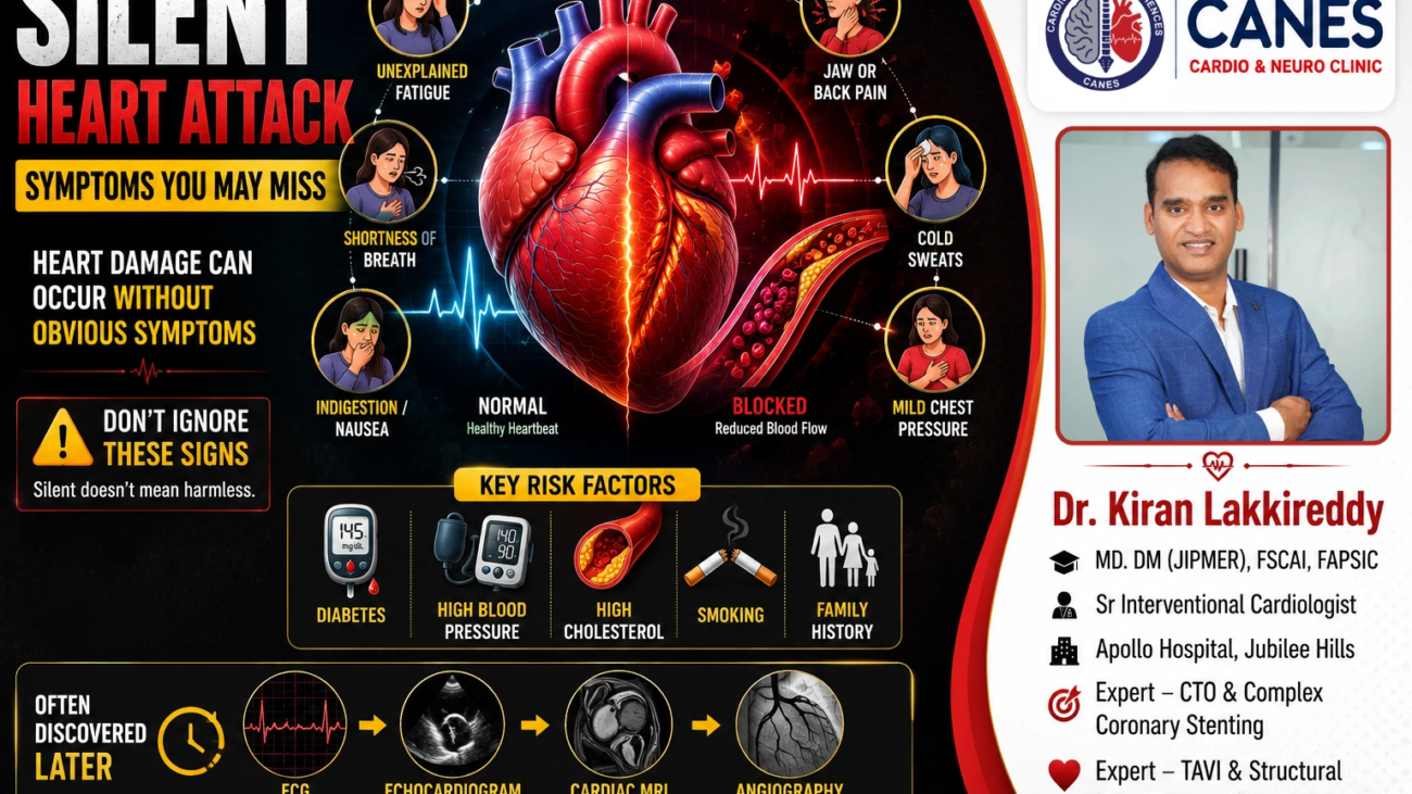
Silent Heart Attack: Symptoms You May Miss, Risk Factors, Diagnosis, Treatment and Prevention
Table of Contents
- Introduction
- What Is a Silent Heart Attack?
- Silent Ischemia vs. Silent Heart Attack: Are They the Same?
- Why Do Some Heart Attacks Go Unnoticed?
- Who Is at Risk for a Silent Heart Attack?
- What Are the Symptoms You Might Miss?
- Who Should Be Evaluated for a Possible Silent Heart Attack?
- How Is a Silent Heart Attack Diagnosed Later?
- Why Are Silent Heart Attacks So Dangerous?
- Does Every Silent Heart Attack Require a Stent?
- Myth vs. Fact: Silent Heart Attacks
- How Can Silent Heart Attacks Be Prevented?
- How Can CANES Cardio & Neuro Clinic Help?
- Conclusion
- FAQs
Book a Consultation
Prevention is better than cure. Talk to our specialists today.
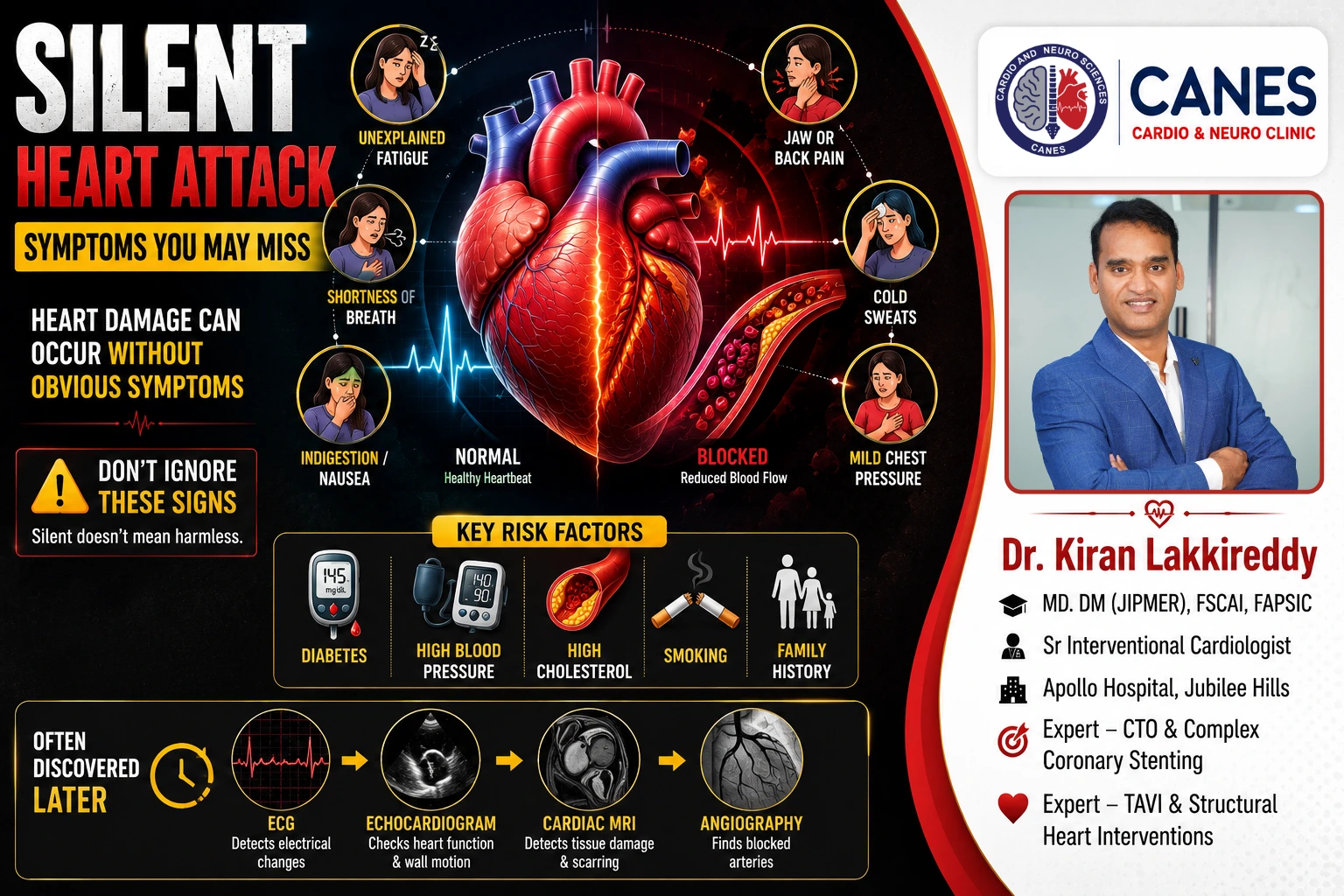
A heart attack is often imagined as a sudden episode of severe chest pain, heavy sweating, and sudden collapse. However, not every heart attack produces obvious or dramatic symptoms. A silent heart attack, also called an unrecognized myocardial infarction, is a heart attack that causes no symptoms, very mild symptoms, or symptoms that are not recognized as being related to the heart. A person may continue their normal daily activities without ever realizing that part of their heart muscle has been permanently injured.
Research suggests that a substantial proportion of heart attacks may go unrecognized because symptoms are absent, mild, or mistaken for another condition. Understanding what a silent heart attack is, who is at risk, and how to identify subtle warning signs can be lifesaving.
Quick Answer
A silent heart attack is a myocardial infarction that occurs without the classic symptoms people commonly associate with a heart attack. While some people experience absolutely no noticeable symptoms, others may have mild or unusual symptoms such as unexplained tiredness, mild chest pressure, indigestion-like discomfort, shortness of breath, or a general feeling that something is not right. Because these symptoms are subtle, they are often mistaken for indigestion, muscle strain, aging, or fatigue. “Silent” does not mean harmless heart-muscle damage still occurs, making comprehensive cardiovascular assessments crucial for preventing future events.
What Is a Silent Heart Attack?
A silent heart attack, or unrecognized myocardial infarction, occurs when blood flow to part of the heart muscle becomes severely reduced or blocked, causing heart muscle injury without triggering the classic, recognizable symptoms of a heart attack.
Detailed Explanation:
Most heart attacks are caused by coronary artery disease, a condition where cholesterol-containing plaque builds up within the walls of the coronary arteries over many years. If this plaque becomes unstable and ruptures, a blood clot quickly forms over the rupture site. This clot interferes with or completely blocks the flow of oxygen-rich blood to the heart muscle. When a portion of the heart muscle is deprived of oxygen for too long, the tissue begins to die a process called myocardial infarction.
During a typical heart attack, this sudden lack of oxygen triggers intense pain signals. However, during a silent heart attack, these symptoms may be completely absent, very mild, or present in ways that seem entirely unrelated to the heart. The person may not recognize their significance, and the event may only be discovered months or years later during routine medical testing. Regardless of what the patient felt, the biological injury to the heart muscle remains the same.
Silent Ischemia vs. Silent Heart Attack: Are They the Same?
No, they are related but distinct conditions. Silent ischemia refers to reduced blood flow to the heart without symptoms, whereas a silent myocardial infarction means that actual, permanent heart-muscle injury (cell death) has already occurred without recognized symptoms.
Detailed Explanation:
Silent Ischemia: Ischemia means inadequate blood and oxygen supply to a tissue. Silent cardiac ischemia occurs when blood flow to the heart muscle is temporarily reduced often due to narrowed arteries without triggering obvious symptoms like angina (chest discomfort). The American Heart Association explicitly describes silent ischemia as reduced blood flow to the heart without obvious symptoms. While it indicates underlying coronary disease, it does not always mean permanent heart muscle death has occurred.
Silent Myocardial Infarction: This is an actual heart attack. A silent myocardial infarction means the ischemia was severe and prolonged enough to cause irreversible injury and scarring to the heart muscle, yet the patient did not recognize it as an acute cardiac episode.
Key Takeaway: A person can experience repeated episodes of silent ischemia without necessarily having a myocardial infarction, but both conditions require rigorous medical management to prevent future emergencies.
Why Do Some Heart Attacks Go Unnoticed?
Heart attacks can be silent because the symptoms may be very mild, they may mimic other minor health issues like indigestion or fatigue, or conditions like diabetes and older age may alter how the body perceives pain.
Detailed Explanation:
There is no single explanation for why a heart attack presents silently, but several factors influence symptom recognition:
- Symptoms May Be Mild: Not every myocardial infarction causes severe, crushing chest pain. Some heart attacks begin gradually, causing only mild discomfort that a patient assumes will just fade away with rest.
- Symptoms Feel Like Something Else: Possible symptoms may closely resemble common, non-threatening issues such as indigestion, heartburn, a pulled muscle in the back, general tiredness, anxiety, or a viral illness. The American Heart Association notes that silent heart attacks frequently involve unusual symptoms like prolonged fatigue or jaw discomfort.
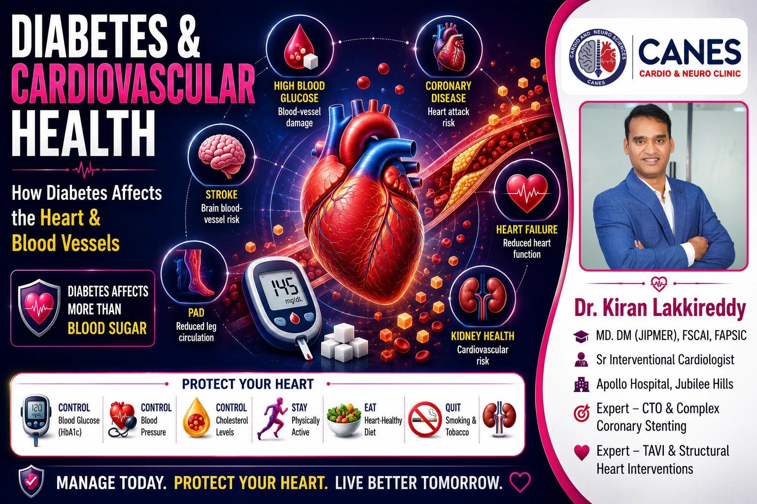
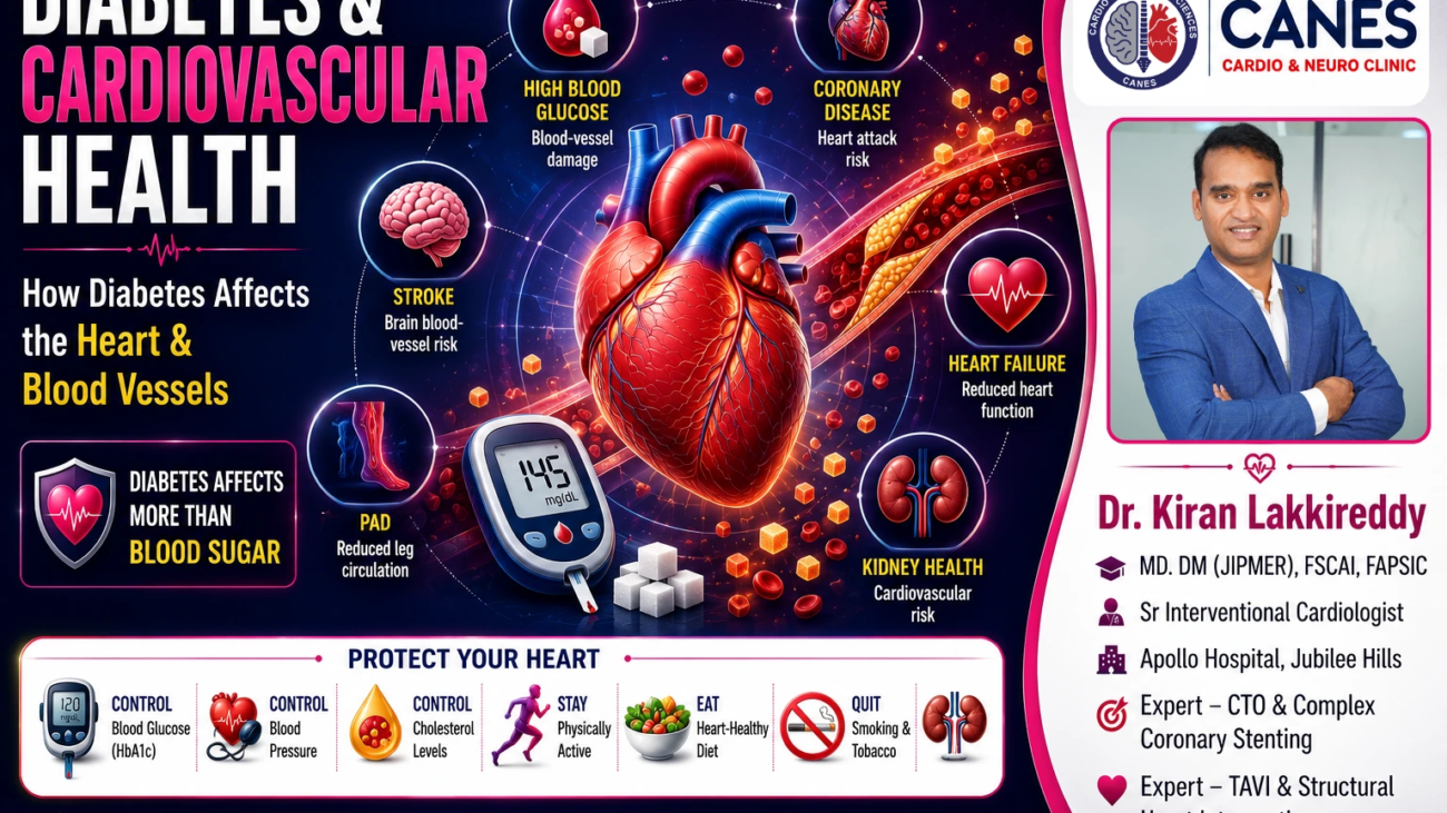
- The Impact of Diabetes: People with diabetes are at a significantly increased cardiovascular risk. Over time, persistently high blood glucose damages blood vessels and nerves. Diabetic autonomic neuropathy can affect pain perception and may contribute to less-recognized symptoms in some people with diabetes.
- Age and Multiple Conditions: Silent heart attacks are reported more frequently among older adults. Because older patients may have several coexisting conditions, mild breathlessness or discomfort is easier to mistakenly attribute to simply “getting older.”
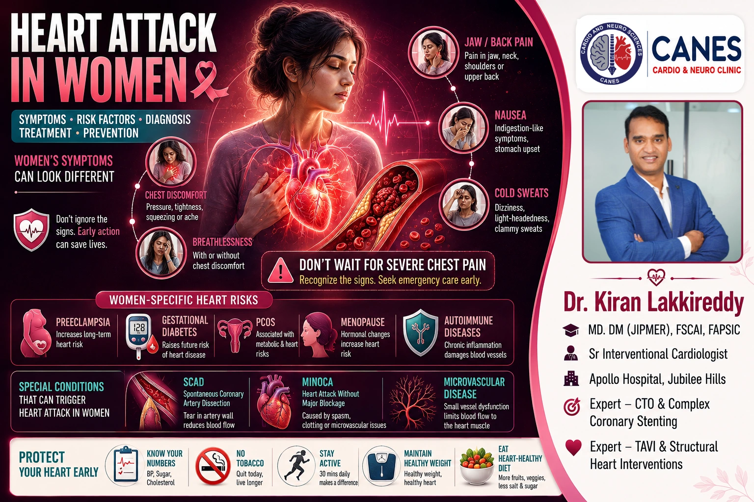
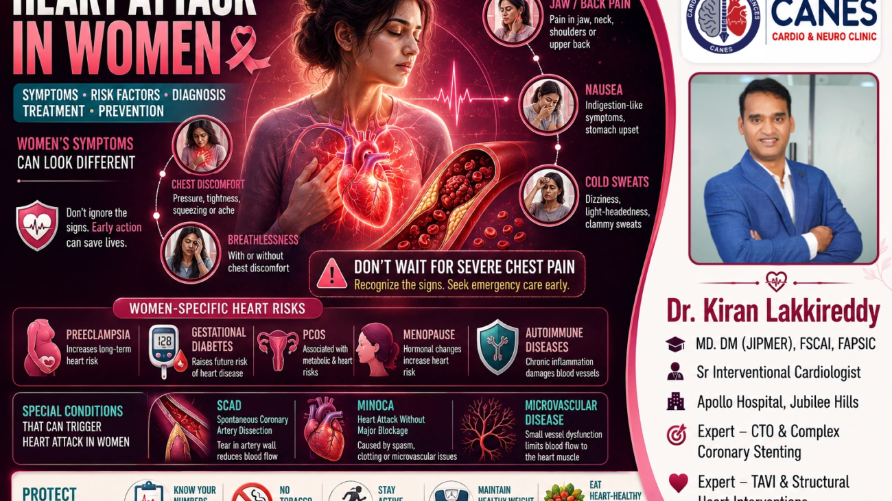
- Gender Differences in Symptoms: While chest pain remains the primary symptom for both men and women, women may experience associated symptoms such as shortness of breath, unusual weakness, nausea, or upper back discomfort, either alongside or instead of prominent chest discomfort. When these occur without prominent chest pain, they may be misinterpreted.
Who Is at Risk for a Silent Heart Attack?
The risk factors for a silent heart attack are identical to those for a symptomatic heart attack. They include diabetes, high blood pressure, high cholesterol, smoking, obesity, physical inactivity, older age, and a strong family history of heart disease.
Detailed Explanation:
Because a silent heart attack is biologically the same as a recognized heart attack, the underlying drivers of the disease are the same. Atherosclerosis (plaque buildup) does not distinguish between silent and symptomatic events.
- Diabetes and Cardiovascular Risk: The relationship between diabetes and heart disease is profound. People with diabetes frequently have high blood pressure, abnormal cholesterol, excess body weight, and kidney disease. This combination substantially multiplies cardiovascular burden.
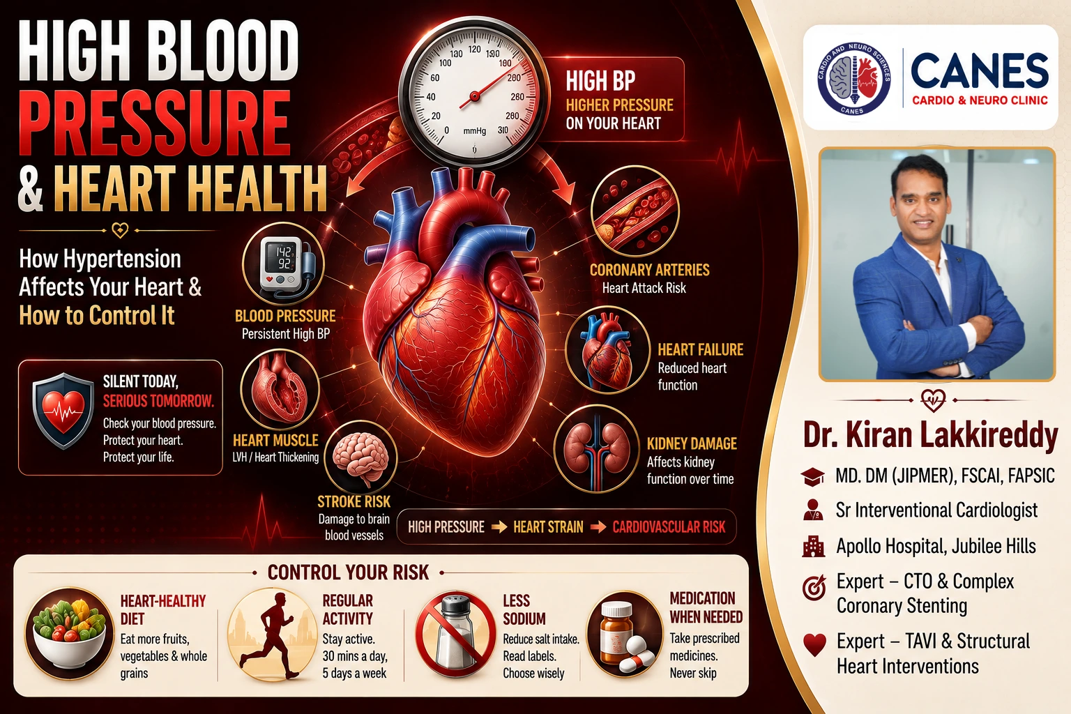
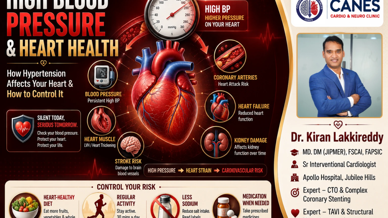
- High Blood Pressure (Hypertension): High blood pressure often causes no noticeable symptoms itself, yet it places immense, constant stress on blood-vessel walls. A person may have hypertension for years without knowing it, silently accelerating plaque formation.
- High Cholesterol: Elevated levels of atherogenic cholesterol particularly LDL (bad) cholesterol contribute directly to the development of plaque.
- Smoking: Smoking damages the endothelial lining of blood vessels, promotes inflammation, and makes the blood more prone to clotting.
What Are the Symptoms You Might Miss?
While some people truly experience no symptoms, others may brush off subtle warning signs such as unexplained fatigue, mild chest heaviness, indigestion, shortness of breath, jaw or back discomfort, cold sweating, or a vague feeling of being unwell.
Detailed Explanation:
The phrase “silent” does not necessarily mean that absolutely nothing was felt at the time; it often means the symptoms were misunderstood. When patients are diagnosed with a previous silent heart attack, they sometimes remember subtle events in hindsight, such as:
- Mild pressure, heaviness, or discomfort in the center of the chest
- A sudden onset of profound, unexplained fatigue
- Shortness of breath doing simple tasks
- Indigestion-like discomfort or mild nausea that doesn’t resolve with antacids
- A dull ache in the jaw, neck, shoulder, or upper back
- Sudden light-headedness or cold sweating
Key Takeaway: Heart attacks can and do occur without typical chest pain. While you shouldn’t assume every bout of fatigue is a heart attack, you should never ignore new, persistent, or unusual symptoms, especially if you have known cardiovascular risk factors.
Who Should Be Evaluated for a Possible Silent Heart Attack?
People with symptoms or risk factors suggesting cardiovascular disease should discuss appropriate evaluation with a healthcare professional, particularly those with diabetes, hypertension, high cholesterol, a smoking history, established coronary disease, or unexplained reductions in exercise tolerance.
Detailed Explanation
Routine screening for silent heart disease is not automatically appropriate for every asymptomatic person. Testing should be individualized according to cardiovascular risk, symptoms, medical history, and clinical findings. If you notice a sudden, unexplained decline in your physical stamina or new, vague chest symptoms, a structured cardiovascular evaluation can help determine if an underlying cardiac event has occurred.
How Is a Silent Heart Attack Diagnosed Later?
A past silent heart attack is usually discovered during medical testing for another issue. Diagnosis relies on evaluating a patient’s medical history alongside tests like an electrocardiogram (ECG), echocardiogram, cardiac MRI, or coronary angiography.
Detailed Explanation
Because the acute event was missed, a silent heart attack is often identified months or years later. Accurate diagnosis requires interpreting these findings alongside the patient’s symptoms, medical history, and other test results.
- Electrocardiogram (ECG/EKG): An ECG may show changes such as pathologic Q waves that can suggest a previous myocardial infarction, although ECG findings alone do not always establish when or whether a prior infarction occurred.
- Cardiac Troponin: If a suspected heart attack is recent, cardiac troponin blood tests can help detect acute myocardial injury. Troponin levels change over time, so interpretation depends on when symptoms or the suspected event occurred.
- Echocardiography: An ultrasound of the heart can show how well the heart is pumping. Areas of the heart wall that are scarred from a previous silent heart attack will not move or contract normally.
- Cardiac MRI: A cardiac magnetic resonance imaging scan provides highly detailed views of heart tissue, excellent for identifying precise patterns of previous myocardial damage.
- Coronary Angiography: If non-invasive tests suggest significant damage or ongoing ischemia, coronary angiography is used to directly visualize the coronary arteries using a specialized dye and X-rays.
Why Are Silent Heart Attacks So Dangerous?
When a heart attack goes unrecognized, the patient may miss the opportunity for time-sensitive treatment during the acute event and may also remain untreated for important long-term cardiovascular risk factors.
Detailed Explanation:
The absence of dramatic symptoms does not protect the heart from physical injury. A myocardial infarction means tissue has died. If a person does not know they had a heart attack, they do not receive the critical emergency treatments that save heart muscle, nor do they start the essential secondary-prevention medications such as antiplatelet therapy, statins, beta-blockers, and other guideline-directed treatments when appropriate.
Because the underlying cardiovascular disease may remain untreated, the risk of future cardiovascular events can remain elevated. Furthermore, if a significant amount of heart muscle was damaged during the silent event, the heart may lose its ability to pump efficiently, eventually leading to symptomatic heart failure characterized by chronic breathlessness and fluid retention.
Does Every Silent Heart Attack Require a Stent?
No. The discovery of an old silent heart attack does not automatically mean a stent is required. Revascularization (stenting or bypass) depends entirely on the patient’s current symptoms, coronary anatomy, and evidence of ongoing ischemia.
Detailed Explanation:
When a cardiologist discovers evidence of a past silent myocardial infarction, the first step is comprehensive risk assessment. A coronary stent is a procedure used to open narrowed or blocked arteries to restore blood flow.
If the patient has ongoing or recurrent symptoms, evidence of significant active ischemia on a stress test, or a high-risk blockage identified on an angiogram, stenting may be highly appropriate. However, if the damaged area is fully scarred with no viable muscle left to save, or if the remaining arteries are relatively clear, the patient may be managed optimally with evidence-based medications and aggressive lifestyle and risk-factor control.
Myth vs. Fact: Silent Heart Attacks
| Common Myth | Medical Fact |
| Every heart attack causes severe, crushing chest pain. | Some heart attacks cause only mild, subtle symptoms, or no recognized symptoms at all. |
| If there was no chest pain, it couldn’t have been a heart attack. | A myocardial infarction can absolutely occur without typical chest pain, presenting instead with fatigue or breathlessness. |
| Silent heart attacks do not damage the heart muscle. | Heart-muscle injury and cell death occur regardless of whether the symptoms were felt or recognized. |
| Silent ischemia and a silent heart attack are the exact same thing. | Silent ischemia is reduced blood flow without symptoms; a silent heart attack involves myocardial injury with irreversible loss of heart muscle cells. |
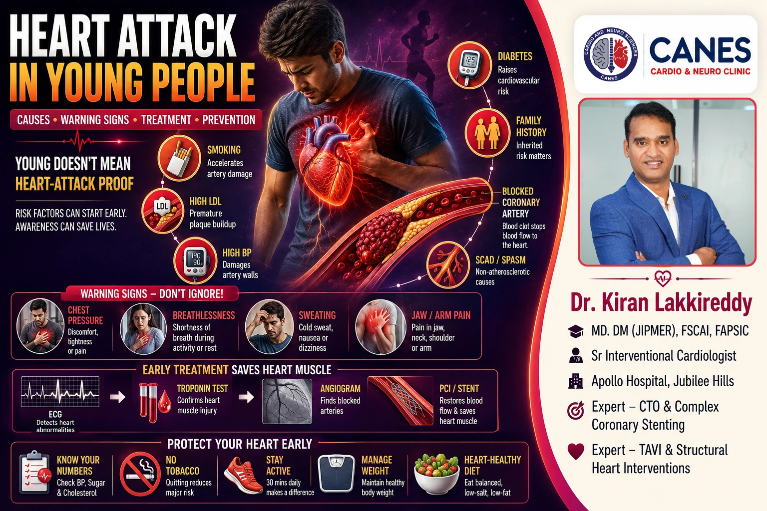
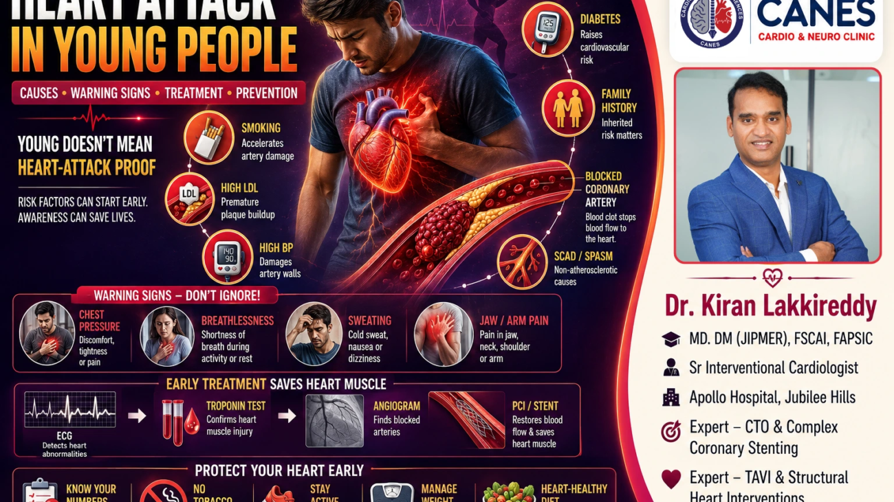
| Only older people experience silent heart attacks. | Silent or unrecognized myocardial infarction can also occur in younger adults, particularly when important cardiovascular risk factors are present. |
How Can Silent Heart Attacks Be Prevented?
Preventing a silent heart attack requires proactive, multifactorial cardiovascular risk management. This includes managing blood pressure, cholesterol, and blood glucose to recommended targets, avoiding tobacco, eating a heart-healthy diet, and maintaining regular physical activity.
Actionable Information:
- Know and Control Your Numbers: High blood pressure and high cholesterol operate silently. Regular checkups are essential to detect hypertension and lipid abnormalities before they cause arterial damage. If you are concerned about your cardiovascular risk, scheduling a comprehensive cardiovascular evaluation can help establish an appropriate baseline for your heart health.
- Manage Diabetes Carefully: For patients with diabetes, good cardiovascular care extends far beyond just controlling blood sugar (HbA1c). It requires aggressive management of blood pressure, cholesterol, kidney health, and weight.
- Avoid All Tobacco: Smoking cessation is one of the most powerful, modifiable steps you can take for cardiovascular protection. Quitting rapidly reduces the risk of clot formation and arterial spasm.
- Stay Physically Active: Regular exercise supports cardiovascular fitness, lowers blood pressure, and improves glucose metabolism. If you have known coronary disease or previous heart damage, discuss safe exercise parameters with your cardiologist.
- Adopt a Heart-Healthy Diet: Emphasize vegetables, fruits, whole grains, lean proteins, and healthy unsaturated fats, while limiting highly processed foods, excess sodium, and foods containing trans fats.
- Attend Regular Follow-Ups: Routine medical reviews allow your physician to monitor changes in your risk profile. Managing blood pressure, cholesterol, diabetes, weight, and tobacco exposure together provides a more comprehensive approach to cardiovascular risk reduction than focusing on a single risk factor.
How Can CANES Cardio & Neuro Clinic Help?
The clinical circumstances surrounding a silent myocardial infarction can vary considerably. One patient may simply have an abnormal ECG discovered during a routine checkup, while another may have a complex history of diabetes, hypertension, known coronary disease, and reduced exercise tolerance. Assessment and treatment must be highly individualized.
At CANES Cardio & Neuro Clinic, patients receive personalized cardiovascular evaluation and understandable guidance regarding coronary disease, myocardial infarction, cardiovascular risk factors, and appropriate follow-up care.
Guided by Dr. Kiran Lakkireddy, an experienced interventional cardiologist specializing in complex cardiology, the clinic focuses on careful assessment, patient education, shared decision-making, and coordination with cardiac rehabilitation teams when indicated.
(Note: A routine clinic appointment is not appropriate for a suspected current heart attack. A person experiencing severe or persistent chest discomfort, sudden significant breathlessness, fainting, loss of consciousness, or other possible heart-attack symptoms should seek immediate emergency medical attention.)
Location: 3rd Floor, Tapasavi Commercial Complex, Hi-Tension Road, Kondapur, Hyderabad
Contact: +91 9014944654
Appointment Booking: Visit CANES Clinic Official Portal
Specialist Profile: Dr. Kiran Lakkireddy Official Website
Key Points to Remember
- A heart attack does not always cause dramatic chest pain: Some myocardial infarctions occur with mild, atypical, or completely unnoticed symptoms.
- Silent does not mean harmless: Heart-muscle damage occurs and can lead to serious long-term complications like heart failure.
- Diabetes increases risk: Silent heart attacks are significantly more common among people with diabetes due to factors like altered pain perception.
- Evidence may be found later: Previous unrecognized events are often revealed via ECG or echocardiography performed for other reasons.
- Prevention requires comprehensive care: Managing blood pressure, cholesterol, diabetes, weight, and tobacco exposure together provides a more comprehensive approach to cardiovascular risk reduction than focusing on a single risk factor.
Conclusion
A silent heart attack is a dangerous cardiovascular event that occurs without sounding the usual alarms. Because symptoms can mimic benign conditions like indigestion or simple fatigue, the physical damage to the heart muscle often goes untreated, leaving the patient at elevated risk for future heart failure or a subsequent, fatal heart attack. Understanding your risk factors especially if you have diabetes, hypertension, or a family history of heart disease and undergoing appropriate, individualized cardiovascular evaluation and managing modifiable risk factors can help reduce the risk of unrecognized cardiovascular disease and future events. For those seeking specialist cardiovascular care in Kondapur, Dr. Kiran Lakkireddy and the team at CANES Cardio & Neuro Clinic provide individualized cardiovascular evaluation and management.
Medical Sources & Guidelines
This article is grounded in established cardiovascular research and evidence-based clinical practice guidelines from leading medical organizations:
- American Heart Association (AHA) — relevant scientific statements on silent ischemia and unrecognized myocardial infarction
- American College of Cardiology (ACC) / AHA — guideline on acute coronary syndromes
- National Heart, Lung, and Blood Institute (NHLBI) — coronary heart disease resources
- Centers for Disease Control and Prevention (CDC) — heart disease resources
- Peer-reviewed research on unrecognized myocardial infarction
Medically Reviewed By: Dr. Kiran Lakkireddy Last Reviewed: August 2026
Medical Disclaimer: This article is for general educational information and does not replace consultation with a qualified healthcare professional. A routine clinic appointment is not appropriate for a suspected medical emergency. If you experience severe or persistent chest discomfort, significant breathlessness, fainting, loss of consciousness, or other possible heart-attack symptoms, seek immediate emergency medical attention.
Frequently Asked Questions (FAQs)
A silent heart attack is a myocardial infarction that occurs without obvious symptoms or with symptoms so mild or unusual that the person does not recognize the event as a heart attack. Heart-muscle damage still occurs despite the lack of recognized symptoms.
Not exactly. Silent ischemia means that the heart muscle experiences temporarily reduced blood and oxygen supply without obvious symptoms. A silent heart attack means that actual, permanent heart-muscle injury and cell death have occurred.
Yes. Some heart attacks cause no recognized symptoms, while others cause mild or atypical symptoms—such as fatigue, nausea, or shortness of breath—instead of classic chest pain.
Silent or unrecognized heart attacks are reported more frequently in people with diabetes. Diabetic autonomic neuropathy may alter pain perception in some people, while other factors can also contribute to less-recognized symptoms.
No. Coronary angiography and stenting are performed when clinically appropriate based on current symptoms, evidence of ongoing ischemia, and overall cardiovascular risk. Some patients are managed highly effectively with medication and risk-factor control alone.
Expert Care
Hyderabad's leading Cardio & Neuro specialists.
![Exercise and Cardiovascular Health: How Physical Activity Supports a Healthier Heart Regular physical activity is one of the most important lifestyle measures for maintaining your long-term well-being. The heart is a muscle, and just like other muscles in the body, it adapts and strengthens in response to regular physical activity. Over time, appropriate exercise can improve cardiovascular fitness and help your heart and circulation work much more efficiently. The connection between exercise and cardiovascular health extends far beyond the heart itself. Regular physical activity can help improve blood pressure, lower cholesterol, boost insulin sensitivity, manage body weight, and heavily support emotional well-being. It is definitively associated with a lower risk of heart disease, stroke, and type 2 diabetes. However, exercise is not identical for every person. Someone who is generally healthy may require a very different program from someone recovering from a heart attack or living with heart failure. At CANES Cardio & Neuro Clinic in Hyderabad, guided by Dr. Kiran Lakkireddy, an experienced interventional cardiologist, our approach emphasizes careful cardiovascular assessment and patient education. This comprehensive guide will explain exactly how physical activity protects your heart and how to begin exercising safely. [Insert Image Here, alt="Exercise and cardiovascular health"] Quick Answer Exercise supports cardiovascular health by strengthening the heart muscle, lowering blood pressure, improving cholesterol levels, and regulating blood sugar. For most adults, current guidelines recommend aiming for 150–300 minutes of moderate-intensity aerobic activity each week (like brisk walking), or 75–150 minutes of vigorous-intensity activity, alongside muscle-strengthening exercises on at least two days per week. However, people with known cardiovascular disease, significant symptoms, or certain uncontrolled medical conditions should discuss an appropriate exercise plan with their healthcare professional before starting. Exercise and Cardiovascular Health at a Glance Cardiovascular Benefit How Exercise Helps Why It Matters Cardiovascular Fitness Trains the heart and lungs to support activity more efficiently. Improves exercise capacity and makes everyday physical activities easier to perform. Blood Pressure Regular activity helps lower and control resting blood pressure. High blood pressure is a major risk factor for heart attack, stroke, and other cardiovascular disease. Cholesterol Improves lipid-related risk factors by balancing blood fats. Crucial for preventing atherosclerotic plaque buildup. Metabolic Health Improves insulin sensitivity and glucose utilization. Essential for people with diabetes or pre-diabetes. Vascular Health Improves circulation and overall endothelial (blood vessel) function. Supports healthy blood-vessel function and circulation. Mental Well-Being Reduces anxiety, improves sleep, and boosts mood. Emotional stress directly impacts cardiovascular strain. What Is the Difference Between Physical Activity and Exercise? Physical activity refers to any bodily movement that burns energy (like gardening or walking), whereas exercise is a structured, planned activity performed specifically to improve fitness, strength, or endurance. Key Takeaway: Movement matters. While structured exercise is highly valuable, simply increasing your everyday physical activity and reducing the hours you spend sitting is crucial for a heart-healthy lifestyle. You do not necessarily need a gym membership to protect your heart. Taking the stairs, cycling for transportation, and completing household chores all contribute to your overall activity levels and provide meaningful cardiovascular benefits. How Does Exercise Affect the Heart? During physical activity, the cardiovascular system increases blood flow, making the heart beat faster to deliver more oxygen; with regular aerobic training, the heart adapts and becomes highly efficient at supporting this demand. Key Takeaway: The goal of exercise is to create a healthy cardiovascular training stimulus, not to push yourself to absolute exhaustion. With regular aerobic training, the cardiovascular system adapts, improving the heart's efficiency and your overall exercise capacity. It pumps more blood with every beat, meaning it doesn't have to work as hard during your normal daily activities. 1. Exercise Can Improve Cardiovascular Fitness Aerobic exercise (such as brisk walking, cycling, swimming, or dancing) helps condition the heart and lungs. As fitness improves, you will notice that walking long distances or climbing stairs requires far less effort. Importantly, physical activity improves cardiometabolic risk factors even if substantial weight loss does not occur. 2. Exercise and Blood Pressure High blood pressure places severe stress on the heart and blood vessels. Regular physical activity naturally lowers blood pressure. While it does not replace prescribed blood-pressure medicine, exercise is an essential part of hypertension management. If your blood pressure is severely uncontrolled, seek medical advice before starting vigorous activity. 3. Exercise and Cholesterol Regular physical activity can improve several lipid-related risk factors, although its effects on LDL (bad) cholesterol are generally modest. Exercise works best alongside a heart-healthy diet and cholesterol-lowering medication when prescribed by your doctor. 4. Exercise and Blood-Sugar Control During exercise, your working muscles consume glucose for energy. Regular physical activity improves insulin sensitivity and helps the body regulate blood sugar more effectively. Because diabetes is a major cardiovascular risk factor, controlling blood sugar through physical activity can contribute to better cardiovascular health. 5. Exercise and Body Weight Physical activity is vital for preventing weight gain. However, the relationship is complex. Current cardiovascular guidelines emphasize that physical activity improves blood pressure, insulin resistance, and cholesterol even if substantial weight loss does not occur. 6. Exercise and Blood-Vessel Health The inner lining of your blood vessels is called the endothelium. Regular exercise improves endothelial function, helping your arteries relax, open up, and respond appropriately to the body's changing demands. Does Sitting Too Much Affect Heart Health? Yes. Spending long periods of time sitting or remaining sedentary is independently associated with poorer cardiovascular health and a higher risk of heart disease. Key Takeaway: "Move more, sit less." Breaking up prolonged periods of sitting with regular, brief movement is a highly useful complement to your structured exercise routine. Even if you meet the weekly exercise recommendations, sitting at a desk for 8 to 10 hours a day can still negatively impact your metabolic health. Breaking up prolonged sitting with standing, stretching, or short periods of walking can help reduce sedentary time and add more movement to your day. Can Exercise Prevent a Heart Attack? Regular physical activity significantly reduces the long-term risk of heart disease and stroke by controlling blood pressure, cholesterol, and diabetes, but it cannot absolutely guarantee that a heart attack will never occur. Key Takeaway: Exercise works best as one foundational component of a comprehensive cardiovascular prevention plan that also includes a healthy diet and avoidance of smoking. Heart-attack risk is driven by a combination of genetics, age, kidney disease, and established atherosclerosis. Exercise actively reduces your modifiable risks by improving metabolic health and reducing the danger of physical inactivity. Can Exercise Ever Trigger a Heart Problem? For most people, the cardiovascular benefits of exercise heavily outweigh the risks; however, sudden, vigorous exertion can temporarily increase cardiac risk in people who are poorly conditioned or have underlying heart disease. Key Takeaway: The safest approach is to start low and build gradually. Do not suddenly attempt heavy weightlifting or intense running if you have been sedentary for years. How Much Exercise Is Recommended? Current guidelines recommend that most healthy adults complete 150 to 300 minutes of moderate-intensity aerobic activity per week, or 75 to 150 minutes of vigorous activity, alongside muscle-strengthening exercises on at least two days per week. Key Takeaway: You do not need to complete your exercise in one long session. Shorter periods of activity throughout the day can still contribute to your weekly total, and any movement is better than none. What Does "Moderate-Intensity" Mean? A practical way to measure intensity is the "talk test." During moderate activity (like brisk walking or water aerobics): Your breathing is noticeably faster. Your heart rate rises. You can generally still hold a conversation. Singing comfortably becomes difficult. What Is "Vigorous-Intensity"? During vigorous activity (like running, fast cycling, or lap swimming): Breathing becomes substantially harder. Speaking more than a few words without pausing for breath becomes very difficult. What Types of Exercise Are Good for Cardiovascular Health? A well-rounded heart-health program combines aerobic exercise to build endurance, resistance training to improve muscle strength, and flexibility/balance exercises to support overall mobility. Aerobic Exercise: Walking, cycling, and swimming train the heart and lungs to support sustained activity. Brisk walking is often the simplest and best place to start. Resistance Training: Using free weights or resistance bands improves muscle strength and body composition, which provides beneficial effects on cardiovascular risk factors. Flexibility and Balance: Flexibility and balance activities can be particularly valuable for older adults, especially those with reduced mobility, to support function and help prevent falls. Who Should Seek Medical Advice Before Starting Exercise? People with known cardiovascular disease, significant symptoms, or certain uncontrolled medical conditions should discuss an appropriate exercise plan with their healthcare professional before starting vigorous or substantially increasing physical activity. You should seek medical advice before starting or substantially increasing exercise if you have: Known coronary artery disease or a previous heart attack. Heart failure or significant rhythm problems. Unexplained chest discomfort or severe breathlessness. Dizziness or fainting with activity. Warning Signs During Exercise Exercise should naturally increase your breathing and heart rate, but you should never ignore potentially serious symptoms. Stop exercising immediately and seek emergency care if you experience: Chest pressure, tightness, or burning pain. Unusual or severe shortness of breath. Dizziness, confusion, or feeling faint. Unusual palpitations or an irregular rapid heartbeat. What Is Cardiac Rehabilitation? Cardiac rehabilitation is a structured, medically supervised program combining supervised exercise, heart-health education, risk-factor management, and emotional support for people recovering from cardiovascular conditions. Key Takeaway: Cardiac rehabilitation provides a medically supervised environment where patients can rebuild physical confidence after a heart attack, angioplasty, bypass surgery, or heart failure diagnosis. After a cardiac event, you should not simply return to your previous exercise routine. A rehabilitation program tailors the exercise specifically to your current heart function, surgical healing, and medical limitations. Myth Versus Fact: Exercise and Heart Health Understanding the realities of physical activity helps you exercise safely and consistently. Common Myth Medical Fact Only intense exercise benefits the heart. Moderate activity, such as brisk walking, provides substantial and proven cardiovascular benefits. Exercise is useless without weight loss. Exercise improves blood pressure, cholesterol, and metabolic health even if substantial weight loss does not occur. People with heart disease should avoid exercise. Many patients with heart disease benefit significantly from medically appropriate physical activity, often through cardiac rehabilitation. Exercise replaces my heart medications. Physical activity complements your prescribed treatment; it never automatically replaces necessary medication. If it makes me breathless, something is wrong. Breathing harder is expected during aerobic activity. Breathlessness that is new, unusually severe, disproportionate to the activity, or accompanied by chest discomfort, dizziness, or fainting warrants medical assessment. Actionable Checklist: What Should You Ask Your Cardiologist? Before starting a new routine, ask your healthcare team these essential questions: [ ] Is it safe for me to begin exercising, and do I need a cardiac stress test first? [ ] What specific heart-rate range should I aim for during activity? [ ] Do my current cardiac medicines affect how my heart rate will respond to exercise? [ ] Are there any specific exercises (like heavy lifting) that I should avoid? [ ] Which specific warning symptoms should make me stop exercising immediately? [ ] Am I a candidate for a medically supervised cardiac rehabilitation program? Cardiovascular Exercise Guidance at CANES Clinic The appropriate type and intensity of physical activity depend entirely on the individual. A healthy young adult requires very different guidance from someone managing coronary artery disease, heart failure, or recovering from a heart attack. At CANES Cardio & Neuro Clinic, patients receive personalized cardiovascular evaluation and understandable guidance regarding heart conditions, risk factors, and safe physical activity. Guided by Dr. Kiran Lakkireddy, an experienced interventional cardiologist specializing in complex cardiology, the clinic focuses on careful assessment, shared decision-making, and coordination with cardiac rehabilitation teams when indicated. (Note: A routine clinic appointment is not appropriate for a suspected cardiovascular emergency. Severe chest discomfort, fainting, or marked breathlessness require immediate emergency medical assessment.) Location: 3rd Floor, Tapasavi Commercial Complex, Hi-Tension Road, Kondapur, Hyderabad Contact: +91 9014944654 Appointment Booking: Visit CANES Clinic Official Portal Specialist Profile: Dr. Kiran Lakkireddy Official Website Key Points to Remember Exercise strengthens the heart: Regular physical activity trains the cardiovascular system to work more efficiently. Benefits beyond fitness: It improves blood pressure, cholesterol, insulin sensitivity, and overall cardiometabolic health. Weight loss is not the only metric: Cardiovascular benefits occur even if the number on the scale does not drastically change. Walking is excellent exercise: Brisk walking is a highly effective, practical form of moderate-intensity aerobic activity. Consistency over intensity: Sustainable physical activity over the long term is far more valuable than occasional, extreme exhaustion. Seek guidance when necessary: People with existing cardiovascular disease or concerning symptoms should consult a doctor before starting an exercise program. Never ignore warning signs: Stop exercising immediately if you experience chest pain, severe breathlessness, or dizziness. Frequently Asked Questions (FAQ) How much exercise should an adult do each week? General guidelines encourage 150 to 300 minutes of moderate-intensity aerobic activity (like brisk walking) or 75 to 150 minutes of vigorous activity per week, plus muscle-strengthening exercises on at least two days. Do I have to exercise for 30 minutes continuously? No. Activity can be spread throughout the week. You do not need to complete your exercise in one long session; shorter periods of activity throughout the day can still contribute to your weekly total. Does exercise improve cholesterol? Yes. Regular physical activity can improve several lipid-related risk factors, although its effects on LDL cholesterol are generally modest. Exercise works alongside a heart-healthy diet and cholesterol-lowering medication when prescribed. Is vigorous exercise better than moderate exercise? Not necessarily. Both provide cardiovascular health benefits. The right intensity depends on your current fitness, age, and any underlying medical conditions. Can I exercise after a heart attack or angioplasty? Many people can and should return to appropriate physical activity after a heart procedure, but the timing and intensity should be guided by their healthcare team, often through a structured cardiac rehabilitation program. Do I need a stress test before exercising? Most healthy adults without symptoms do not need routine cardiac stress testing simply to begin moderate physical activity. Testing may be appropriate when symptoms, known cardiovascular disease, or specific clinical concerns are present. What should I do if I get chest pain while exercising? Stop exercising immediately. Chest pressure, squeezing, or pain can be a severe warning sign. If symptoms persist or are accompanied by breathlessness or nausea, seek urgent emergency medical assessment. Is strength training important for heart health? Exercise and Cardiovascular Health: How Physical Activity Supports a Healthier Heart Regular physical activity is one of the most important lifestyle measures for maintaining your long-term well-being. The heart is a muscle, and just like other muscles in the body, it adapts and strengthens in response to regular physical activity. Over time, appropriate exercise can improve cardiovascular fitness and help your heart and circulation work much more efficiently. The connection between exercise and cardiovascular health extends far beyond the heart itself. Regular physical activity can help improve blood pressure, lower cholesterol, boost insulin sensitivity, manage body weight, and heavily support emotional well-being. It is definitively associated with a lower risk of heart disease, stroke, and type 2 diabetes. However, exercise is not identical for every person. Someone who is generally healthy may require a very different program from someone recovering from a heart attack or living with heart failure. At CANES Cardio & Neuro Clinic in Hyderabad, guided by Dr. Kiran Lakkireddy, an experienced interventional cardiologist, our approach emphasizes careful cardiovascular assessment and patient education. This comprehensive guide will explain exactly how physical activity protects your heart and how to begin exercising safely. [Insert Image Here, alt="Exercise and cardiovascular health"] Quick Answer Exercise supports cardiovascular health by strengthening the heart muscle, lowering blood pressure, improving cholesterol levels, and regulating blood sugar. For most adults, current guidelines recommend aiming for 150–300 minutes of moderate-intensity aerobic activity each week (like brisk walking), or 75–150 minutes of vigorous-intensity activity, alongside muscle-strengthening exercises on at least two days per week. However, people with known cardiovascular disease, significant symptoms, or certain uncontrolled medical conditions should discuss an appropriate exercise plan with their healthcare professional before starting. Exercise and Cardiovascular Health at a Glance Cardiovascular Benefit How Exercise Helps Why It Matters Cardiovascular Fitness Trains the heart and lungs to support activity more efficiently. Improves exercise capacity and makes everyday physical activities easier to perform. Blood Pressure Regular activity helps lower and control resting blood pressure. High blood pressure is a major risk factor for heart attack, stroke, and other cardiovascular disease. Cholesterol Improves lipid-related risk factors by balancing blood fats. Crucial for preventing atherosclerotic plaque buildup. Metabolic Health Improves insulin sensitivity and glucose utilization. Essential for people with diabetes or pre-diabetes. Vascular Health Improves circulation and overall endothelial (blood vessel) function. Supports healthy blood-vessel function and circulation. Mental Well-Being Reduces anxiety, improves sleep, and boosts mood. Emotional stress directly impacts cardiovascular strain. What Is the Difference Between Physical Activity and Exercise? Physical activity refers to any bodily movement that burns energy (like gardening or walking), whereas exercise is a structured, planned activity performed specifically to improve fitness, strength, or endurance. Key Takeaway: Movement matters. While structured exercise is highly valuable, simply increasing your everyday physical activity and reducing the hours you spend sitting is crucial for a heart-healthy lifestyle. You do not necessarily need a gym membership to protect your heart. Taking the stairs, cycling for transportation, and completing household chores all contribute to your overall activity levels and provide meaningful cardiovascular benefits. How Does Exercise Affect the Heart? During physical activity, the cardiovascular system increases blood flow, making the heart beat faster to deliver more oxygen; with regular aerobic training, the heart adapts and becomes highly efficient at supporting this demand. Key Takeaway: The goal of exercise is to create a healthy cardiovascular training stimulus, not to push yourself to absolute exhaustion. With regular aerobic training, the cardiovascular system adapts, improving the heart's efficiency and your overall exercise capacity. It pumps more blood with every beat, meaning it doesn't have to work as hard during your normal daily activities. 1. Exercise Can Improve Cardiovascular Fitness Aerobic exercise (such as brisk walking, cycling, swimming, or dancing) helps condition the heart and lungs. As fitness improves, you will notice that walking long distances or climbing stairs requires far less effort. Importantly, physical activity improves cardiometabolic risk factors even if substantial weight loss does not occur. 2. Exercise and Blood Pressure High blood pressure places severe stress on the heart and blood vessels. Regular physical activity naturally lowers blood pressure. While it does not replace prescribed blood-pressure medicine, exercise is an essential part of hypertension management. If your blood pressure is severely uncontrolled, seek medical advice before starting vigorous activity. 3. Exercise and Cholesterol Regular physical activity can improve several lipid-related risk factors, although its effects on LDL (bad) cholesterol are generally modest. Exercise works best alongside a heart-healthy diet and cholesterol-lowering medication when prescribed by your doctor. 4. Exercise and Blood-Sugar Control During exercise, your working muscles consume glucose for energy. Regular physical activity improves insulin sensitivity and helps the body regulate blood sugar more effectively. Because diabetes is a major cardiovascular risk factor, controlling blood sugar through physical activity can contribute to better cardiovascular health. 5. Exercise and Body Weight Physical activity is vital for preventing weight gain. However, the relationship is complex. Current cardiovascular guidelines emphasize that physical activity improves blood pressure, insulin resistance, and cholesterol even if substantial weight loss does not occur. 6. Exercise and Blood-Vessel Health The inner lining of your blood vessels is called the endothelium. Regular exercise improves endothelial function, helping your arteries relax, open up, and respond appropriately to the body's changing demands. Does Sitting Too Much Affect Heart Health? Yes. Spending long periods of time sitting or remaining sedentary is independently associated with poorer cardiovascular health and a higher risk of heart disease. Key Takeaway: "Move more, sit less." Breaking up prolonged periods of sitting with regular, brief movement is a highly useful complement to your structured exercise routine. Even if you meet the weekly exercise recommendations, sitting at a desk for 8 to 10 hours a day can still negatively impact your metabolic health. Breaking up prolonged sitting with standing, stretching, or short periods of walking can help reduce sedentary time and add more movement to your day. Can Exercise Prevent a Heart Attack? Regular physical activity significantly reduces the long-term risk of heart disease and stroke by controlling blood pressure, cholesterol, and diabetes, but it cannot absolutely guarantee that a heart attack will never occur. Key Takeaway: Exercise works best as one foundational component of a comprehensive cardiovascular prevention plan that also includes a healthy diet and avoidance of smoking. Heart-attack risk is driven by a combination of genetics, age, kidney disease, and established atherosclerosis. Exercise actively reduces your modifiable risks by improving metabolic health and reducing the danger of physical inactivity. Can Exercise Ever Trigger a Heart Problem? For most people, the cardiovascular benefits of exercise heavily outweigh the risks; however, sudden, vigorous exertion can temporarily increase cardiac risk in people who are poorly conditioned or have underlying heart disease. Key Takeaway: The safest approach is to start low and build gradually. Do not suddenly attempt heavy weightlifting or intense running if you have been sedentary for years. How Much Exercise Is Recommended? Current guidelines recommend that most healthy adults complete 150 to 300 minutes of moderate-intensity aerobic activity per week, or 75 to 150 minutes of vigorous activity, alongside muscle-strengthening exercises on at least two days per week. Key Takeaway: You do not need to complete your exercise in one long session. Shorter periods of activity throughout the day can still contribute to your weekly total, and any movement is better than none. What Does "Moderate-Intensity" Mean? A practical way to measure intensity is the "talk test." During moderate activity (like brisk walking or water aerobics): Your breathing is noticeably faster. Your heart rate rises. You can generally still hold a conversation. Singing comfortably becomes difficult. What Is "Vigorous-Intensity"? During vigorous activity (like running, fast cycling, or lap swimming): Breathing becomes substantially harder. Speaking more than a few words without pausing for breath becomes very difficult. What Types of Exercise Are Good for Cardiovascular Health? A well-rounded heart-health program combines aerobic exercise to build endurance, resistance training to improve muscle strength, and flexibility/balance exercises to support overall mobility. Aerobic Exercise: Walking, cycling, and swimming train the heart and lungs to support sustained activity. Brisk walking is often the simplest and best place to start. Resistance Training: Using free weights or resistance bands improves muscle strength and body composition, which provides beneficial effects on cardiovascular risk factors. Flexibility and Balance: Flexibility and balance activities can be particularly valuable for older adults, especially those with reduced mobility, to support function and help prevent falls. Who Should Seek Medical Advice Before Starting Exercise? People with known cardiovascular disease, significant symptoms, or certain uncontrolled medical conditions should discuss an appropriate exercise plan with their healthcare professional before starting vigorous or substantially increasing physical activity. You should seek medical advice before starting or substantially increasing exercise if you have: Known coronary artery disease or a previous heart attack. Heart failure or significant rhythm problems. Unexplained chest discomfort or severe breathlessness. Dizziness or fainting with activity. Warning Signs During Exercise Exercise should naturally increase your breathing and heart rate, but you should never ignore potentially serious symptoms. Stop exercising immediately and seek emergency care if you experience: Chest pressure, tightness, or burning pain. Unusual or severe shortness of breath. Dizziness, confusion, or feeling faint. Unusual palpitations or an irregular rapid heartbeat. What Is Cardiac Rehabilitation? Cardiac rehabilitation is a structured, medically supervised program combining supervised exercise, heart-health education, risk-factor management, and emotional support for people recovering from cardiovascular conditions. Key Takeaway: Cardiac rehabilitation provides a medically supervised environment where patients can rebuild physical confidence after a heart attack, angioplasty, bypass surgery, or heart failure diagnosis. After a cardiac event, you should not simply return to your previous exercise routine. A rehabilitation program tailors the exercise specifically to your current heart function, surgical healing, and medical limitations. Myth Versus Fact: Exercise and Heart Health Understanding the realities of physical activity helps you exercise safely and consistently. Common Myth Medical Fact Only intense exercise benefits the heart. Moderate activity, such as brisk walking, provides substantial and proven cardiovascular benefits. Exercise is useless without weight loss. Exercise improves blood pressure, cholesterol, and metabolic health even if substantial weight loss does not occur. People with heart disease should avoid exercise. Many patients with heart disease benefit significantly from medically appropriate physical activity, often through cardiac rehabilitation. Exercise replaces my heart medications. Physical activity complements your prescribed treatment; it never automatically replaces necessary medication. If it makes me breathless, something is wrong. Breathing harder is expected during aerobic activity. Breathlessness that is new, unusually severe, disproportionate to the activity, or accompanied by chest discomfort, dizziness, or fainting warrants medical assessment. Actionable Checklist: What Should You Ask Your Cardiologist? Before starting a new routine, ask your healthcare team these essential questions: [ ] Is it safe for me to begin exercising, and do I need a cardiac stress test first? [ ] What specific heart-rate range should I aim for during activity? [ ] Do my current cardiac medicines affect how my heart rate will respond to exercise? [ ] Are there any specific exercises (like heavy lifting) that I should avoid? [ ] Which specific warning symptoms should make me stop exercising immediately? [ ] Am I a candidate for a medically supervised cardiac rehabilitation program? Cardiovascular Exercise Guidance at CANES Clinic The appropriate type and intensity of physical activity depend entirely on the individual. A healthy young adult requires very different guidance from someone managing coronary artery disease, heart failure, or recovering from a heart attack. At CANES Cardio & Neuro Clinic, patients receive personalized cardiovascular evaluation and understandable guidance regarding heart conditions, risk factors, and safe physical activity. Guided by Dr. Kiran Lakkireddy, an experienced interventional cardiologist specializing in complex cardiology, the clinic focuses on careful assessment, shared decision-making, and coordination with cardiac rehabilitation teams when indicated. (Note: A routine clinic appointment is not appropriate for a suspected cardiovascular emergency. Severe chest discomfort, fainting, or marked breathlessness require immediate emergency medical assessment.) Location: 3rd Floor, Tapasavi Commercial Complex, Hi-Tension Road, Kondapur, Hyderabad Contact: +91 9014944654 Appointment Booking: Visit CANES Clinic Official Portal Specialist Profile: Dr. Kiran Lakkireddy Official Website Key Points to Remember Exercise strengthens the heart: Regular physical activity trains the cardiovascular system to work more efficiently. Benefits beyond fitness: It improves blood pressure, cholesterol, insulin sensitivity, and overall cardiometabolic health. Weight loss is not the only metric: Cardiovascular benefits occur even if the number on the scale does not drastically change. Walking is excellent exercise: Brisk walking is a highly effective, practical form of moderate-intensity aerobic activity. Consistency over intensity: Sustainable physical activity over the long term is far more valuable than occasional, extreme exhaustion. Seek guidance when necessary: People with existing cardiovascular disease or concerning symptoms should consult a doctor before starting an exercise program. Never ignore warning signs: Stop exercising immediately if you experience chest pain, severe breathlessness, or dizziness. Frequently Asked Questions (FAQ) How much exercise should an adult do each week? General guidelines encourage 150 to 300 minutes of moderate-intensity aerobic activity (like brisk walking) or 75 to 150 minutes of vigorous activity per week, plus muscle-strengthening exercises on at least two days. Do I have to exercise for 30 minutes continuously? No. Activity can be spread throughout the week. You do not need to complete your exercise in one long session; shorter periods of activity throughout the day can still contribute to your weekly total. Does exercise improve cholesterol? Yes. Regular physical activity can improve several lipid-related risk factors, although its effects on LDL cholesterol are generally modest. Exercise works alongside a heart-healthy diet and cholesterol-lowering medication when prescribed. Is vigorous exercise better than moderate exercise? Not necessarily. Both provide cardiovascular health benefits. The right intensity depends on your current fitness, age, and any underlying medical conditions. Can I exercise after a heart attack or angioplasty? Many people can and should return to appropriate physical activity after a heart procedure, but the timing and intensity should be guided by their healthcare team, often through a structured cardiac rehabilitation program. Do I need a stress test before exercising? Most healthy adults without symptoms do not need routine cardiac stress testing simply to begin moderate physical activity. Testing may be appropriate when symptoms, known cardiovascular disease, or specific clinical concerns are present. What should I do if I get chest pain while exercising? Stop exercising immediately. Chest pressure, squeezing, or pain can be a severe warning sign. If symptoms persist or are accompanied by breathlessness or nausea, seek urgent emergency medical assessment. Is strength training important for heart health? Yes. Appropriate resistance training complements aerobic exercise by improving muscle strength, physical function, and several cardiometabolic risk factors.](https://drkiranlakkireddy.com/wp-content/uploads/2026/08/exercise-and-cardiovascular-heart-health-1300x731.webp)
![Exercise and Cardiovascular Health: How Physical Activity Supports a Healthier Heart Regular physical activity is one of the most important lifestyle measures for maintaining your long-term well-being. The heart is a muscle, and just like other muscles in the body, it adapts and strengthens in response to regular physical activity. Over time, appropriate exercise can improve cardiovascular fitness and help your heart and circulation work much more efficiently. The connection between exercise and cardiovascular health extends far beyond the heart itself. Regular physical activity can help improve blood pressure, lower cholesterol, boost insulin sensitivity, manage body weight, and heavily support emotional well-being. It is definitively associated with a lower risk of heart disease, stroke, and type 2 diabetes. However, exercise is not identical for every person. Someone who is generally healthy may require a very different program from someone recovering from a heart attack or living with heart failure. At CANES Cardio & Neuro Clinic in Hyderabad, guided by Dr. Kiran Lakkireddy, an experienced interventional cardiologist, our approach emphasizes careful cardiovascular assessment and patient education. This comprehensive guide will explain exactly how physical activity protects your heart and how to begin exercising safely. [Insert Image Here, alt="Exercise and cardiovascular health"] Quick Answer Exercise supports cardiovascular health by strengthening the heart muscle, lowering blood pressure, improving cholesterol levels, and regulating blood sugar. For most adults, current guidelines recommend aiming for 150–300 minutes of moderate-intensity aerobic activity each week (like brisk walking), or 75–150 minutes of vigorous-intensity activity, alongside muscle-strengthening exercises on at least two days per week. However, people with known cardiovascular disease, significant symptoms, or certain uncontrolled medical conditions should discuss an appropriate exercise plan with their healthcare professional before starting. Exercise and Cardiovascular Health at a Glance Cardiovascular Benefit How Exercise Helps Why It Matters Cardiovascular Fitness Trains the heart and lungs to support activity more efficiently. Improves exercise capacity and makes everyday physical activities easier to perform. Blood Pressure Regular activity helps lower and control resting blood pressure. High blood pressure is a major risk factor for heart attack, stroke, and other cardiovascular disease. Cholesterol Improves lipid-related risk factors by balancing blood fats. Crucial for preventing atherosclerotic plaque buildup. Metabolic Health Improves insulin sensitivity and glucose utilization. Essential for people with diabetes or pre-diabetes. Vascular Health Improves circulation and overall endothelial (blood vessel) function. Supports healthy blood-vessel function and circulation. Mental Well-Being Reduces anxiety, improves sleep, and boosts mood. Emotional stress directly impacts cardiovascular strain. What Is the Difference Between Physical Activity and Exercise? Physical activity refers to any bodily movement that burns energy (like gardening or walking), whereas exercise is a structured, planned activity performed specifically to improve fitness, strength, or endurance. Key Takeaway: Movement matters. While structured exercise is highly valuable, simply increasing your everyday physical activity and reducing the hours you spend sitting is crucial for a heart-healthy lifestyle. You do not necessarily need a gym membership to protect your heart. Taking the stairs, cycling for transportation, and completing household chores all contribute to your overall activity levels and provide meaningful cardiovascular benefits. How Does Exercise Affect the Heart? During physical activity, the cardiovascular system increases blood flow, making the heart beat faster to deliver more oxygen; with regular aerobic training, the heart adapts and becomes highly efficient at supporting this demand. Key Takeaway: The goal of exercise is to create a healthy cardiovascular training stimulus, not to push yourself to absolute exhaustion. With regular aerobic training, the cardiovascular system adapts, improving the heart's efficiency and your overall exercise capacity. It pumps more blood with every beat, meaning it doesn't have to work as hard during your normal daily activities. 1. Exercise Can Improve Cardiovascular Fitness Aerobic exercise (such as brisk walking, cycling, swimming, or dancing) helps condition the heart and lungs. As fitness improves, you will notice that walking long distances or climbing stairs requires far less effort. Importantly, physical activity improves cardiometabolic risk factors even if substantial weight loss does not occur. 2. Exercise and Blood Pressure High blood pressure places severe stress on the heart and blood vessels. Regular physical activity naturally lowers blood pressure. While it does not replace prescribed blood-pressure medicine, exercise is an essential part of hypertension management. If your blood pressure is severely uncontrolled, seek medical advice before starting vigorous activity. 3. Exercise and Cholesterol Regular physical activity can improve several lipid-related risk factors, although its effects on LDL (bad) cholesterol are generally modest. Exercise works best alongside a heart-healthy diet and cholesterol-lowering medication when prescribed by your doctor. 4. Exercise and Blood-Sugar Control During exercise, your working muscles consume glucose for energy. Regular physical activity improves insulin sensitivity and helps the body regulate blood sugar more effectively. Because diabetes is a major cardiovascular risk factor, controlling blood sugar through physical activity can contribute to better cardiovascular health. 5. Exercise and Body Weight Physical activity is vital for preventing weight gain. However, the relationship is complex. Current cardiovascular guidelines emphasize that physical activity improves blood pressure, insulin resistance, and cholesterol even if substantial weight loss does not occur. 6. Exercise and Blood-Vessel Health The inner lining of your blood vessels is called the endothelium. Regular exercise improves endothelial function, helping your arteries relax, open up, and respond appropriately to the body's changing demands. Does Sitting Too Much Affect Heart Health? Yes. Spending long periods of time sitting or remaining sedentary is independently associated with poorer cardiovascular health and a higher risk of heart disease. Key Takeaway: "Move more, sit less." Breaking up prolonged periods of sitting with regular, brief movement is a highly useful complement to your structured exercise routine. Even if you meet the weekly exercise recommendations, sitting at a desk for 8 to 10 hours a day can still negatively impact your metabolic health. Breaking up prolonged sitting with standing, stretching, or short periods of walking can help reduce sedentary time and add more movement to your day. Can Exercise Prevent a Heart Attack? Regular physical activity significantly reduces the long-term risk of heart disease and stroke by controlling blood pressure, cholesterol, and diabetes, but it cannot absolutely guarantee that a heart attack will never occur. Key Takeaway: Exercise works best as one foundational component of a comprehensive cardiovascular prevention plan that also includes a healthy diet and avoidance of smoking. Heart-attack risk is driven by a combination of genetics, age, kidney disease, and established atherosclerosis. Exercise actively reduces your modifiable risks by improving metabolic health and reducing the danger of physical inactivity. Can Exercise Ever Trigger a Heart Problem? For most people, the cardiovascular benefits of exercise heavily outweigh the risks; however, sudden, vigorous exertion can temporarily increase cardiac risk in people who are poorly conditioned or have underlying heart disease. Key Takeaway: The safest approach is to start low and build gradually. Do not suddenly attempt heavy weightlifting or intense running if you have been sedentary for years. How Much Exercise Is Recommended? Current guidelines recommend that most healthy adults complete 150 to 300 minutes of moderate-intensity aerobic activity per week, or 75 to 150 minutes of vigorous activity, alongside muscle-strengthening exercises on at least two days per week. Key Takeaway: You do not need to complete your exercise in one long session. Shorter periods of activity throughout the day can still contribute to your weekly total, and any movement is better than none. What Does "Moderate-Intensity" Mean? A practical way to measure intensity is the "talk test." During moderate activity (like brisk walking or water aerobics): Your breathing is noticeably faster. Your heart rate rises. You can generally still hold a conversation. Singing comfortably becomes difficult. What Is "Vigorous-Intensity"? During vigorous activity (like running, fast cycling, or lap swimming): Breathing becomes substantially harder. Speaking more than a few words without pausing for breath becomes very difficult. What Types of Exercise Are Good for Cardiovascular Health? A well-rounded heart-health program combines aerobic exercise to build endurance, resistance training to improve muscle strength, and flexibility/balance exercises to support overall mobility. Aerobic Exercise: Walking, cycling, and swimming train the heart and lungs to support sustained activity. Brisk walking is often the simplest and best place to start. Resistance Training: Using free weights or resistance bands improves muscle strength and body composition, which provides beneficial effects on cardiovascular risk factors. Flexibility and Balance: Flexibility and balance activities can be particularly valuable for older adults, especially those with reduced mobility, to support function and help prevent falls. Who Should Seek Medical Advice Before Starting Exercise? People with known cardiovascular disease, significant symptoms, or certain uncontrolled medical conditions should discuss an appropriate exercise plan with their healthcare professional before starting vigorous or substantially increasing physical activity. You should seek medical advice before starting or substantially increasing exercise if you have: Known coronary artery disease or a previous heart attack. Heart failure or significant rhythm problems. Unexplained chest discomfort or severe breathlessness. Dizziness or fainting with activity. Warning Signs During Exercise Exercise should naturally increase your breathing and heart rate, but you should never ignore potentially serious symptoms. Stop exercising immediately and seek emergency care if you experience: Chest pressure, tightness, or burning pain. Unusual or severe shortness of breath. Dizziness, confusion, or feeling faint. Unusual palpitations or an irregular rapid heartbeat. What Is Cardiac Rehabilitation? Cardiac rehabilitation is a structured, medically supervised program combining supervised exercise, heart-health education, risk-factor management, and emotional support for people recovering from cardiovascular conditions. Key Takeaway: Cardiac rehabilitation provides a medically supervised environment where patients can rebuild physical confidence after a heart attack, angioplasty, bypass surgery, or heart failure diagnosis. After a cardiac event, you should not simply return to your previous exercise routine. A rehabilitation program tailors the exercise specifically to your current heart function, surgical healing, and medical limitations. Myth Versus Fact: Exercise and Heart Health Understanding the realities of physical activity helps you exercise safely and consistently. Common Myth Medical Fact Only intense exercise benefits the heart. Moderate activity, such as brisk walking, provides substantial and proven cardiovascular benefits. Exercise is useless without weight loss. Exercise improves blood pressure, cholesterol, and metabolic health even if substantial weight loss does not occur. People with heart disease should avoid exercise. Many patients with heart disease benefit significantly from medically appropriate physical activity, often through cardiac rehabilitation. Exercise replaces my heart medications. Physical activity complements your prescribed treatment; it never automatically replaces necessary medication. If it makes me breathless, something is wrong. Breathing harder is expected during aerobic activity. Breathlessness that is new, unusually severe, disproportionate to the activity, or accompanied by chest discomfort, dizziness, or fainting warrants medical assessment. Actionable Checklist: What Should You Ask Your Cardiologist? Before starting a new routine, ask your healthcare team these essential questions: [ ] Is it safe for me to begin exercising, and do I need a cardiac stress test first? [ ] What specific heart-rate range should I aim for during activity? [ ] Do my current cardiac medicines affect how my heart rate will respond to exercise? [ ] Are there any specific exercises (like heavy lifting) that I should avoid? [ ] Which specific warning symptoms should make me stop exercising immediately? [ ] Am I a candidate for a medically supervised cardiac rehabilitation program? Cardiovascular Exercise Guidance at CANES Clinic The appropriate type and intensity of physical activity depend entirely on the individual. A healthy young adult requires very different guidance from someone managing coronary artery disease, heart failure, or recovering from a heart attack. At CANES Cardio & Neuro Clinic, patients receive personalized cardiovascular evaluation and understandable guidance regarding heart conditions, risk factors, and safe physical activity. Guided by Dr. Kiran Lakkireddy, an experienced interventional cardiologist specializing in complex cardiology, the clinic focuses on careful assessment, shared decision-making, and coordination with cardiac rehabilitation teams when indicated. (Note: A routine clinic appointment is not appropriate for a suspected cardiovascular emergency. Severe chest discomfort, fainting, or marked breathlessness require immediate emergency medical assessment.) Location: 3rd Floor, Tapasavi Commercial Complex, Hi-Tension Road, Kondapur, Hyderabad Contact: +91 9014944654 Appointment Booking: Visit CANES Clinic Official Portal Specialist Profile: Dr. Kiran Lakkireddy Official Website Key Points to Remember Exercise strengthens the heart: Regular physical activity trains the cardiovascular system to work more efficiently. Benefits beyond fitness: It improves blood pressure, cholesterol, insulin sensitivity, and overall cardiometabolic health. Weight loss is not the only metric: Cardiovascular benefits occur even if the number on the scale does not drastically change. Walking is excellent exercise: Brisk walking is a highly effective, practical form of moderate-intensity aerobic activity. Consistency over intensity: Sustainable physical activity over the long term is far more valuable than occasional, extreme exhaustion. Seek guidance when necessary: People with existing cardiovascular disease or concerning symptoms should consult a doctor before starting an exercise program. Never ignore warning signs: Stop exercising immediately if you experience chest pain, severe breathlessness, or dizziness. Frequently Asked Questions (FAQ) How much exercise should an adult do each week? General guidelines encourage 150 to 300 minutes of moderate-intensity aerobic activity (like brisk walking) or 75 to 150 minutes of vigorous activity per week, plus muscle-strengthening exercises on at least two days. Do I have to exercise for 30 minutes continuously? No. Activity can be spread throughout the week. You do not need to complete your exercise in one long session; shorter periods of activity throughout the day can still contribute to your weekly total. Does exercise improve cholesterol? Yes. Regular physical activity can improve several lipid-related risk factors, although its effects on LDL cholesterol are generally modest. Exercise works alongside a heart-healthy diet and cholesterol-lowering medication when prescribed. Is vigorous exercise better than moderate exercise? Not necessarily. Both provide cardiovascular health benefits. The right intensity depends on your current fitness, age, and any underlying medical conditions. Can I exercise after a heart attack or angioplasty? Many people can and should return to appropriate physical activity after a heart procedure, but the timing and intensity should be guided by their healthcare team, often through a structured cardiac rehabilitation program. Do I need a stress test before exercising? Most healthy adults without symptoms do not need routine cardiac stress testing simply to begin moderate physical activity. Testing may be appropriate when symptoms, known cardiovascular disease, or specific clinical concerns are present. What should I do if I get chest pain while exercising? Stop exercising immediately. Chest pressure, squeezing, or pain can be a severe warning sign. If symptoms persist or are accompanied by breathlessness or nausea, seek urgent emergency medical assessment. Is strength training important for heart health? Exercise and Cardiovascular Health: How Physical Activity Supports a Healthier Heart Regular physical activity is one of the most important lifestyle measures for maintaining your long-term well-being. The heart is a muscle, and just like other muscles in the body, it adapts and strengthens in response to regular physical activity. Over time, appropriate exercise can improve cardiovascular fitness and help your heart and circulation work much more efficiently. The connection between exercise and cardiovascular health extends far beyond the heart itself. Regular physical activity can help improve blood pressure, lower cholesterol, boost insulin sensitivity, manage body weight, and heavily support emotional well-being. It is definitively associated with a lower risk of heart disease, stroke, and type 2 diabetes. However, exercise is not identical for every person. Someone who is generally healthy may require a very different program from someone recovering from a heart attack or living with heart failure. At CANES Cardio & Neuro Clinic in Hyderabad, guided by Dr. Kiran Lakkireddy, an experienced interventional cardiologist, our approach emphasizes careful cardiovascular assessment and patient education. This comprehensive guide will explain exactly how physical activity protects your heart and how to begin exercising safely. [Insert Image Here, alt="Exercise and cardiovascular health"] Quick Answer Exercise supports cardiovascular health by strengthening the heart muscle, lowering blood pressure, improving cholesterol levels, and regulating blood sugar. For most adults, current guidelines recommend aiming for 150–300 minutes of moderate-intensity aerobic activity each week (like brisk walking), or 75–150 minutes of vigorous-intensity activity, alongside muscle-strengthening exercises on at least two days per week. However, people with known cardiovascular disease, significant symptoms, or certain uncontrolled medical conditions should discuss an appropriate exercise plan with their healthcare professional before starting. Exercise and Cardiovascular Health at a Glance Cardiovascular Benefit How Exercise Helps Why It Matters Cardiovascular Fitness Trains the heart and lungs to support activity more efficiently. Improves exercise capacity and makes everyday physical activities easier to perform. Blood Pressure Regular activity helps lower and control resting blood pressure. High blood pressure is a major risk factor for heart attack, stroke, and other cardiovascular disease. Cholesterol Improves lipid-related risk factors by balancing blood fats. Crucial for preventing atherosclerotic plaque buildup. Metabolic Health Improves insulin sensitivity and glucose utilization. Essential for people with diabetes or pre-diabetes. Vascular Health Improves circulation and overall endothelial (blood vessel) function. Supports healthy blood-vessel function and circulation. Mental Well-Being Reduces anxiety, improves sleep, and boosts mood. Emotional stress directly impacts cardiovascular strain. What Is the Difference Between Physical Activity and Exercise? Physical activity refers to any bodily movement that burns energy (like gardening or walking), whereas exercise is a structured, planned activity performed specifically to improve fitness, strength, or endurance. Key Takeaway: Movement matters. While structured exercise is highly valuable, simply increasing your everyday physical activity and reducing the hours you spend sitting is crucial for a heart-healthy lifestyle. You do not necessarily need a gym membership to protect your heart. Taking the stairs, cycling for transportation, and completing household chores all contribute to your overall activity levels and provide meaningful cardiovascular benefits. How Does Exercise Affect the Heart? During physical activity, the cardiovascular system increases blood flow, making the heart beat faster to deliver more oxygen; with regular aerobic training, the heart adapts and becomes highly efficient at supporting this demand. Key Takeaway: The goal of exercise is to create a healthy cardiovascular training stimulus, not to push yourself to absolute exhaustion. With regular aerobic training, the cardiovascular system adapts, improving the heart's efficiency and your overall exercise capacity. It pumps more blood with every beat, meaning it doesn't have to work as hard during your normal daily activities. 1. Exercise Can Improve Cardiovascular Fitness Aerobic exercise (such as brisk walking, cycling, swimming, or dancing) helps condition the heart and lungs. As fitness improves, you will notice that walking long distances or climbing stairs requires far less effort. Importantly, physical activity improves cardiometabolic risk factors even if substantial weight loss does not occur. 2. Exercise and Blood Pressure High blood pressure places severe stress on the heart and blood vessels. Regular physical activity naturally lowers blood pressure. While it does not replace prescribed blood-pressure medicine, exercise is an essential part of hypertension management. If your blood pressure is severely uncontrolled, seek medical advice before starting vigorous activity. 3. Exercise and Cholesterol Regular physical activity can improve several lipid-related risk factors, although its effects on LDL (bad) cholesterol are generally modest. Exercise works best alongside a heart-healthy diet and cholesterol-lowering medication when prescribed by your doctor. 4. Exercise and Blood-Sugar Control During exercise, your working muscles consume glucose for energy. Regular physical activity improves insulin sensitivity and helps the body regulate blood sugar more effectively. Because diabetes is a major cardiovascular risk factor, controlling blood sugar through physical activity can contribute to better cardiovascular health. 5. Exercise and Body Weight Physical activity is vital for preventing weight gain. However, the relationship is complex. Current cardiovascular guidelines emphasize that physical activity improves blood pressure, insulin resistance, and cholesterol even if substantial weight loss does not occur. 6. Exercise and Blood-Vessel Health The inner lining of your blood vessels is called the endothelium. Regular exercise improves endothelial function, helping your arteries relax, open up, and respond appropriately to the body's changing demands. Does Sitting Too Much Affect Heart Health? Yes. Spending long periods of time sitting or remaining sedentary is independently associated with poorer cardiovascular health and a higher risk of heart disease. Key Takeaway: "Move more, sit less." Breaking up prolonged periods of sitting with regular, brief movement is a highly useful complement to your structured exercise routine. Even if you meet the weekly exercise recommendations, sitting at a desk for 8 to 10 hours a day can still negatively impact your metabolic health. Breaking up prolonged sitting with standing, stretching, or short periods of walking can help reduce sedentary time and add more movement to your day. Can Exercise Prevent a Heart Attack? Regular physical activity significantly reduces the long-term risk of heart disease and stroke by controlling blood pressure, cholesterol, and diabetes, but it cannot absolutely guarantee that a heart attack will never occur. Key Takeaway: Exercise works best as one foundational component of a comprehensive cardiovascular prevention plan that also includes a healthy diet and avoidance of smoking. Heart-attack risk is driven by a combination of genetics, age, kidney disease, and established atherosclerosis. Exercise actively reduces your modifiable risks by improving metabolic health and reducing the danger of physical inactivity. Can Exercise Ever Trigger a Heart Problem? For most people, the cardiovascular benefits of exercise heavily outweigh the risks; however, sudden, vigorous exertion can temporarily increase cardiac risk in people who are poorly conditioned or have underlying heart disease. Key Takeaway: The safest approach is to start low and build gradually. Do not suddenly attempt heavy weightlifting or intense running if you have been sedentary for years. How Much Exercise Is Recommended? Current guidelines recommend that most healthy adults complete 150 to 300 minutes of moderate-intensity aerobic activity per week, or 75 to 150 minutes of vigorous activity, alongside muscle-strengthening exercises on at least two days per week. Key Takeaway: You do not need to complete your exercise in one long session. Shorter periods of activity throughout the day can still contribute to your weekly total, and any movement is better than none. What Does "Moderate-Intensity" Mean? A practical way to measure intensity is the "talk test." During moderate activity (like brisk walking or water aerobics): Your breathing is noticeably faster. Your heart rate rises. You can generally still hold a conversation. Singing comfortably becomes difficult. What Is "Vigorous-Intensity"? During vigorous activity (like running, fast cycling, or lap swimming): Breathing becomes substantially harder. Speaking more than a few words without pausing for breath becomes very difficult. What Types of Exercise Are Good for Cardiovascular Health? A well-rounded heart-health program combines aerobic exercise to build endurance, resistance training to improve muscle strength, and flexibility/balance exercises to support overall mobility. Aerobic Exercise: Walking, cycling, and swimming train the heart and lungs to support sustained activity. Brisk walking is often the simplest and best place to start. Resistance Training: Using free weights or resistance bands improves muscle strength and body composition, which provides beneficial effects on cardiovascular risk factors. Flexibility and Balance: Flexibility and balance activities can be particularly valuable for older adults, especially those with reduced mobility, to support function and help prevent falls. Who Should Seek Medical Advice Before Starting Exercise? People with known cardiovascular disease, significant symptoms, or certain uncontrolled medical conditions should discuss an appropriate exercise plan with their healthcare professional before starting vigorous or substantially increasing physical activity. You should seek medical advice before starting or substantially increasing exercise if you have: Known coronary artery disease or a previous heart attack. Heart failure or significant rhythm problems. Unexplained chest discomfort or severe breathlessness. Dizziness or fainting with activity. Warning Signs During Exercise Exercise should naturally increase your breathing and heart rate, but you should never ignore potentially serious symptoms. Stop exercising immediately and seek emergency care if you experience: Chest pressure, tightness, or burning pain. Unusual or severe shortness of breath. Dizziness, confusion, or feeling faint. Unusual palpitations or an irregular rapid heartbeat. What Is Cardiac Rehabilitation? Cardiac rehabilitation is a structured, medically supervised program combining supervised exercise, heart-health education, risk-factor management, and emotional support for people recovering from cardiovascular conditions. Key Takeaway: Cardiac rehabilitation provides a medically supervised environment where patients can rebuild physical confidence after a heart attack, angioplasty, bypass surgery, or heart failure diagnosis. After a cardiac event, you should not simply return to your previous exercise routine. A rehabilitation program tailors the exercise specifically to your current heart function, surgical healing, and medical limitations. Myth Versus Fact: Exercise and Heart Health Understanding the realities of physical activity helps you exercise safely and consistently. Common Myth Medical Fact Only intense exercise benefits the heart. Moderate activity, such as brisk walking, provides substantial and proven cardiovascular benefits. Exercise is useless without weight loss. Exercise improves blood pressure, cholesterol, and metabolic health even if substantial weight loss does not occur. People with heart disease should avoid exercise. Many patients with heart disease benefit significantly from medically appropriate physical activity, often through cardiac rehabilitation. Exercise replaces my heart medications. Physical activity complements your prescribed treatment; it never automatically replaces necessary medication. If it makes me breathless, something is wrong. Breathing harder is expected during aerobic activity. Breathlessness that is new, unusually severe, disproportionate to the activity, or accompanied by chest discomfort, dizziness, or fainting warrants medical assessment. Actionable Checklist: What Should You Ask Your Cardiologist? Before starting a new routine, ask your healthcare team these essential questions: [ ] Is it safe for me to begin exercising, and do I need a cardiac stress test first? [ ] What specific heart-rate range should I aim for during activity? [ ] Do my current cardiac medicines affect how my heart rate will respond to exercise? [ ] Are there any specific exercises (like heavy lifting) that I should avoid? [ ] Which specific warning symptoms should make me stop exercising immediately? [ ] Am I a candidate for a medically supervised cardiac rehabilitation program? Cardiovascular Exercise Guidance at CANES Clinic The appropriate type and intensity of physical activity depend entirely on the individual. A healthy young adult requires very different guidance from someone managing coronary artery disease, heart failure, or recovering from a heart attack. At CANES Cardio & Neuro Clinic, patients receive personalized cardiovascular evaluation and understandable guidance regarding heart conditions, risk factors, and safe physical activity. Guided by Dr. Kiran Lakkireddy, an experienced interventional cardiologist specializing in complex cardiology, the clinic focuses on careful assessment, shared decision-making, and coordination with cardiac rehabilitation teams when indicated. (Note: A routine clinic appointment is not appropriate for a suspected cardiovascular emergency. Severe chest discomfort, fainting, or marked breathlessness require immediate emergency medical assessment.) Location: 3rd Floor, Tapasavi Commercial Complex, Hi-Tension Road, Kondapur, Hyderabad Contact: +91 9014944654 Appointment Booking: Visit CANES Clinic Official Portal Specialist Profile: Dr. Kiran Lakkireddy Official Website Key Points to Remember Exercise strengthens the heart: Regular physical activity trains the cardiovascular system to work more efficiently. Benefits beyond fitness: It improves blood pressure, cholesterol, insulin sensitivity, and overall cardiometabolic health. Weight loss is not the only metric: Cardiovascular benefits occur even if the number on the scale does not drastically change. Walking is excellent exercise: Brisk walking is a highly effective, practical form of moderate-intensity aerobic activity. Consistency over intensity: Sustainable physical activity over the long term is far more valuable than occasional, extreme exhaustion. Seek guidance when necessary: People with existing cardiovascular disease or concerning symptoms should consult a doctor before starting an exercise program. Never ignore warning signs: Stop exercising immediately if you experience chest pain, severe breathlessness, or dizziness. Frequently Asked Questions (FAQ) How much exercise should an adult do each week? General guidelines encourage 150 to 300 minutes of moderate-intensity aerobic activity (like brisk walking) or 75 to 150 minutes of vigorous activity per week, plus muscle-strengthening exercises on at least two days. Do I have to exercise for 30 minutes continuously? No. Activity can be spread throughout the week. You do not need to complete your exercise in one long session; shorter periods of activity throughout the day can still contribute to your weekly total. Does exercise improve cholesterol? Yes. Regular physical activity can improve several lipid-related risk factors, although its effects on LDL cholesterol are generally modest. Exercise works alongside a heart-healthy diet and cholesterol-lowering medication when prescribed. Is vigorous exercise better than moderate exercise? Not necessarily. Both provide cardiovascular health benefits. The right intensity depends on your current fitness, age, and any underlying medical conditions. Can I exercise after a heart attack or angioplasty? Many people can and should return to appropriate physical activity after a heart procedure, but the timing and intensity should be guided by their healthcare team, often through a structured cardiac rehabilitation program. Do I need a stress test before exercising? Most healthy adults without symptoms do not need routine cardiac stress testing simply to begin moderate physical activity. Testing may be appropriate when symptoms, known cardiovascular disease, or specific clinical concerns are present. What should I do if I get chest pain while exercising? Stop exercising immediately. Chest pressure, squeezing, or pain can be a severe warning sign. If symptoms persist or are accompanied by breathlessness or nausea, seek urgent emergency medical assessment. Is strength training important for heart health? Yes. Appropriate resistance training complements aerobic exercise by improving muscle strength, physical function, and several cardiometabolic risk factors.](https://drkiranlakkireddy.com/wp-content/uploads/2026/08/exercise-and-cardiovascular-heart-health.webp)