

DEVICE CLOSURE OF ACYANOTIC CONGENITAL HEART DISEASE
Home > Services > Device Closure of Acyanotic Congenital Heart Disease
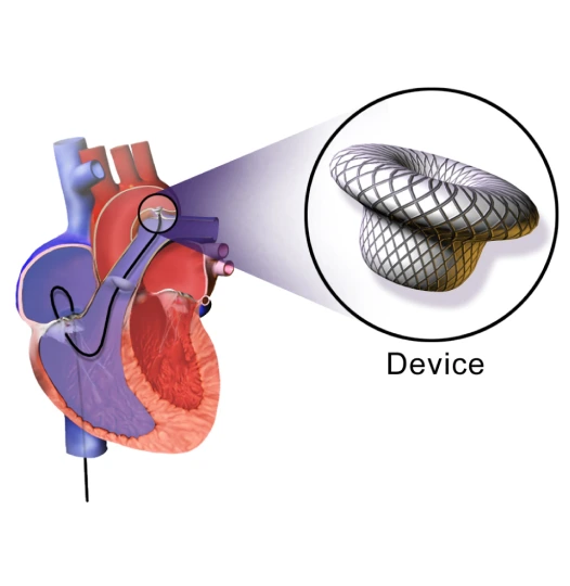
Acyanotic congenital heart diseases are structural defects in the heart that are present from birth, most commonly taking the form of a “hole in the heart” such as an Atrial Septal Defect (ASD), Ventricular Septal Defect (VSD), or Patent Ductus Arteriosus (PDA). These holes cause oxygen-rich blood to abnormally mix with oxygen-poor blood, forcing the heart and lungs to work much harder than they should. If left untreated, this can lead to frequent lung infections, heart enlargement, arrhythmias, and eventually heart failure. Historically, repairing these defects required open-heart surgery. Today, expert interventional cardiologists can permanently seal these holes using a minimally invasive, catheter-based device closure. This advanced technique safely plugs the defect from the inside, restoring normal blood flow without the need for a major chest incision, leaving no surgical scar, and offering a rapid recovery.
1. Indications:
Device closure is the preferred, gold-standard treatment for patients with a suitable heart anatomy who require closure of a congenital defect. Common indications include:
- Diagnosis of an Atrial Septal Defect (ASD), Ventricular Septal Defect (VSD), or Patent Ductus Arteriosus (PDA) causing a significant abnormal blood flow (left-to-right shunt)
- Symptoms such as extreme fatigue, shortness of breath during exercise, or a rapid, fluttering heartbeat (palpitations)
- Frequent respiratory infections or poor physical growth and weight gain in children
- Echocardiogram results showing enlargement of the right side of the heart or high pressure in the lungs (pulmonary hypertension)
- Patients who strongly prefer a minimally invasive alternative to open-heart surgery
- Patients whose defects have a distinct “rim” of tissue around the hole, allowing the closure device to securely grip the edges
Closing these defects early prevents permanent damage to the heart muscle and lungs, ensuring a normal, healthy life expectancy.
2. Procedure:
A device closure procedure is performed in an advanced cardiac cath lab. Depending on the patient’s age and the complexity of the hole, it is done under local anesthesia with sedation for adults, or general anesthesia for children. A small puncture is made in a blood vessel in the groin (the femoral vein or artery).
Through this small access point, a thin, flexible catheter is gently navigated up to the heart. The cardiologist uses continuous X-ray imaging (fluoroscopy) and a specialized internal ultrasound—either a Transesophageal Echo (TEE) or Intracardiac Echo (ICE)—to see the hole in high-definition 3D. A special measuring balloon is often used to determine the exact millimeter size of the defect.
Once sized, a tiny, collapsible closure device (typically made of flexible nitinol wire mesh shaped like a double-umbrella) is pushed through the catheter. As it exits the catheter, it springs open, sandwiching the hole securely from both sides and instantly stopping the abnormal blood flow. Once the doctor confirms it is perfectly positioned and stable, the device is released, and the catheter is removed. Over the next few months, the body’s natural heart tissue will permanently grow over the mesh device. The procedure takes 1 to 2 hours, and patients are usually discharged the next day.
3. Safety:
Catheter-based device closure is highly successful and exceptionally safe when performed by a specialized interventional cardiologist. By avoiding the heart-lung bypass machine and a cracked breastbone, patients experience significantly less pain, a lower risk of infection, and return to normal activities within days rather than months.
Continuous monitoring of vital signs and heart rhythm is strictly maintained throughout the procedure. Possible but uncommon risks include:
- Bleeding, bruising, or a hematoma at the catheter insertion site in the groin
- Dislodgment or movement of the device immediately after placement (a rare event that requires the doctor to retrieve it with a catheter or through surgery)
- Irregular heartbeats (arrhythmias), such as atrial fibrillation or heart block, which may be temporary or occasionally require a pacemaker
- A small residual leak around the edges of the device (most small leaks close on their own over time)
- A rare risk of blood clots or infection on the device (endocarditis), which is why patients are prescribed preventative antibiotics for dental work for a few months after the procedure