

CT Coronary Angiogram in Hyderabad
Home > Services > CT Coronary Angiogram in Hyderabad
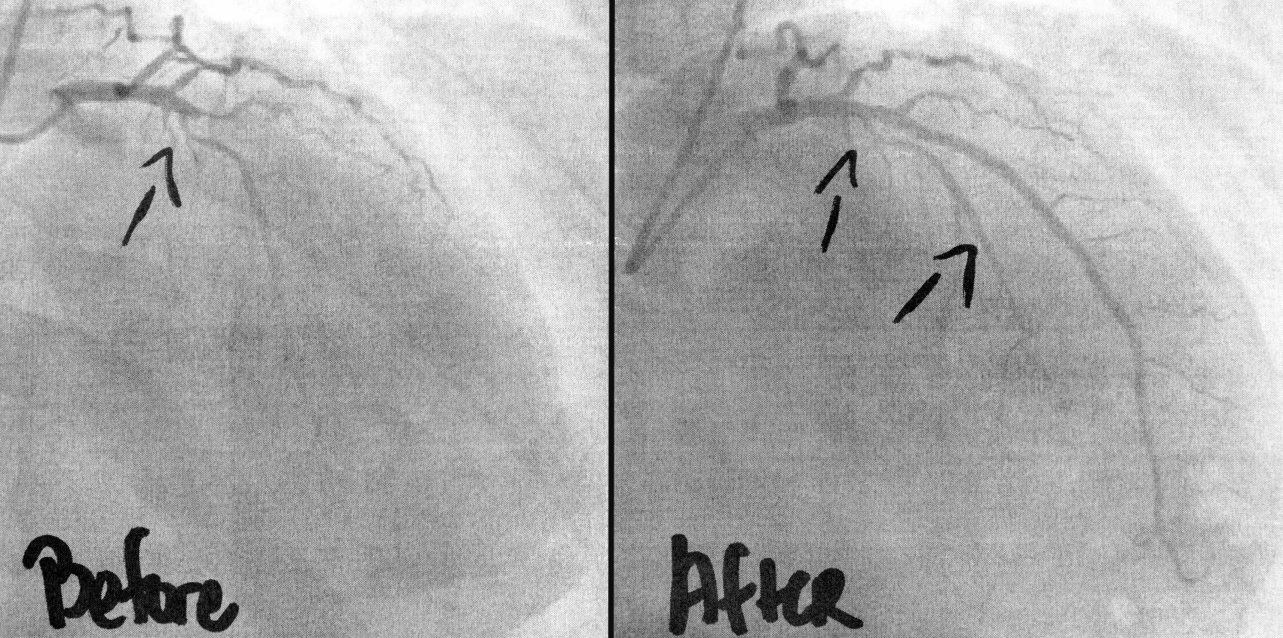
A CT Coronary Angiogram, also known as Coronary CT Angiography or CCTA, is a non-invasive imaging test used to obtain detailed pictures of the coronary arteries that supply blood to the heart muscle. The test combines a modern CT scanner with intravenous contrast material to show the inside of the coronary arteries and identify plaque, narrowing, or other abnormalities.
In Hyderabad, a CT Coronary Angiogram may be recommended when a patient has chest discomfort, unexplained breathlessness, cardiovascular risk factors, or an inconclusive result from another cardiac investigation. Unlike a conventional coronary angiogram, CCTA usually does not require a catheter to be inserted into an artery. It is primarily a diagnostic test and can provide detailed information about coronary anatomy without an invasive procedure.
What Is a CT Coronary Angiogram?
The coronary arteries are small blood vessels that run over the surface of the heart and provide oxygen-rich blood to the heart muscle. Over time, cholesterol, calcium, and other substances can accumulate in their walls and form atherosclerotic plaque. As plaque increases, the artery may become narrowed and blood flow may be affected.
During CCTA, an iodine-based contrast material is injected through a vein in the arm. The contrast makes the coronary arteries appear clearly on CT images. The scanner takes multiple images very quickly while the examination is synchronized with the patient’s ECG. Computer software then reconstructs these images into detailed views of the coronary arteries and heart.
CCTA can show both calcified and non-calcified plaque and can estimate the severity and location of coronary narrowing. This is different from a Treadmill Stress Test, which evaluates the heart’s response to exercise but does not directly show the coronary arteries.
Why Is CCTA Recommended?
A cardiologist may recommend CCTA when symptoms raise concern about coronary artery disease and detailed anatomical information is required. Chest pressure, heaviness, or discomfort during activity can have several possible causes. CCTA can help determine whether significant plaque or narrowing is present in the coronary arteries.
The test may also be considered when previous investigations, such as an ECG, TMT, or stress imaging study, do not provide a clear answer. In appropriately selected patients, seeing the coronary arteries directly can help determine whether symptoms are likely to be related to coronary disease or whether another cause should be investigated.
CCTA may also provide useful information about certain congenital coronary artery abnormalities, selected bypass grafts, and other structural features around the heart. Whether it is the most appropriate test depends on symptoms, age, heart rhythm, kidney function, previous cardiac procedures, and the probability of coronary artery disease.
What Can a CT Coronary Angiogram Show?
One of the major advantages of CCTA is its ability to show the coronary artery wall and plaque, not simply the open channel through which blood flows. Plaque may be calcified, non-calcified, or mixed. The report can describe where plaque is located and whether it produces mild, moderate, or severe narrowing.
A CCTA can also show whether the major coronary arteries have no significant visible plaque. In the appropriate clinical situation, this can be reassuring and may help the cardiologist consider other explanations for chest symptoms.
The scan may identify anatomical variations in how the coronary arteries originate or travel around the heart. It may also provide information about the aorta, heart chambers, and nearby structures visible within the scanned area.
However, CT images must be interpreted carefully. Heavy coronary calcium, very rapid or irregular heart rhythm, movement during scanning, obesity, or very small vessels can sometimes reduce image quality.
How Is CCTA Different From a Calcium Score?
A coronary calcium score and CT Coronary Angiogram are both CT-based cardiac tests, but they answer different questions. A calcium-score scan is generally performed without contrast and measures calcified plaque in the coronary arteries. It is mainly used in selected patients to help estimate future cardiovascular risk and guide preventive treatment decisions.
CCTA uses intravenous contrast and is designed to show the coronary artery lumen and plaque in much greater anatomical detail. It can identify both calcified and non-calcified plaque and assess whether the artery is narrowed.
A calcium score therefore should not be considered a substitute for CCTA when a patient has symptoms that require anatomical assessment of the coronary arteries. The cardiologist chooses the appropriate investigation according to the clinical situation.
How Should You Prepare?
Preparation is important because clear coronary images depend partly on having a suitable heart rate and minimizing movement. You may be instructed to avoid caffeine before the test because caffeine can increase heart rate. Specific fasting instructions may also be provided by the imaging centre.
Tell the clinical team about your current medicines, kidney disease, diabetes, asthma, allergies, previous reactions to contrast material, and any possibility of pregnancy. Because iodinated contrast is used, kidney function may need to be checked before the scan in patients with relevant risk factors.
Medicines may sometimes be given before or during CCTA to improve image quality. A beta blocker can be used in suitable patients to slow the heart rate. Nitroglycerin may also be given to widen the coronary arteries and improve visualization. These medicines are not appropriate for everyone, so the clinical team checks blood pressure, medical history, and contraindications before administering them.
Do not stop your regular medicines unless your doctor or imaging team specifically advises you to do so.
What Happens During the Scan?
Before scanning, ECG electrodes are placed on the chest so the CT scanner can synchronize image acquisition with the heartbeat. An intravenous cannula is inserted into a vein, usually in the arm, for contrast administration.
You lie on the scanning table, usually with your arms positioned above your head. If required, medicine is given to control the heart rate. Nitroglycerin may also be administered when appropriate.
When contrast is injected, you may temporarily feel warmth spreading through the body or notice a metallic taste. These sensations usually disappear quickly. The table then moves through the CT scanner while images are obtained. You will normally be asked to hold your breath for a few seconds so that chest movement does not blur the coronary arteries.
The actual image acquisition is usually fast, although preparation, heart-rate control, IV placement, and post-scan observation make the overall appointment longer.
Understanding Your CCTA Results
The report usually describes each major coronary artery and whether plaque or narrowing is present. If disease is identified, the report may describe plaque type, location, and estimated severity.
Mild plaque does not necessarily mean an angioplasty or stent is required. Many patients can be managed with cardiovascular risk reduction and appropriate medicines. More significant narrowing, particularly when it matches the patient’s symptoms, may require additional assessment to determine whether blood flow is being affected.
The cardiologist interprets CCTA together with symptoms, cholesterol levels, diabetes, blood pressure, family history, ECG findings, and other investigations. A CT finding should not be considered in isolation.
If significant or uncertain disease is identified, further evaluation may include stress imaging, myocardial perfusion imaging, or invasive coronary angiography.
CCTA vs Invasive Coronary Angiography
CCTA usually requires only an intravenous injection and CT scanning. Invasive coronary angiography requires a catheter to be introduced through an artery, usually at the wrist or groin, and advanced toward the heart.
The major advantage of invasive angiography is that treatment can sometimes be performed during the same procedure. If a suitable significant narrowing is confirmed, balloon angioplasty or stenting may be possible.
CCTA is primarily diagnostic. It can often provide detailed coronary anatomy without arterial catheterization, but it does not replace invasive angiography in every patient. The best test depends on the clinical situation and likelihood that immediate treatment may be required.
Safety and Limitations
CCTA uses X-rays and therefore involves ionising radiation. Modern CT technology and ECG-based scanning techniques are designed to obtain diagnostic images while keeping radiation exposure as low as reasonably achievable.
Iodinated contrast is generally well tolerated, but allergic-type reactions can occasionally occur. Kidney function also needs consideration, particularly in patients with significant kidney disease. Women who are pregnant or may be pregnant should tell the medical team before the examination.
CCTA may be less accurate when there is extensive coronary calcification, uncontrolled rapid heart rate, irregular rhythm, motion, or certain previously placed coronary stents. The cardiologist may recommend a different investigation when these factors are expected to significantly limit image quality.
Frequently Asked Questions:-
Is a CT Coronary Angiogram painful?
The scan itself is not painful. You will feel a small needle for the IV line and may experience temporary warmth when contrast is injected.
Can CCTA detect heart blockages?
Yes. CCTA can show coronary plaque and narrowing and can estimate how severely an artery appears narrowed.
Does CCTA use contrast?
Yes. Coronary CT angiography normally uses intravenous iodine-based contrast so that the coronary arteries can be seen clearly.
Does the test use radiation?
Yes. CCTA uses X-rays. Modern protocols aim to achieve the necessary image quality with the lowest reasonable radiation exposure.
Do I need to avoid caffeine?
You may be instructed to avoid coffee, tea, energy drinks, and other caffeinated products before the scan because caffeine can increase heart rate. Follow the instructions from your imaging centre.
Is CCTA the same as a conventional coronary angiogram?
No. CCTA is a non-invasive CT-based diagnostic test, while conventional coronary angiography requires arterial catheterization and may allow angioplasty or stenting.
What happens if a significant narrowing is found?
Your cardiologist will consider the severity of the finding, symptoms, and other test results. Additional functional testing or invasive coronary angiography may be recommended when appropriate.
CT Coronary Angiogram in Hyderabad
If you have symptoms suggestive of coronary artery disease or your cardiologist needs detailed information about coronary plaque and narrowing, a CT Coronary Angiogram may be an appropriate investigation.
Dr. Kiran Lakkireddy’s cardiac practice in Hyderabad provides evaluation of heart-related symptoms and guidance on appropriate diagnostic testing. The decision to undergo CCTA should be based on your symptoms, cardiovascular risk, kidney function, heart rhythm, previous investigations, and overall clinical condition.