

CATH STUDY FOR CONGENITAL HEART DISEASES
Home > Services > Cath Study Congenital Heart Disease
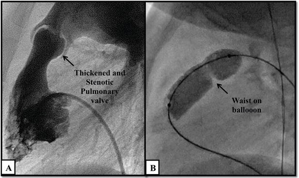
A cardiac catheterization study (often called a “cath study”) is a highly specialized, minimally invasive diagnostic procedure used to precisely evaluate structural heart defects present at birth (congenital heart diseases). While non-invasive tests like echocardiograms provide excellent pictures of the heart’s anatomy, they cannot always measure exact internal pressures or oxygen levels. A cath study provides a real-time, highly detailed “inside view” by measuring exact blood pressure, oxygen saturation, and blood flow dynamics (hemodynamics) directly within the heart’s chambers and the lungs. This critical data helps pediatric cardiologists and adult congenital heart specialists understand the exact severity of the defect, determine if a patient is a candidate for surgery, or evaluate how well the heart is functioning after a previous repair.
1. Indications:
A diagnostic cath study is recommended when doctors need definitive, internal measurements that cannot be obtained through standard imaging to plan the safest course of treatment. Common indications include:
Detailed mapping of complex congenital anomalies, such as Tetralogy of Fallot, single ventricle defects, or complex septal defects (holes in the heart)
Measuring pulmonary artery pressures to diagnose pulmonary hypertension, which can determine if a patient is a safe candidate for specific corrective surgeries (like the Fontan procedure)
Performing an “oximetry run” to draw blood samples from various heart chambers, identifying exactly where oxygen-rich and oxygen-poor blood are abnormally mixing (shunting)
Evaluating the success of a previous open-heart surgery, checking for new leaks, narrowing, or post-surgical complications
Serving as a diagnostic prelude immediately before an interventional procedure such as a balloon valvotomy or placing a closure device performed during the same session
By providing exact physiological data, a cath study takes the guesswork out of treating complex congenital heart disease.
2. Procedure:
A cath study is performed in an advanced pediatric or adult congenital cardiac cath lab. Depending on the patient’s age and medical condition, it is done under local anesthesia with deep sedation or, more commonly for infants and young children, under general anesthesia so they remain completely still and pain-free.
The doctor makes a small puncture in a major blood vessel, typically in the groin (the femoral vein or artery). In newborns, the umbilical vessels in the belly button or a vein in the neck may be used instead.
Through this tiny access point, thin, flexible tubes (catheters) are gently navigated up to the heart. The cardiologist uses continuous X-ray imaging (fluoroscopy) to track the catheter’s position perfectly. Once inside the heart, the doctor records precise pressure readings in each chamber and takes small blood samples to check oxygen levels. Contrast dye is often injected (an angiogram) to record high-definition X-ray video of blood flowing through the heart and lungs, revealing the exact size and shape of the defects. Once all data is collected, the catheters are carefully removed, and pressure is applied to the access site to stop any bleeding. The diagnostic portion usually takes 1 to 2 hours.
3. Safety:
A cardiac cath study is an incredibly safe and routine procedure when performed by a specialized congenital cardiology team. It avoids the need for a surgical incision and allows for a rapid recovery, with most patients going home the same day or the next morning.
Continuous, rigorous monitoring of vital signs and heart rhythm is strictly maintained throughout the entire procedure. While major complications are rare, possible risks include:
- Bleeding, bruising, or a hematoma at the catheter insertion site (groin, neck, or umbilicus)
- Temporary, irregular heartbeats (arrhythmias) as the catheter touches the sensitive internal walls of the heart muscle, which usually resolve on their own once the catheter is moved
- A temporary decline in kidney function due to the contrast dye used for X-ray imaging (preventative IV fluids are given to flush the kidneys)
- Allergic reaction to the contrast dye or anesthesia medications
- A rare risk of injury or tearing to a blood vessel or the heart wall
- Decreased pulse in the leg or arm used for access, particularly in very small infants, which usually improves with blood-thinning medication