

Deep Vein Thrombosis Treatment in Hyderabad
Home > Services > Deep Vein Thrombosis Treatment in Hyderabad
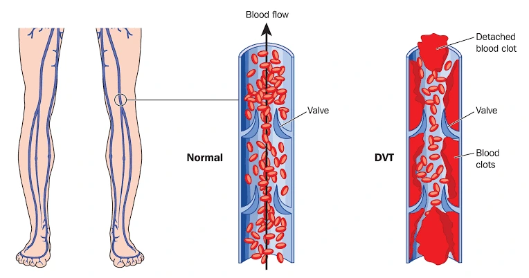
Deep vein thrombosis, commonly called DVT, is a condition in which a blood clot forms in a deep vein, most often in the lower leg, thigh, or pelvis. It can also occur in the arm. DVT is important because part of the clot can break away, travel through the bloodstream, and block an artery in the lungs. This complication is called pulmonary embolism, or PE, and it can be life-threatening.
Some people with DVT develop obvious leg swelling and pain, while others have mild symptoms or no symptoms at all. Early diagnosis is important because treatment can reduce the risk of clot extension, pulmonary embolism, and long-term complications such as post-thrombotic syndrome.
What Is Deep Vein Thrombosis?
Blood normally flows through the veins back toward the heart. A clot can form when blood flow becomes slow, the inner lining of a vein is injured, or the blood becomes more likely to clot. These three mechanisms are often described together as Virchow’s triad.
DVT most commonly affects the deep veins of the legs. A clot limited to the calf may behave differently from a clot that extends into the popliteal, femoral, or iliac veins. Proximal DVT is generally more concerning because it carries a greater risk of pulmonary embolism and often requires anticoagulant treatment.
What Causes DVT?
DVT can occur after surgery, hospitalization, major injury, prolonged bed rest, or long periods of reduced mobility. Long-distance travel can also contribute when several risk factors are present and a person remains seated for many hours.
Other risk factors include active cancer, previous DVT or pulmonary embolism, pregnancy and the postpartum period, obesity, increasing age, estrogen-containing medicines, and some inherited or acquired clotting disorders. Central venous catheters can increase the risk of upper-extremity DVT.
Sometimes DVT occurs without an obvious temporary trigger, which can affect decisions about the duration of anticoagulation.
What Are the Symptoms of DVT?
The most typical symptoms are swelling, pain or tenderness, warmth, and redness or discoloration in the affected leg or arm. Leg DVT often causes swelling on one side rather than both sides. The calf or thigh may feel tight, heavy, or painful, especially while walking or standing.
However, DVT cannot be diagnosed from symptoms alone. Muscle injury, infection, venous insufficiency, and other conditions can cause similar symptoms. Importantly, about half of people with DVT may have no symptoms.
New unexplained one-sided leg swelling or pain, particularly in a person with recent surgery, hospitalization, immobility, cancer, pregnancy, or a previous clot, should be assessed promptly.
Why Can DVT Be Dangerous?
The most serious early complication of DVT is pulmonary embolism. This happens when part of the clot breaks free and travels through the right side of the heart into the pulmonary arteries.
Warning symptoms of pulmonary embolism include sudden unexplained breathlessness, chest pain that may worsen with deep breathing, rapid heartbeat, coughing, coughing up blood, dizziness, or fainting. Severe breathlessness, collapse, confusion, or very low blood pressure can indicate a serious PE and requires emergency hospital care.
DVT can also cause long-term damage to the veins. Some patients later develop post-thrombotic syndrome, with persistent leg swelling, heaviness, pain, skin discoloration, or, in severe cases, venous ulcers.
How Is DVT Diagnosed?
Diagnosis begins with the patient’s symptoms, medical history, examination, and assessment of the clinical probability of DVT. Because signs and symptoms are not specific, objective testing is usually required.
A D-dimer blood test may be useful in selected patients with a low or intermediate probability of DVT. A negative result can sometimes help exclude a clot without additional imaging. D-dimer levels can also rise for many other reasons, so an elevated result does not by itself confirm DVT.
Compression ultrasonography, often performed as a venous Doppler ultrasound, is the most commonly used imaging test for suspected leg DVT. It evaluates the deep veins and helps identify clot formation. Repeat ultrasound or additional imaging may occasionally be needed when clinical suspicion remains high despite an initially negative study.
For suspected DVT involving the pelvic veins, abdomen, or other difficult-to-image areas, CT venography, MR venography, or other imaging may be considered depending on the clinical situation.
How Is Deep Vein Thrombosis Treated?
Anticoagulation is the main treatment for most patients with acute DVT. These medicines are often called blood thinners, although they do not literally thin the blood. They reduce the blood’s ability to form additional clot and help prevent the existing clot from extending while the body gradually breaks it down.
Several anticoagulant options are available, including direct oral anticoagulants, heparin-based medicines, and warfarin. The choice depends on factors such as kidney function, pregnancy, cancer, bleeding risk, other medicines, and whether a procedure is planned.
The duration of treatment is individualized. A clot caused by a major temporary risk factor may require a defined treatment period, while an unprovoked DVT, recurrent DVT, or persistent risk factor may lead to consideration of extended anticoagulation. The decision balances the risk of another clot against the risk of bleeding.
When Are Thrombolysis or Thrombectomy Considered?
Most patients with DVT do not need a clot-removal procedure. Anticoagulation alone is sufficient in many cases.
Catheter-directed thrombolysis or mechanical thrombectomy may be considered in selected patients with extensive acute DVT, particularly when a large clot affects the iliac or femoral veins and causes severe symptoms, threatens the limb, or creates concern for major long-term venous problems. Because these treatments also have bleeding and procedural risks, they are not routine treatments for every DVT.
In rare cases of severe venous obstruction with threatened circulation to the limb, urgent specialist intervention may be required.
Are Compression Stockings Needed?
Compression stockings may help selected patients with persistent swelling or discomfort after DVT, but they are not automatically required for everyone. Their use should be individualized according to symptoms and medical advice.
Patients are often encouraged to resume safe mobility once anticoagulation has been started and the treating team considers it appropriate. Prolonged unnecessary bed rest is generally avoided.
Can DVT Be Prevented?
Prevention is especially important during hospitalization, after major surgery, or during periods of reduced mobility. Depending on individual risk, prevention may include early walking, leg exercises, mechanical compression devices, or preventive-dose anticoagulant medicines.
During long journeys, people at risk can benefit from regular movement and calf exercises. People with previous DVT should discuss travel, surgery, pregnancy, and interruption of anticoagulants with their treating doctor.
Frequently Asked Questions:-
Can DVT go away on its own?
The body can gradually break down a clot, but suspected DVT should not simply be left untreated. Anticoagulation is used in many patients to prevent the clot from growing or travelling to the lungs while natural clot resolution occurs.
Is every swollen leg caused by DVT?
No. Leg swelling can result from many conditions. However, sudden one-sided swelling, especially with pain or risk factors for clotting, should be evaluated promptly.
Can DVT happen again?
Yes. A previous DVT increases the risk of another venous thromboembolism. Recurrence risk depends on what caused the first clot, whether persistent risk factors remain, and whether anticoagulation has been stopped.
Can walking make a DVT move to the lungs?
Appropriate walking is generally encouraged after treatment is started and the patient is clinically stable. Patients should follow the advice of their treating team, particularly if symptoms are severe or an intervention is being considered.
Does every DVT need lifelong blood thinners?
No. The duration of anticoagulation varies. Some patients need a limited course, while others with recurrent or unprovoked clotting may benefit from extended treatment.
When should emergency care be sought?
A person with possible or confirmed DVT who develops sudden breathlessness, significant chest pain, fainting, coughing up blood, or sudden severe deterioration should seek emergency hospital care because these symptoms may indicate pulmonary embolism.
Deep Vein Thrombosis Treatment in Hyderabad
New one-sided leg swelling, pain, warmth, or discoloration should be evaluated promptly, especially when there has been recent surgery, hospitalization, prolonged immobility, cancer, pregnancy, or a previous blood clot.
Dr. Kiran Lakkireddy’s cardiac practice in Hyderabad provides cardiovascular evaluation for patients with suspected or established thromboembolic disease, including assessment of pulmonary embolism risk and associated cardiopulmonary effects when appropriate. DVT diagnosis commonly relies on clinical assessment, D-dimer testing in selected patients, and venous Doppler ultrasonography. Treatment may include anticoagulation and referral for specialist vascular or catheter-based intervention when clinically indicated.
Sudden chest pain, breathlessness, fainting, or coughing up blood requires urgent hospital assessment rather than a routine consultation.