

Cardiomyopathy Evaluation and Treatment in Hyderabad
Home > Services > Cardiomyopathy Evaluation and Treatment in Hyderabad
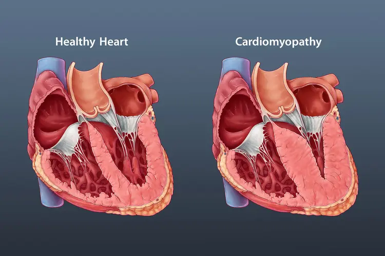
Cardiomyopathy is a group of diseases that affect the heart muscle and can make it harder for the heart to pump blood effectively. Depending on the type, the heart muscle may become enlarged and weak, abnormally thick, or unusually stiff. These changes can lead to breathlessness, fatigue, swelling, palpitations, fainting, heart failure, or abnormal heart rhythms. Some people have no symptoms in the early stages and are diagnosed only after an ECG, echocardiogram, family screening, or another cardiac test.
In Hyderabad, cardiomyopathy evaluation may be recommended when symptoms suggest heart muscle disease, when an echocardiogram shows abnormal chamber size or wall thickness, or when there is a family history of cardiomyopathy or sudden cardiac death. The three major forms covered on this page are Dilated Cardiomyopathy (DCM), Hypertrophic Cardiomyopathy (HCM), and Restrictive Cardiomyopathy (RCM).
What Is Cardiomyopathy?
The heart is a muscular pump. Cardiomyopathy changes the structure or function of that muscle. In dilated cardiomyopathy, one or both ventricles can enlarge and the pumping action becomes weaker. In hypertrophic cardiomyopathy, the heart muscle becomes abnormally thick, sometimes obstructing blood flow out of the heart. In restrictive cardiomyopathy, the ventricular walls become stiff and cannot relax normally, so the chambers do not fill properly.
The causes are different for each patient. Cardiomyopathy may be inherited, related to long-standing high blood pressure, previous heart muscle injury, viral or inflammatory disease, alcohol or certain toxins, pregnancy-related changes, metabolic conditions, or infiltrative diseases. In some people, no clear cause is found despite careful investigation.
Dilated Cardiomyopathy
Dilated Cardiomyopathy is characterized by enlargement of the heart’s main pumping chamber, usually the left ventricle, together with reduced contraction. As the ventricle stretches, the heart may not pump enough blood to meet the body’s needs. Over time this can lead to heart failure, valve leakage, rhythm abnormalities, or blood clot formation.
Symptoms can include breathlessness during activity or at rest, unusual tiredness, swelling of the feet or legs, reduced exercise capacity, palpitations, or episodes of dizziness. Some people are diagnosed after an abnormal ECG or echocardiogram before significant symptoms develop.
Treatment depends on the cause and severity. Medicines used for heart failure can reduce symptoms, improve pumping efficiency, and lower the risk of progression. Selected patients may need treatment for arrhythmias, blood clots, or valve problems. If the heart’s electrical activation is significantly uncoordinated, cardiac resynchronisation therapy may be considered. An implantable cardioverter-defibrillator may be recommended in selected higher-risk patients to reduce the risk of sudden cardiac death.
Hypertrophic Cardiomyopathy
Hypertrophic Cardiomyopathy causes abnormal thickening of the heart muscle, most often involving the left ventricle and the wall between the two ventricles. HCM is frequently inherited and can occur in several members of the same family. The degree of thickening and its effect on blood flow vary widely.
Some patients remain symptom-free, while others develop exertional breathlessness, chest discomfort, palpitations, dizziness, or fainting. In obstructive HCM, thickened muscle can narrow the pathway through which blood leaves the left ventricle. The mitral valve may also move abnormally and contribute to obstruction or leakage.
Evaluation includes assessing the degree and location of thickening, whether obstruction is present, heart function, rhythm abnormalities, and individual risk factors for sudden cardiac death. Treatment may involve medicines that slow the heart rate and improve filling. Selected symptomatic patients with significant obstruction despite medical treatment may require specialist therapies such as septal reduction procedures. Patients at increased risk of dangerous ventricular rhythms may be considered for an implantable defibrillator.
Restrictive Cardiomyopathy
Restrictive Cardiomyopathy is less common. In this condition, the ventricles become stiff and cannot relax normally between heartbeats. Pumping strength may initially appear relatively preserved, but impaired relaxation means the heart cannot fill efficiently. Pressure can then build up behind the heart, causing congestion and symptoms of heart failure.
Patients may develop breathlessness, swelling, fatigue, abdominal fullness, or exercise intolerance. Restrictive cardiomyopathy can occur because of abnormal material or scar tissue within the heart muscle. Conditions such as cardiac amyloidosis, iron overload, sarcoidosis, and certain other systemic diseases may cause a restrictive pattern.
Because treatment depends strongly on the underlying cause, identifying the specific disease is especially important. Management may include medicines to control congestion, treatment of arrhythmias, anticoagulation when indicated, and disease-specific therapy when an infiltrative or systemic cause is identified.
How Is Cardiomyopathy Diagnosed?
Evaluation starts with symptoms, medical history, family history, and physical examination. The doctor will ask about breathlessness, fainting, palpitations, swelling, exercise tolerance, previous infections, alcohol or medicine exposure, and relatives with cardiomyopathy, heart failure, or sudden unexplained death.
A 12-lead ECG can show rhythm abnormalities or electrical patterns that suggest heart muscle disease. A 2D Echocardiogram is one of the most important tests because it shows chamber size, wall thickness, pumping function, valve function, and blood flow. In HCM, echocardiography can also assess obstruction to blood leaving the left ventricle.
Cardiac MRI can provide more detailed assessment of heart muscle structure and may identify scar or fibrosis. Holter Monitoring may be used to look for intermittent arrhythmias. Blood tests can help identify thyroid disease, iron abnormalities, kidney or liver problems, or other possible causes. In selected patients, coronary imaging or additional specialist investigations may be required.
Because some cardiomyopathies are inherited, family screening and genetic counselling or testing may be considered when the clinical pattern suggests a genetic condition.
Understanding Your Results
A diagnosis of cardiomyopathy is not based on one measurement alone. Ejection fraction is important in dilated cardiomyopathy, but chamber size, symptoms, rhythm findings, valve function, scar, and the underlying cause also influence treatment. In hypertrophic cardiomyopathy, the amount of wall thickening is important, but the presence of obstruction, fainting history, rhythm abnormalities, family history, and other risk markers also matter.
The cardiologist therefore combines ECG, echocardiography, MRI, rhythm monitoring, symptoms, and family history to understand both the type of cardiomyopathy and the patient’s individual risk. Follow-up is often necessary because heart structure, symptoms, and rhythm can change over time.
Treatment and Long-Term Care
Treatment is individualized. Many patients need medicines to reduce the workload on the heart, improve heart failure symptoms, control heart rate or rhythm, remove excess fluid, or reduce the risk of blood clots. The exact combination depends on the type of cardiomyopathy and associated conditions.
Lifestyle advice is also individualized. Patients may be advised about salt intake, alcohol, body weight, blood pressure, exercise, and pregnancy depending on their condition. Exercise is generally beneficial for cardiovascular health, but people with certain cardiomyopathies may need personalized advice about intensity, competitive sports, or strenuous activity.
Advanced treatment can include pacemakers, cardiac resynchronisation therapy, implantable cardioverter-defibrillators, catheter-based procedures, septal myectomy for selected HCM patients, ventricular assist devices, or heart transplantation in severe end-stage disease.
Frequently Asked Questions:-
Is cardiomyopathy the same as heart failure?
No. Cardiomyopathy is disease of the heart muscle. It can cause heart failure, but not every person with cardiomyopathy has heart failure at diagnosis.
Can cardiomyopathy be inherited?
Yes. Some forms, particularly hypertrophic cardiomyopathy and some dilated cardiomyopathies, can run in families. Family screening may therefore be recommended.
Can cardiomyopathy cause sudden cardiac arrest?
Certain cardiomyopathies can increase the risk of dangerous ventricular arrhythmias and sudden cardiac death. Risk varies considerably, so individualized assessment is important.
Can cardiomyopathy be cured?
Some causes may improve substantially when the underlying trigger is treated, while inherited or chronic forms often require long-term monitoring and treatment. The goal is to control symptoms, improve heart function where possible, and reduce complications.
Which test is most important for cardiomyopathy?
Echocardiography is usually central to the initial assessment. ECG, Cardiac MRI, Holter Monitoring, blood tests, genetic evaluation, and other investigations may be added depending on the suspected type and cause.
Can a person with cardiomyopathy exercise?
Many patients can remain physically active, but the safest type and intensity of exercise depend on the cardiomyopathy, symptoms, rhythm risk, and treatment. Individual advice from the cardiology team is recommended.
Cardiomyopathy Evaluation and Treatment in Hyderabad
If you have unexplained breathlessness, reduced exercise tolerance, swelling, recurrent palpitations, fainting, an abnormal echocardiogram, or a family history of cardiomyopathy or sudden cardiac death, a detailed cardiac evaluation can help determine whether heart muscle disease is present.
Dr. Kiran Lakkireddy’s cardiac practice in Hyderabad provides evaluation of cardiomyopathy and related symptoms, with testing selected according to the suspected type, severity, family history, and clinical findings. Dilated, hypertrophic, and restrictive cardiomyopathies require different approaches, so identifying the exact pattern and underlying cause is an important part of planning treatment and long-term follow-up.