Carotid & Cerebral Angiogram
Home > Services > Carotid & Cerebral Angiogram
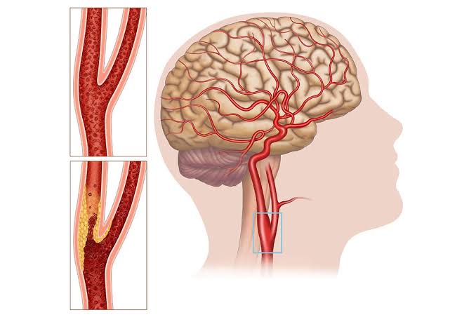
A carotid and cerebral angiogram is a minimally invasive diagnostic procedure that uses X-rays and a specialized contrast dye to evaluate the blood flow in the arteries of your neck (carotid arteries) and brain (cerebral arteries). When these vital blood vessels become narrowed by cholesterol plaque or are affected by structural abnormalities, it significantly increases the risk of a stroke or brain hemorrhage. This highly detailed imaging test provides the doctor with a clear, real-time map of the blood vessels supplying your brain, helping to pinpoint exact blockages, aneurysms, or malformations so the safest and most effective treatment plan can be developed.
1. Indications:
A carotid and cerebral angiogram is recommended for patients who have symptoms of restricted blood flow to the brain or suspected vascular abnormalities. Common indications include:
- Symptoms of a transient ischemic attack (TIA) or “mini-stroke” (e.g., sudden weakness, slurred speech, facial drooping)
- History of a recent stroke
- A carotid bruit (an abnormal whooshing sound in the neck heard by a doctor with a stethoscope)
- Suspicion or diagnosis of brain aneurysms or arteriovenous malformations (AVMs)
- Unexplained severe headaches, dizziness, or sudden blurred vision
- Abnormal findings on non-invasive tests like a Carotid Doppler ultrasound, CT angiogram, or MR angiogram
- Planning for a therapeutic procedure such as carotid artery stenting or neurosurgery
Early diagnosis of neck and brain vessel abnormalities is critical in preventing life-threatening strokes and permanent neurological damage.
2. Procedure:
A carotid and cerebral angiogram is performed in a cath lab under local anesthesia with strict sterile precautions. You will be awake and able to communicate with the doctor, but relaxed. A small puncture is made in an artery, usually in the groin (femoral artery) or the wrist (radial artery).
Through this puncture, a thin, flexible catheter is gently advanced through your blood vessels and guided up to the carotid or vertebral arteries in the neck using continuous X-ray imaging. Once the catheter is perfectly positioned, a contrast dye is injected. This dye highlights the blood vessels on the monitors, creating a detailed “roadmap” of the circulation inside your head and neck. You may feel a brief, warm sensation in your head, neck, or face as the dye is injected, which is completely normal and passes quickly.
If a severe narrowing or abnormality is identified, the doctor can determine if interventions like carotid stenting or aneurysm coiling are necessary. The diagnostic procedure itself typically takes about 45 minutes to an hour.
3. Safety:
Carotid and cerebral angiograms are safe and highly effective diagnostic tools when performed by experienced specialists in a well-equipped cath lab. Continuous monitoring of your heart rate, blood pressure, oxygen levels, and neurological status is maintained throughout the procedure.
Possible but uncommon risks include:
- Bleeding or hematoma at the puncture site
- Vessel injury or dissection
- Contrast allergy
- Kidney function changes due to the dye
- Clot formation or embolism
- Stroke or TIA (a very rare risk, minimized by the use of specialized techniques and catheters)