

Valvular Heart Disease Treatment in Hyderabad
Home > Services >
Valvular Heart Disease Treatment in Hyderabad
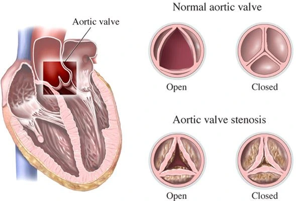
Valvular heart disease refers to problems that affect one or more of the four heart valves: the aortic, mitral, tricuspid, and pulmonary valves. These valves open and close with every heartbeat to keep blood moving in the correct direction. When a valve becomes narrowed and does not open properly, the condition is called stenosis. When a valve does not close completely and allows blood to leak backward, it is called regurgitation.
In Hyderabad, valvular heart disease evaluation may be needed when a person develops unexplained breathlessness, reduced exercise capacity, chest discomfort, palpitations, dizziness, fainting, or swelling of the legs or abdomen. Some valve problems cause few symptoms in the early stages, so regular follow-up is important once a murmur or structural valve abnormality has been identified.
What Is Valvular Heart Disease?
The heart contains four valves that control blood flow between the chambers and into the major arteries. The mitral and tricuspid valves lie between the upper and lower chambers, while the aortic and pulmonary valves control blood leaving the ventricles. For the heart to work efficiently, each valve must open fully and close tightly.
Valve stenosis makes it harder for blood to move forward. The heart has to generate higher pressure to push blood through the narrowed opening, which can eventually cause thickening of the heart muscle, enlargement of chambers, reduced exercise tolerance, and heart failure. Valve regurgitation creates the opposite problem: blood leaks backward each time the heart contracts, increasing the amount of work the heart must do and sometimes causing progressive chamber enlargement and weakening.
A valve abnormality can be mild and remain stable for years, or it can gradually become severe. Symptoms alone do not always reflect severity, which is why echocardiographic monitoring is important.
Aortic Stenosis
Aortic stenosis occurs when the aortic valve becomes narrowed and cannot open fully. In older adults, this commonly develops because calcium accumulates on the valve over time. Some people are born with a bicuspid aortic valve and may develop narrowing earlier in life.
As aortic stenosis progresses, the left ventricle has to pump against increasing resistance. Patients may develop exertional breathlessness, chest discomfort, dizziness, fainting, or declining exercise capacity. Severe symptomatic aortic stenosis usually requires valve replacement rather than medicines alone.
Treatment options include surgical aortic valve replacement and transcatheter aortic valve implantation or replacement, commonly called TAVI or TAVR. The choice depends on age, anatomy, surgical risk, life expectancy, associated coronary disease, and discussion by the heart team.
Mitral Stenosis
Mitral stenosis is narrowing of the mitral valve between the left atrium and left ventricle. Rheumatic heart disease remains an important cause in many parts of the world. As the opening becomes smaller, pressure rises in the left atrium and lungs, which can cause breathlessness, reduced exercise tolerance, palpitations, or atrial fibrillation.
Treatment depends on severity, symptoms, valve anatomy, and pulmonary pressure. Medicines may help control heart rate or congestion but do not physically open a severely narrowed valve. Selected patients with suitable rheumatic mitral stenosis may benefit from balloon mitral valvotomy, while others require surgery.
Mitral Regurgitation
Mitral regurgitation occurs when the mitral valve does not close tightly and blood leaks backward from the left ventricle into the left atrium. The problem may arise from mitral valve prolapse, degenerative valve disease, damage after a heart attack, cardiomyopathy, or enlargement of the ventricle.
Mild regurgitation may cause no symptoms. More significant leakage can lead to breathlessness, fatigue, palpitations, atrial fibrillation, and eventually heart failure. Echocardiography helps determine how severe the leak is and whether the left ventricle is beginning to enlarge or weaken.
When intervention is required, mitral valve repair is often preferred when a durable repair is feasible because it preserves the patient’s own valve. Valve replacement may be required when repair is not possible. Selected high-risk patients may also be considered for transcatheter mitral therapies depending on anatomy and the type of regurgitation.
Aortic and Tricuspid Regurgitation
Aortic regurgitation occurs when the aortic valve allows blood to leak back into the left ventricle after each heartbeat. Over time, the ventricle may enlarge to handle the extra volume. Patients can remain symptom-free for years, but progressive disease may eventually cause breathlessness, reduced exercise tolerance, or heart failure. Surgery may be needed when regurgitation becomes severe and symptoms or changes in ventricular size or function develop.
Tricuspid regurgitation affects the valve on the right side of the heart and can occur with pulmonary hypertension, right-heart enlargement, or atrial fibrillation. More advanced disease may cause leg swelling, abdominal distension, liver congestion, or fatigue. Treatment focuses on the cause and severity, and selected patients may require surgical or transcatheter valve intervention.
How Is Valvular Heart Disease Diagnosed?
Evaluation begins with symptoms, medical history, and examination. A heart murmur heard through a stethoscope may be the first clue, although a murmur alone does not determine how severe a valve problem is.
A 2D Echocardiogram is the main investigation for most valve disorders. It shows valve structure and movement, measures blood flow with Doppler, estimates the severity of stenosis or regurgitation, and assesses chamber size, heart pumping function, and pulmonary pressures. Repeat echocardiography may be recommended over time to monitor progression even when symptoms are mild.
Transesophageal Echocardiography, or TEE, can provide more detailed images when a standard echo does not answer the clinical question or when the valve anatomy needs to be studied before repair or intervention. CT imaging, Cardiac MRI, stress testing, or cardiac catheterization may also be used in selected patients when additional anatomical or functional information is required.
How Is Valvular Heart Disease Treated?
Treatment depends on which valve is affected, whether the problem is stenosis or regurgitation, how severe it is, and whether the heart has begun to change in response. Mild disease may only require regular follow-up and management of blood pressure and other cardiovascular risk factors.
Medicines can help control symptoms such as fluid retention, blood pressure, heart rate, or atrial fibrillation, but they do not directly reverse a severely narrowed valve or stop a significant mechanical leak. When valve disease becomes severe, intervention may be needed before permanent heart damage develops.
Valve repair preserves the patient’s own valve and is particularly important for selected mitral and tricuspid regurgitation. Valve replacement may involve a mechanical or biological prosthetic valve.
The timing of intervention is important and depends on symptoms, echocardiographic severity, ventricular response, age, anatomy, and overall health.
Living With Heart Valve Disease
Patients with known valve disease need regular follow-up even when they feel well. Changes in walking distance, breathlessness, swelling, palpitations, chest discomfort, or fainting should be reported because they may indicate progression.
The frequency of follow-up and repeat echocardiography depends on the valve involved and the severity of disease. Regular monitoring helps the cardiologist identify changes in valve function or heart response before symptoms become advanced.
Frequently Asked Questions:-
Can valve disease be present without symptoms?
Yes. Mild or even significant valve disease can sometimes be present before obvious symptoms develop. Regular clinical review and echocardiography help monitor severity and heart function.
Can medicines cure a narrowed or leaking heart valve?
Medicines can control symptoms and related conditions, but they do not mechanically open a severely stenotic valve or stop an important valve leak. Severe disease may require repair or replacement.
What is the difference between TAVI and open-heart valve surgery?
TAVI or TAVR places a new aortic valve through a catheter, usually through an artery in the groin, without traditional open-heart surgery. Surgical valve replacement involves removing the diseased valve and implanting a new valve through surgery. The best option depends on the individual patient.
Can a leaking mitral valve be repaired?
Yes. Many forms of mitral regurgitation can be repaired, particularly when the valve anatomy is suitable. Repair is often preferred over replacement when a durable result is expected.
How often is an echocardiogram needed?
The interval depends on the valve involved and the severity of disease. Mild disease may need less frequent monitoring, while moderate or severe disease usually requires closer follow-up.
When should valve disease be treated urgently?
New fainting, severe breathlessness, chest pain, rapidly worsening swelling, or sudden deterioration in someone with known valve disease should be assessed promptly.
Valvular Heart Disease Treatment in Hyderabad
If you have a heart murmur, known valve stenosis or regurgitation, unexplained breathlessness, reduced exercise tolerance, palpitations, fainting, or swelling, cardiology evaluation can determine which valve is affected and how severe the problem is.
Dr. Kiran Lakkireddy’s cardiac practice in Hyderabad provides evaluation of valvular heart disease using 2D Echocardiography, TEE, and other cardiac tests when appropriate. Treatment may range from regular monitoring and medicines to valve repair, surgical valve replacement, or transcatheter procedures such as TAVI/TAVR depending on the valve, severity, symptoms, and overall clinical condition.