

COARCTATION OF AORTA STENTING
Home > Services > Coarctation of Aorta Stenting
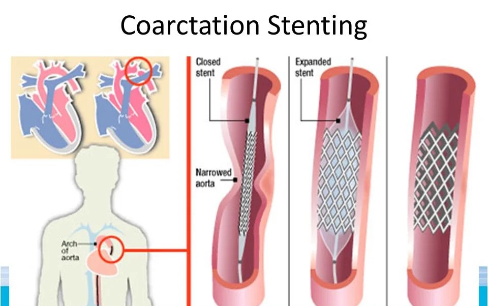
Coarctation of the aorta is a congenital heart defect characterized by a narrowing (pinching) of the aorta, the large main blood vessel that carries oxygen-rich blood from the heart to the rest of the body. This severe narrowing acts like a kink in a garden hose, forcing the heart to pump much harder to push blood through. As a result, patients often experience dangerously high blood pressure in their upper body and weak, poorly oxygenated blood flow to their lower body. In the past, correcting this required major open-heart surgery. Today, interventional cardiologists can successfully treat many cases using a minimally invasive stenting procedure. By safely expanding a permanent metal mesh tube inside the narrowed segment, the aorta is held wide open, instantly normalizing blood pressure and restoring healthy circulation throughout the body.
1. Indications:
Stenting for coarctation of the aorta is typically recommended for older children, adolescents, and adults whose anatomy is suitable for a catheter-based approach. Common indications include:
- A severe narrowing of the aorta causing a significant blood pressure difference (gradient) between the arms and the legs
- Severe, difficult-to-control high blood pressure (hypertension) in the upper body caused by the blockage
- Symptoms such as frequent headaches, shortness of breath, chest pain, or leg cramping and weakness during physical activity
- Heart enlargement or thickened heart muscle (left ventricular hypertrophy) due to the heart working overtime
- Re-coarctation, a condition where the aorta narrows again years after a previous open-heart surgical repair
- Patients who prefer a minimally invasive alternative to a highly invasive open-chest surgery
By permanently opening the pathway, this procedure relieves the extreme workload on the heart and prevents long-term complications like heart failure or aortic aneurysms.
2. Procedure:
A coarctation stenting procedure is performed in an advanced cardiac cath lab. It is usually done under general anesthesia (so you are completely asleep and still) or deep sedation, depending on the patient’s age and the complexity of the narrowing. A small puncture is made in a major artery in the groin (the femoral artery).
Through this small access point, a thin, flexible catheter is gently navigated up to the aorta using continuous X-ray imaging (fluoroscopy) and contrast dye (an angiogram) to precisely map the narrowed area.
First, a tiny, deflated balloon is advanced over a guidewire and carefully positioned exactly across the pinched segment of the aorta. The doctor firmly inflates the balloon to gently stretch the tough, narrowed tissue. Next, a specialized, heavy-duty metal stent—which is tightly compressed over another balloon—is moved into place. The balloon is inflated, forcing the stent to expand outwardly. The stent locks firmly into the walls of the aorta, acting as a permanent, strong scaffold that holds the vessel wide open. The balloons and catheters are then completely removed. The procedure typically takes 2 to 3 hours, and most patients go home the next day.
3. Safety:
Catheter-based stenting for aortic coarctation is a highly successful and safe procedure that offers a far shorter recovery time and less pain compared to traditional open-heart surgery.
Continuous, rigorous monitoring of vital signs is maintained throughout the procedure. While complications are uncommon, possible risks include:
- Bleeding, bruising, or a hematoma at the catheter insertion site in the groin
- Injury or tearing of the aortic wall (aortic dissection or rupture), which is rare but can be life-threatening and require emergency surgery or a covered stent to repair
- The formation of a bulge in the weakened aortic wall (aneurysm) over time
- Dislodgment or shifting of the stent immediately after it is placed (stent migration)
- Temporary decline in kidney function due to the contrast dye used for X-ray imaging
- The potential need for the stent to be further expanded (re-dilated) with a balloon years later, especially if the procedure is performed on a growing child