

CT Aortogram & Peripheral Angiogram in Hyderabad
Home > Services > CT Aortogram & Peripheral Angiogram in Hyderabad
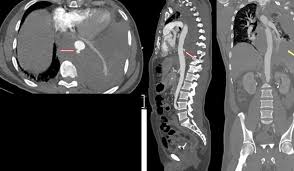
A CT Aortogram and Peripheral Angiogram is a non-invasive imaging test used to examine the aorta and the arteries that carry blood to different parts of the body, particularly the abdomen, pelvis, and legs. The test uses computed tomography, commonly called CT, together with an iodine-based contrast material injected through a vein. As the contrast travels through the arteries, the CT scanner creates detailed images that allow doctors to assess blood-vessel structure and blood flow.
In Hyderabad, CT angiography may be recommended when there is concern about narrowing or blockage of the peripheral arteries, an aortic aneurysm, aortic dissection, or another vascular abnormality. It may also be used to plan treatment before angioplasty, stenting, vascular surgery, or other procedures. Although the name “angiogram” is often used, this page refers to CT angiography, not invasive catheter angiography.
What Is a CT Aortogram and Peripheral Angiogram?
The aorta is the largest artery in the body. It begins at the heart, travels through the chest and abdomen, and gives rise to major arteries that supply the organs, pelvis, and legs. A CT aortogram creates detailed images of this vessel and its major branches.
Peripheral CT angiography focuses on arteries outside the heart and brain, most commonly the arteries supplying the legs. The scan can show whether blood vessels are open, narrowed, completely blocked, enlarged, or affected by other structural abnormalities.
During the examination, contrast material is injected into a vein, usually in the arm. The CT scan is timed so that images are captured while the contrast is passing through the arteries. Computer processing can then reconstruct the images in multiple planes and three-dimensional views. These images help the radiologist and treating specialist understand the location and extent of vascular disease.
Why Is This Test Recommended?
One common reason for peripheral CT angiography is suspected peripheral artery disease, often called PAD. PAD usually develops when atherosclerotic plaque builds up inside the arteries and reduces blood flow, particularly to the legs. Some patients experience calf, thigh, or buttock discomfort when walking that improves with rest. Others may notice cold feet, reduced pulses, wounds that heal slowly, or pain in the foot even while resting when disease is more advanced.
A CT aortogram may be requested when the doctor suspects an aneurysm, which is an abnormal widening of the aorta, or when there is concern about a tear within the wall of the aorta known as a dissection. It may also be used to evaluate the size and shape of a known aneurysm and to help plan or follow treatment.
The test can also provide detailed arterial mapping before a vascular intervention. If angioplasty, stenting, endovascular repair, or bypass surgery is being considered, knowing the exact location, length, and severity of disease can help the treatment team plan the most appropriate approach.
What Can CT Angiography Show?
CT angiography can show areas where arteries have become narrowed because of plaque and can identify complete arterial blockages. It can also demonstrate calcification within the vessel walls, the length of diseased arterial segments, and the condition of arteries above and below a blockage.
In the aorta, the scan can evaluate aneurysms, dissections, vessel-wall abnormalities, and changes following previous surgical or endovascular treatment. It may also show abnormalities involving major branches of the aorta that supply the kidneys, abdominal organs, or lower limbs.
The images provide both anatomical and planning information. For example, a patient with leg symptoms may have disease at several different levels, from the abdominal aorta and pelvic arteries down to vessels in the thigh or lower leg. CT angiography can help show how widespread the disease is rather than evaluating only one small area.
How Should You Prepare?
Preparation may vary according to the imaging centre and the patient’s medical condition. Because iodine-containing contrast is usually required, the clinical team may ask about kidney function, previous contrast reactions, diabetes, thyroid problems, allergies, and current medicines before the scan.
A blood test to check kidney function may be required, particularly in older adults or patients with known kidney disease, diabetes, dehydration, or other risk factors. People who have previously experienced a significant reaction to iodinated contrast should inform the radiology team before the appointment because additional precautions or an alternative investigation may sometimes be considered.
You may be instructed not to eat for several hours before the scan. Water may still be allowed depending on the centre’s protocol. Follow the specific instructions given when your appointment is scheduled rather than applying general fasting rules on your own.
Women who are pregnant or could be pregnant should inform the doctor or radiology team because CT uses X-rays.
What Happens During the Scan?
When you arrive, the clinical team reviews the relevant medical information and prepares you for the examination. A small intravenous cannula is inserted into a vein, usually in the arm. You then lie on the CT examination table, which moves through the opening of the scanner.
The contrast material is injected through the IV while the scan is being timed. Many patients experience a temporary feeling of warmth or a metallic taste when contrast is injected. These sensations usually disappear quickly.
During image acquisition, you may be asked to remain still and hold your breath briefly. The actual CT scanning portion is usually fast, although preparation, IV placement, positioning, and image checking make the complete appointment longer.
After the scan, the images are reconstructed and reviewed. Three-dimensional images may be created to help demonstrate the course of the aorta and peripheral arteries clearly.
Understanding Your Results
The report may describe whether the aorta is normal in size or enlarged, whether an aneurysm or dissection is present, and whether major branches are affected. In peripheral arterial studies, the report may describe plaque, calcification, narrowing, or complete blockage in different arterial segments.
The severity and location of disease are important when deciding what happens next. A mild narrowing in a patient with few symptoms may be managed differently from a long arterial blockage in someone with severe walking limitation or a non-healing wound.
An abnormal CT angiogram does not automatically mean that angioplasty or surgery is necessary. The images must be considered together with symptoms, physical examination, pulse assessment, Doppler or ABI findings, kidney function, diabetes status, and overall cardiovascular health. Treatment may involve lifestyle measures, medicines, supervised exercise, endovascular treatment, surgery, or a combination depending on the individual situation.
CT Angiography vs Catheter Angiography
CT angiography and catheter angiography both produce images of blood vessels, but they are different procedures. CT angiography usually requires only an IV line in the arm. It does not normally require a catheter to be inserted directly into an artery.
Catheter angiography is more invasive. A catheter is introduced into an artery and advanced to the area being examined. One major advantage of catheter angiography is that treatment such as balloon angioplasty or stent placement can sometimes be performed during the same procedure.
CT angiography is primarily diagnostic. When the main question is to understand arterial anatomy and plan treatment, it may provide the information needed without an invasive arterial procedure. If treatment is likely to be required immediately, catheter angiography may sometimes be preferred.
Safety and Limitations
CT angiography uses X-rays, so it involves exposure to ionising radiation. Modern CT protocols are designed to obtain the necessary diagnostic information while limiting radiation exposure as much as reasonably possible.
Iodinated contrast is generally well tolerated, but temporary warmth, nausea, itching, or mild allergic-type reactions can occasionally occur. Serious contrast reactions are uncommon. Patients with significantly reduced kidney function require particular assessment because contrast may not be appropriate in every situation.
The test also has limitations. Heavy arterial calcification can sometimes make narrowing harder to assess accurately, and movement can reduce image quality. Very small distal vessels may be less clearly evaluated than larger arteries. CT angiography also shows anatomy but does not measure every aspect of how much a narrowing affects blood flow.
For this reason, CT findings may sometimes need to be interpreted together with tests such as Doppler ultrasound, ankle-brachial index, or catheter angiography.
Frequently Asked Questions:-
Is CT angiography painful?
The scan itself is not painful. You will feel a small needle when the IV cannula is inserted and may experience temporary warmth when contrast is injected.
Does the test use radiation?
Yes. CT uses X-rays and therefore involves ionising radiation. The scan is performed only when the expected diagnostic benefit justifies the exposure.
Is contrast always required?
CT angiography generally requires intravenous iodine-based contrast because the contrast makes the arteries clearly visible on CT images.
Do I need a kidney function test?
Some patients require a recent kidney function test before receiving contrast. This is especially important when kidney disease or other relevant risk factors are present.
Can this test detect blockages in the legs?
Yes. Peripheral CT angiography can identify narrowing and blockage in arteries supplying the pelvis and legs and can help determine how extensive the disease is.
Can CT angiography detect an aortic aneurysm?
Yes. CT angiography can provide detailed information about the size, location, shape, and extent of an aortic aneurysm.
Is CT angiography the same as invasive angiography?
No. CT angiography usually uses contrast through a vein and CT imaging. Invasive catheter angiography requires arterial catheter access and may also allow treatment during the same procedure.
What happens if a significant blockage is found?
The next step depends on symptoms, severity, location, and overall health. Your doctor may recommend medical treatment, further vascular assessment, angioplasty, stenting, or surgery when appropriate.
CT Aortogram and Peripheral Angiogram in Hyderabad
If you have symptoms suggesting reduced circulation to the legs, a known or suspected aortic condition, or need detailed arterial mapping before vascular treatment, CT angiography may provide valuable information.
Dr. Kiran Lakkireddy’s cardiac practice in Hyderabad provides evaluation of cardiovascular symptoms and guidance on appropriate diagnostic imaging. Whether a CT Aortogram or Peripheral Angiogram is suitable should be decided after reviewing your symptoms, kidney function, previous investigations, and overall vascular risk.