

IMAGE GUIDED (IVUS & OCT) GUIDED THEN CORONARY STENTING
Home > Services > Image Guided (IVUS & OCT) Guided Coronary Then Stenting
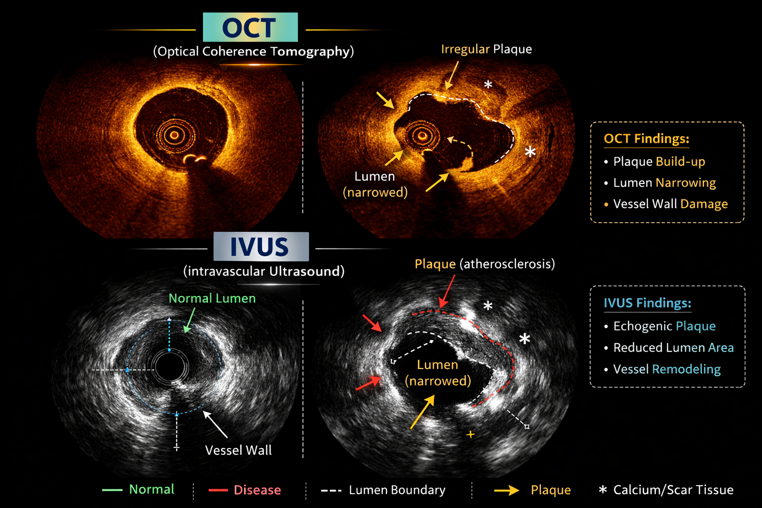
While a traditional X-ray angiogram provides a reliable 2D silhouette of the heart’s arteries, it cannot always show the complete picture of what is happening inside the artery walls. Image-guided coronary stenting uses state-of-the-art miniature cameras—specifically Intravascular Ultrasound (IVUS) and Optical Coherence Tomography (OCT)—to see the blockage from the inside out in high-definition 3D. IVUS uses sound waves, while OCT uses near-infrared light, to provide a 360-degree, microscopic view of the artery wall and the cholesterol plaque. This advanced visualization allows the cardiologist to choose the exact size of the stent needed, prepare the artery perfectly, and ensure the stent is flawlessly expanded against the vessel wall, dramatically reducing the risk of future complications.
1. Indications:
Image-guided stenting using IVUS or OCT is recommended for ensuring precision in complex or high-stakes coronary interventions. Common indications include:
- Treating critical blockages, such as those in the Left Main Coronary Artery
- Complex anatomy, including bifurcation lesions (where the artery branches) or very long blockages
- Evaluating heavily calcified (hardened) plaque to determine if advanced tools like Rotablation or IVL are needed before stenting
- Accurately measuring the exact diameter and length of the artery to select the perfect stent size
- Investigating the cause of a previously placed stent that has failed or narrowed (in-stent restenosis or stent thrombosis)
- Confirming that a newly placed stent is fully expanded and flush against the artery wall with no gaps (optimal stent apposition)
Using internal imaging ensures the highest possible precision, leading to better, longer-lasting results for the patient.
2. Procedure:
Image-guided stenting is performed seamlessly during a standard angioplasty procedure in the cath lab under local anesthesia. You will remain awake and comfortable.
Once the catheter is in position at the heart, the doctor advances a microscopic, flexible imaging wire across the blockage.
If IVUS is used, a tiny ultrasound probe on the tip of the wire emits sound waves to create a real-time cross-sectional image of the artery, revealing the thickness of the plaque and the true size of the vessel.
If OCT is used, a specialized wire emits near-infrared light, capturing microscopic, ultra-high-resolution images of the artery wall in seconds.
The doctor analyzes these internal images on a monitor to map the exact treatment strategy. After the balloon angioplasty and stent placement, the imaging wire is often passed through the artery one more time. This crucial final step acts as a quality control check, allowing the doctor to visually confirm that the stent is perfectly expanded and fully open before finishing the procedure.
3. Safety:
IVUS and OCT are highly safe, widely used technologies that represent the gold standard for complex coronary interventions. By adding just a few minutes to the procedure, these imaging tools significantly increase the long-term safety and success rate of the stent, greatly reducing the chances of a future heart attack or the need for a repeat procedure.
The risks are nearly identical to those of a standard coronary angiogram and angioplasty. Possible but uncommon risks include:
- Bleeding, bruising, or a hematoma at the catheter insertion site (wrist or groin)
- A temporary, very minor spasm of the coronary artery as the imaging wire is passed through
- Injury or micro-dissection of the artery caused by the imaging catheter
- Temporary decline in kidney function due to the contrast dye used during the OCT process (IV fluids are administered to protect the kidneys)