

VSD DEVICE CLOSURE
Home > Services > VSD Device Closure
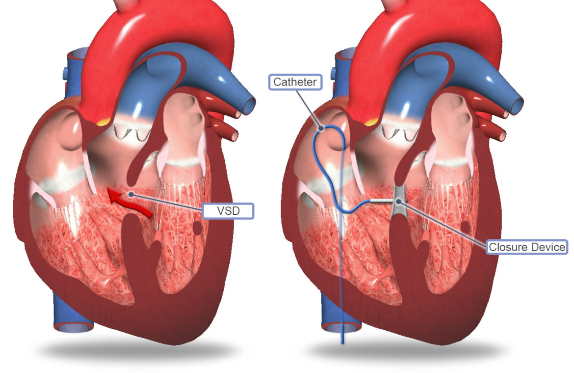
A Ventricular Septal Defect (VSD) is a congenital heart condition characterized by a hole in the septum the muscular wall separating the two lower pumping chambers of the heart (the ventricles). This opening allows oxygen-rich blood from the left ventricle to abnormally mix with oxygen-poor blood in the right ventricle. As a result, the heart has to pump extra blood into the lungs, causing the heart and lungs to work much harder than normal. Over time, this can lead to heart enlargement, high blood pressure in the lungs, and heart failure. While open-heart surgery was once the only option, today, many VSDs can be safely and permanently sealed using a minimally invasive, catheter-based device closure. This advanced technique plugs the hole from the inside, restoring healthy circulation instantly with a much faster, more comfortable recovery than traditional surgery.
1. Indications:
VSD device closure is a highly specialized procedure recommended for patients whose specific heart anatomy makes them suitable candidates for a catheter-based approach. Common indications include:
- A confirmed Ventricular Septal Defect (most commonly a muscular VSD or select perimembranous VSDs) causing a significant left-to-right blood shunt
- Symptoms such as extreme fatigue, rapid breathing, or excessive sweating during physical exertion or feeding
- Poor physical growth, failure to thrive, or frequent respiratory infections in infants and children
- Echocardiogram results showing enlargement or strain on the left side of the heart
- Rising blood pressure in the lungs (pulmonary hypertension) caused by the excess blood flow
- A VSD that has appropriate tissue “rims” for a closure device to safely grip without interfering with the heart’s valves or electrical system
Closing a VSD early relieves the heavy burden on the heart muscle, allowing children to grow normally and preventing irreversible heart and lung damage in adults.
2. Procedure:
A VSD device closure is performed in an advanced cardiac cath lab. Because the procedure requires absolute stillness and precision, it is typically performed under general anesthesia, meaning the patient is completely asleep and comfortable. A small puncture is made in a blood vessel in the groin (the femoral artery or vein, and sometimes a blood vessel in the neck).
Through this access point, a thin, flexible catheter is gently navigated up into the heart. The cardiologist uses continuous X-ray imaging (fluoroscopy) combined with a detailed internal ultrasound—usually a Transesophageal Echocardiogram (TEE)—to visualize the hole in high-definition 3D and measure it precisely.
Once the exact size and location are confirmed, a specialized, collapsible closure device (made of a flexible, memory-shaped wire mesh formed into two connected discs) is pushed through the catheter. As it exits the catheter, the discs spring open. One disc sits flat on the left side of the septum and the other on the right, tightly sandwiching the hole and permanently sealing the leak. Once the doctor verifies the device is secure and the heart’s valves are functioning perfectly, the device is released, and the catheter is removed. The procedure takes about 2 to 3 hours, and natural heart tissue will grow over the device within a few months, permanently embedding it into the wall.
3. Safety:
Catheter-based VSD device closure is a highly successful and safe alternative to open-heart surgery for the right anatomical candidates. It entirely avoids a large chest incision and the heart-lung bypass machine, resulting in less pain, no surgical scarring, and a much shorter hospital stay.
Continuous, rigorous monitoring of vital signs and heart rhythm is strictly maintained throughout the procedure. While complications are uncommon, possible risks include:
- Bleeding, bruising, or a hematoma at the catheter insertion site in the groin or neck
- Irregular heartbeats or heart block (interference with the heart’s electrical system, as the VSD is often located very close to the heart’s natural wiring), which occasionally requires a permanent pacemaker
- Dislodgment or shifting of the device after it is released, requiring retrieval
- A tiny residual leak around the device (which usually seals on its own over time)
- A rare risk of blood clots or infection on the device (patients are prescribed preventative antibiotics for dental work for six months following the closure)