

Transesophageal Echocardiography in Hyderabad
Home > Services > Transesophageal Echocardiography in Hyderabad
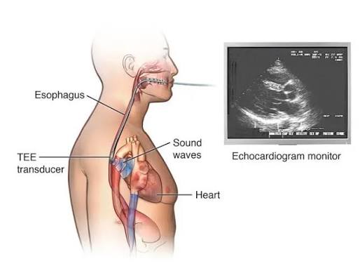
Transesophageal Echocardiography, commonly called TEE, is an advanced ultrasound test that produces detailed images of the heart and nearby blood vessels. Unlike a standard 2D echocardiogram, where the ultrasound probe is placed on the chest, TEE uses a thin flexible probe that passes through the mouth into the esophagus. Because the esophagus lies very close to the heart, the probe can obtain clearer views of structures that may be difficult to assess from the chest.
TEE is commonly advised when a routine echocardiogram does not provide enough detail or when the cardiologist needs a closer look at the heart valves, upper chambers, left atrial appendage, aorta, congenital heart defects, or possible blood clots. In Hyderabad, it may be used when evaluating valve disease, atrial fibrillation, suspected heart-valve infection, structural heart disease, or before selected cardiac procedures.
What Is Transesophageal Echocardiography?
TEE works on the same basic principle as other forms of echocardiography. The probe sends high-frequency sound waves toward the heart and receives the returning echoes. These signals are converted into moving images that allow the cardiologist to study cardiac anatomy and function in real time.
The important difference is the probe position. With a transthoracic echo, sound waves must travel through the chest wall and lungs. In TEE, the probe sits inside the esophagus, just behind the heart. This close position can provide detailed images of the mitral and aortic valves, left atrium, left atrial appendage, interatrial septum, prosthetic valves, and portions of the aorta.
Why Is TEE Recommended?
A cardiologist may recommend TEE when a routine echocardiogram is incomplete or when certain structures need closer examination. One common reason is heart valve disease. TEE can help define the mechanism and severity of valve narrowing or leakage and can provide useful information about artificial or previously repaired valves.
TEE is also frequently used in patients with atrial fibrillation when the doctor needs to check for a blood clot in the left atrium or left atrial appendage before cardioversion or another procedure. Detecting a clot is important because restoring normal rhythm in the presence of a clot may increase stroke risk.
Another important use is suspected infective endocarditis. TEE can provide detailed views of the valves and may show infection-related abnormalities that are not clearly seen on a standard echo. It may also help assess congenital heart abnormalities, selected aortic conditions, or possible cardiac causes of stroke.
What Can a TEE Show?
TEE provides detailed information about the heart’s structure and function. It may show whether a valve is narrowed, leaking, damaged, infected, or functioning abnormally after surgery or replacement. Because it gives a close view of the left atrium and left atrial appendage, it is particularly useful when looking for blood clots associated with atrial fibrillation.
The test can also assess the wall between the upper chambers and may help identify conditions such as an atrial septal defect or patent foramen ovale when relevant. Portions of the aorta can be examined for conditions such as dilation, aneurysm, or dissection.
In structural heart disease, TEE may be used before or during selected procedures involving valves, septal defects, or the left atrial appendage. The detailed images can help the treating team understand anatomy and plan treatment.
How Should You Prepare for TEE?
Preparation is more important for TEE than for a routine transthoracic echo because the probe passes through the throat and sedation is often used. Patients are generally instructed not to eat or drink for a specific period before the procedure. The exact fasting time should be confirmed with the hospital or cardiology team.
Tell your doctor about all medicines you take, including blood thinners, diabetes medicines, and heart medications. Do not stop prescribed treatment unless you have specifically been advised to do so. The team should also know about swallowing problems, esophageal disease, previous stomach or esophageal surgery, bleeding problems, loose teeth, dentures, allergies, and previous reactions to sedation.
Before the test, follow the fasting instructions, carry relevant previous cardiac reports, and arrange for an adult to accompany you if sedation will be given.
What Happens During the Procedure?
Before the test begins, the clinical team reviews your medical history and monitors your ECG, blood pressure, and oxygen level. An intravenous line is usually placed so that medication can be given. A local anesthetic spray is used to numb the throat, and sedative medication may be given to help you relax.
A mouth guard is placed to protect the teeth and probe. The flexible TEE probe is then gently passed through the mouth into the esophagus. Once it is in position, the cardiologist obtains images from several angles by making small adjustments to the probe. The probe remains in the esophagus and does not enter the heart.
The imaging portion is usually completed within a relatively short period, although the total appointment is longer because preparation and recovery from sedation are also required.
What Will You Feel During TEE?
The throat-numbing medicine may have an unusual taste, and you may notice temporary numbness in the mouth and throat. Some patients feel a brief gagging sensation or pressure while the probe is introduced. Sedation generally makes the examination more comfortable and may make you drowsy.
TEE should not normally cause significant pain. After the probe is removed, mild throat irritation or soreness can occur for a short time.
Understanding Your TEE Results
The TEE report describes the structures examined and any important findings. Depending on why the test was requested, the cardiologist may comment on valve anatomy, valve leakage or narrowing, heart pumping function, blood clots, infection-related changes, congenital defects, prosthetic valves, or the aorta.
An abnormal TEE does not automatically mean that surgery or another procedure is required. The importance of a finding depends on its severity, the patient’s symptoms, medical history, and other investigations. In some cases, TEE provides reassurance or confirms that a planned procedure can proceed. In others, it may identify a problem that requires treatment or further imaging.
Safety, Risks, and Recovery
TEE is generally safe when performed by an appropriately trained team, but it is more invasive than a routine transthoracic echo. Temporary sore throat or difficulty swallowing immediately afterward is relatively common. Less common complications include reactions to sedative medicines, minor bleeding, breathing-related problems, or injury to the mouth, throat, or esophagus.
After the procedure, you are usually monitored until the effect of sedation has reduced. Because the throat may remain numb for a period, do not eat or drink until the medical team confirms that swallowing is safe.
If sedation has been used, arrange for someone to take you home and follow the hospital’s advice regarding driving and normal activities. Persistent throat pain, significant difficulty swallowing, bleeding, chest pain, or breathing difficulty should be reported promptly.
When May Another Test Be Needed?
TEE provides excellent detail of many heart structures, but it does not answer every clinical question. Depending on the findings, the cardiologist may recommend a standard 2D Echo for follow-up, CT imaging for the aorta, Cardiac MRI for structural or tissue assessment, or coronary imaging when coronary artery disease is suspected.
Frequently Asked Questions:-
Is TEE painful?
TEE is usually performed with throat-numbing medicine and often sedation. You may feel pressure or a brief gagging sensation, but significant pain is not expected.
Do I need to fast before TEE?
Yes, fasting is usually required. Follow the exact instructions given by the hospital or cardiology team performing your test.
Why is TEE clearer than a regular echo?
The esophagus lies directly behind the heart, allowing the ultrasound probe to obtain close views of several important cardiac structures.
Can TEE detect a blood clot?
TEE is particularly useful for looking for clots in the left atrium and left atrial appendage, especially in patients with atrial fibrillation.
Can I drive after TEE?
If sedation has been given, you should not drive yourself home. Arrange for another adult to accompany you.
Is TEE the same as an endoscopy?
No. TEE uses an ultrasound probe to image the heart, while gastrointestinal endoscopy uses a camera to examine the digestive tract.
Transesophageal Echocardiography in Hyderabad
If a routine echocardiogram has not provided enough information, or if detailed assessment of the heart valves, left atrial appendage, congenital anatomy, prosthetic valves, or aorta is required, TEE may be considered.
Dr. Kiran Lakkireddy’s cardiac practice in Hyderabad provides cardiology evaluation and guidance on appropriate diagnostic testing. Whether TEE is suitable should be decided after reviewing your symptoms, medical history, previous investigations, and the clinical question that needs to be answered.