

Pericardial Disorders Treatment in Hyderabad
Home > Services > Pericardial Disorders Treatment in Hyderabad
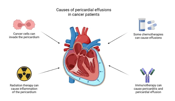
The pericardium is a thin, double-layered sac that surrounds the heart and allows it to move with minimal friction. Pericardial disorders develop when this sac becomes inflamed, collects excess fluid, becomes thickened or scarred, or begins to restrict the normal filling of the heart. Two important conditions affecting the pericardium are pericardial effusion and constrictive pericarditis.
Pericardial effusion means an abnormal accumulation of fluid within the space around the heart. Constrictive pericarditis occurs when the pericardium becomes stiff, scarred, thickened, or sometimes calcified and prevents the heart chambers from filling normally. These conditions can range from mild problems requiring monitoring to serious cardiac conditions needing urgent treatment.
What Is Pericardial Effusion?
A small amount of fluid normally exists between the layers of the pericardium and helps reduce friction as the heart beats. Pericardial effusion develops when an excessive amount of fluid collects in this space.
The effect of an effusion depends not only on the amount of fluid but also on how quickly it accumulates. A slowly developing effusion may become relatively large before significantly affecting the heart because the pericardium has time to stretch. In contrast, fluid that accumulates rapidly can increase pressure around the heart even when the total volume is smaller.
If the pressure becomes high enough to prevent the heart chambers from filling properly, a dangerous condition called cardiac tamponade can occur. Cardiac tamponade reduces the heart’s ability to pump blood effectively and requires emergency treatment.
What Causes Pericardial Effusion?
Pericardial effusion can occur for several reasons. Inflammation of the pericardium, called pericarditis, is one important cause. Viral and other infections, kidney disease, autoimmune or inflammatory disorders, cancer, thyroid disease, trauma, and complications following cardiac surgery or procedures may also lead to fluid accumulation.
In some patients, the exact cause cannot be identified even after investigation. Determining the underlying cause is important because treatment differs depending on why the fluid has developed.
An effusion caused by inflammation may improve when the inflammation is controlled, while an effusion related to kidney disease, infection, cancer, or another condition requires treatment directed toward that problem. Large, recurrent, or unexplained effusions generally require more detailed evaluation.
What Are the Symptoms of Pericardial Effusion?
Small pericardial effusions may produce no symptoms and may be discovered incidentally during a 2D Echocardiogram or another imaging test.
As fluid increases, patients may experience shortness of breath, chest discomfort or pressure, difficulty breathing while lying flat, cough, tiredness, palpitations, or a feeling of heaviness or fullness in the chest.
Symptoms become more concerning when the accumulated fluid begins to significantly compress the heart. Severe breathlessness, dizziness, fainting, confusion, rapid heartbeat, low blood pressure, or sudden deterioration may indicate cardiac tamponade. This is a life-threatening complication and requires immediate hospital treatment.
What Is Constrictive Pericarditis?
Constrictive pericarditis is a condition in which the pericardium becomes abnormally stiff and loses its normal flexibility. The rigid pericardium prevents the heart chambers, particularly the ventricles, from expanding normally as they fill with blood.
As a result, pressure can build up in the veins returning blood to the heart. Patients may develop symptoms that resemble right-sided heart failure, including swelling of the legs, abdominal swelling, fluid accumulation in the abdomen, fatigue, reduced exercise capacity, and breathlessness.
Some patients may also experience decreased appetite or abdominal fullness because of congestion in the liver and abdominal circulation. Unlike heart failure caused mainly by weak heart muscle, the fundamental problem in constrictive pericarditis is restriction of cardiac filling by the abnormal pericardium.
What Causes Constrictive Pericarditis?
Constrictive pericarditis may develop after prolonged or recurrent inflammation of the pericardium. Previous cardiac surgery, chest radiation, tuberculosis and other infections, and systemic inflammatory diseases are among the possible causes. In some patients, no clear underlying cause is found.
Over time, the pericardium may become fibrotic, thickened, or calcified. However, visible thickening is not present in every patient with constriction. Therefore, diagnosis should not depend on pericardial thickness alone.
Doctors usually combine the clinical presentation with echocardiography and advanced imaging to determine whether the pericardium is genuinely restricting cardiac filling.
How Are Pericardial Disorders Diagnosed?
Evaluation begins with the patient’s symptoms, previous illnesses, cardiac procedures, infections, medicines, and medical history. Examination may reveal abnormal heart sounds, elevated neck veins, low blood pressure, leg swelling, abdominal fluid retention, or other signs of impaired cardiac filling.
A 2D Echocardiogram is one of the most important investigations for pericardial disease. It can detect fluid around the heart, estimate its amount, assess its effect on heart filling, and evaluate cardiac pumping function.
Doppler echocardiography can also provide important clues in constrictive pericarditis by showing characteristic changes in ventricular filling and blood flow during breathing.
Cardiac CT may help identify pericardial thickening and calcification. Cardiac MRI can provide detailed information about pericardial inflammation, thickening, fluid, scar tissue, and the movement of the heart within the pericardium.
In difficult cases, cardiac catheterization may be recommended to directly measure pressures inside the heart and help distinguish constrictive pericarditis from conditions such as restrictive cardiomyopathy.
How Is Pericardial Effusion Treated?
Treatment depends on the size of the effusion, how rapidly it developed, whether symptoms are present, its underlying cause, and whether cardiac tamponade is developing.
A small and stable effusion without significant pressure on the heart may sometimes be monitored while the underlying condition is treated. If inflammation is responsible, medical treatment may be used according to the specific diagnosis.
When an effusion is large, causing significant symptoms, progressively increasing, or producing cardiac tamponade, drainage may be necessary.
Pericardiocentesis is a procedure in which a needle and catheter are carefully guided into the pericardial space to remove accumulated fluid. In selected cases, the drained fluid may be tested to investigate infection, malignancy, inflammation, or another possible cause.
Recurrent or difficult-to-manage effusions may occasionally require a surgical pericardial window or another procedure that allows continued drainage and reduces repeated fluid accumulation.
How Is Constrictive Pericarditis Treated?
Treatment depends on whether the constriction is related to active and potentially reversible inflammation or whether permanent pericardial scarring has developed.
Some patients with transient inflammatory constriction may improve with medical treatment and careful monitoring. Diuretic medicines can help reduce leg swelling and abdominal fluid retention in congested patients, but they do not remove a permanently stiff pericardium.
For patients with established chronic and symptomatic constrictive pericarditis, pericardiectomy is the definitive treatment. This is an operation in which the diseased pericardium is surgically removed to allow the heart to fill more freely.
Because pericardiectomy is major cardiac surgery, patients need careful assessment before the procedure. Symptoms, cardiac function, imaging findings, liver and kidney function, previous cardiac surgery or radiation, and overall surgical risk are considered when planning treatment.
Pericardial Effusion vs Constrictive Pericarditis
Although both conditions involve the pericardium, the mechanism is different.
With pericardial effusion, the main problem is excess fluid accumulating around the heart. With constrictive pericarditis, the main problem is a stiff and non-compliant pericardium restricting normal heart filling.
In some patients, fluid accumulation and constrictive physiology can occur together. This is known as effusive-constrictive pericarditis. A complete cardiac assessment is therefore important when symptoms or imaging findings suggest more than a simple effusion.
Frequently Asked Questions:-
Is pericardial effusion dangerous?
Not every pericardial effusion is dangerous. Small and stable effusions may only require monitoring and treatment of their underlying cause. A rapidly accumulating or large effusion can become serious if it compresses the heart and causes cardiac tamponade.
Can pericardial effusion improve with medicines?
Yes. Some effusions improve when the underlying inflammation or medical condition is treated. Other effusions may persist, become larger, or recur and may require drainage.
Is cardiac tamponade the same as pericardial effusion?
No. Pericardial effusion means that excess fluid has collected around the heart. Cardiac tamponade occurs when that fluid creates enough pressure to interfere with the heart’s ability to fill and pump blood. Cardiac tamponade is a medical emergency.
Can constrictive pericarditis cause heart failure symptoms?
Yes. Because the stiff pericardium restricts heart filling, patients can develop leg swelling, abdominal fluid accumulation, breathlessness, fatigue, and reduced exercise capacity.
Can a 2D Echo diagnose constrictive pericarditis?
A 2D Echocardiogram with Doppler is an important investigation and can show findings suggesting constrictive physiology. However, some patients require additional Cardiac CT, Cardiac MRI, or invasive pressure measurements to confirm the diagnosis.
Is surgery always required for constrictive pericarditis?
No. Some patients with temporary inflammation-related constriction may improve with medical treatment. Established chronic symptomatic constrictive pericarditis may require pericardiectomy for definitive treatment.
Pericardial Disorders Treatment in Hyderabad
Patients with unexplained breathlessness, persistent chest discomfort, swelling of the legs or abdomen, known fluid around the heart, or symptoms suggesting restricted cardiac filling may require detailed evaluation for pericardial disease.
Evaluation may include ECG, 2D Echocardiography, Doppler assessment, Cardiac CT, Cardiac MRI, blood tests, and additional investigations depending on the suspected cause and severity.
Dr. Kiran Lakkireddy provides cardiac evaluation in Hyderabad for patients with conditions including pericardial effusion and constrictive pericarditis. Management is individualized and may range from observation and medical treatment to pericardial drainage or referral for surgical pericardiectomy when clinically required.
Patients with a known pericardial effusion who develop severe breathlessness, fainting, very low blood pressure, confusion, or sudden deterioration require urgent hospital evaluation because cardiac tamponade can be life-threatening.