● Lifestyle & Prevention
Successful TAVI Procedure for Mixed Aortic Valve Disease
Table of Contents
- Introduction
- What Is Mixed Aortic Valve Disease?
- Why Can Aortic Stenosis Be Mistaken for a Lung Problem?
- How Was the Diagnosis Established?
- How Was the TAVI Procedure Performed?
- Why Was the Transfemoral Approach Important?
- What Made This a Successful TAVI Procedure?
- Benefits and Limitations of TAVI
- Who May Be Considered for TAVI?
- Myth vs. Fact: TAVI Procedures
- Actionable Checklist: Post-Procedure Care
- Consultation and Appointment Details
- FAQs
Book a Consultation
Prevention is better than cure. Talk to our specialists today.
Severe breathlessness in an older adult is not always caused by the lungs.
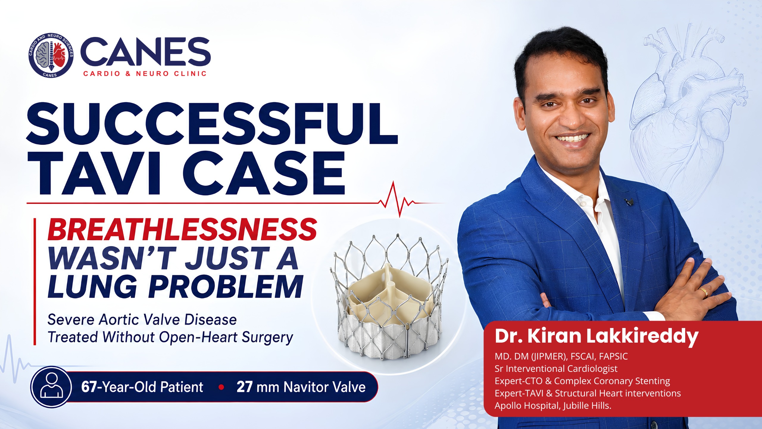
A 67-year-old woman who had developed progressively worsening shortness of breath over ten days was initially treated for a suspected worsening of chronic lung disease. Further assessment, however, revealed a serious heart-valve problem: severe calcific aortic stenosis accompanied by moderate aortic regurgitation and moderate mitral regurgitation.
After comprehensive cardiac evaluation and CT-based procedural planning, she underwent a successful Transcatheter Aortic Valve Implantation, or TAVI. A 27 mm Navitor valve was implanted through an artery in the leg without opening the chest. The procedure was completed without immediate complications, and the patient experienced a marked improvement in breathing.
TAVI, also known as TAVR, is a minimally invasive treatment in which a replacement valve is positioned inside the diseased aortic valve through a catheter. Unlike conventional surgical valve replacement, it usually does not require a large chest incision or open-heart surgery. Treatment decisions for severe aortic valve disease should be individualized through shared decision-making involving the patient and the multidisciplinary Heart Team.
Quick Answer
This case involved the successful treatment of severe symptomatic aortic stenosis with accompanying valve leakage using a TAVI procedure. Following CT planning, a 27 mm Navitor valve was implanted through the right femoral artery. Final angiography and echocardiography showed satisfactory valve position and function, and the patient remained stable with significant symptomatic improvement.
TAVI Case at a Glance
Case feature | Finding |
|---|---|
Patient | 67-year-old woman |
Main symptom | Progressive breathlessness for approximately 10 days |
Initial clinical concern | Suspected COPD exacerbation |
Blood-test findings | Elevated NT-proBNP and mildly elevated troponin |
Aortic valve finding | Severe calcific aortic stenosis |
Associated valve disease | Moderate aortic and mitral regurgitation |
Coronary arteries | Normal coronary angiogram |
Pre-procedure assessment | Echocardiography, angiography and CT-TAVI planning |
Treatment | Transfemoral TAVI |
Valve implanted | 27 mm Navitor valve |
Pre-dilatation | Balloon aortic valvuloplasty using a 22 mm balloon |
Implantation depth | Approximately 3-4 mm |
Immediate outcome | Good valve positioning with no reported procedural complication |
Clinical result | Hemodynamic stability and substantial improvement in breathlessness |
What Is Mixed Aortic Valve Disease?
Mixed aortic valve disease means that the aortic valve suffers from both narrowing (aortic stenosis), which restricts blood flow outward, and leaking (aortic regurgitation), which allows blood to flow backward into the heart.
Key Takeaway: When both problems are present, the heart may face a combined burden of pressure overload and volume overload.
Detailed Explanation
Aortic stenosis restricts blood leaving the left ventricle. Aortic regurgitation allows some blood to return to the ventricle after it has been pumped forward. The 2025 ESC/EACTS valvular heart-disease guidelines emphasize that mixed valve disease should be evaluated according to symptoms, functional limitation, cardiac damage, anatomical suitability, and the expected risks and benefits of intervention. They also note that even patients with combined moderate aortic stenosis and moderate regurgitation can experience outcomes comparable to those with isolated severe aortic stenosis.
What about the mitral valve leakage?
The patient also had moderate mitral regurgitation, meaning that the mitral valve allowed blood to flow backwards from the left ventricle into the left atrium. This did not prevent treatment of the dominant severe aortic-valve obstruction. However, the mitral regurgitation should continue to be reassessed after the TAVI procedure through clinical review and follow-up echocardiography.
Key clinical principle: In multivalve disease, treatment should focus on the lesion producing the greatest hemodynamic burden while maintaining a plan to reassess the remaining valves.
Why Can Aortic Stenosis Be Mistaken for a Lung Problem?
Severe aortic stenosis can cause shortness of breath that mimics a lung condition because the narrowed valve makes it harder for the heart to pump blood, causing pressure to back up and rise inside the lungs.
Key Takeaway: Breathlessness should not automatically be attributed to age or lung disease, especially when accompanied by other cardiac symptoms.
Detailed Explanation
Breathlessness is common to both cardiac and respiratory disease. It may be caused by COPD, asthma, pneumonia, heart failure, or valve disease. Severe aortic stenosis can cause shortness of breath because the narrowed valve restricts blood flow into the aorta, causing pressure to rise inside the heart and lungs, particularly as the disease progresses. Shortness of breath and chest discomfort are recognized symptoms of clinically significant aortic stenosis.
In this case, elevated NT-proBNP, mildly elevated troponin, and persistent symptoms prompted a more detailed cardiac evaluation.
Warning signs that warrant cardiac assessment
Breathlessness should not automatically be attributed to age or lung disease when it is accompanied by:
Reduced exercise tolerance
Chest pressure or discomfort
Dizziness or fainting
An abnormal heart murmur
Swelling of the feet
Difficulty breathing while lying down
Unexplained fatigue
Raised cardiac biomarkers
Symptoms that do not improve with appropriate lung treatment
Severe breathlessness at rest, chest pain, fainting, confusion, or blue discoloration requires emergency assessment rather than a routine clinic visit.
How Was the Diagnosis Established?
A successful TAVI procedure relies on meticulous diagnostics, including echocardiography to measure valve severity, coronary angiography to check for blockages, and a specialized CT scan to plan the exact valve size and delivery route.
Key Takeaway: The diagnostic process must establish the severity of the disease and confirm whether the patient’s specific anatomy is suitable for catheter-based treatment.
Detailed Explanation
A successful TAVI procedure begins well before the valve is implanted:
- Clinical evaluation: The cardiology team reviewed the pattern and speed of symptom progression, examined the patient, and considered whether the respiratory presentation could have a cardiac cause.
- Echocardiography: Echocardiography identified severe calcific aortic stenosis, moderate aortic regurgitation, and moderate mitral regurgitation. An echocardiogram helps measure the severity of valve narrowing, evaluate regurgitation, and assess left-ventricular function.
- Coronary angiography: The coronary angiogram was normal. This was an important part of procedural planning because significant coronary disease may affect whether a patient requires angioplasty or a combined treatment strategy.
- CT-TAVI planning: Cardiac CT was used to assess the anatomy and confirm suitability for a 27 mm Navitor valve. CT planning commonly evaluates the aortic annulus dimensions, valve calcification, coronary-artery height, femoral access vessels, and the most appropriate valve size.
How Was the TAVI Procedure Performed?
The TAVI procedure was performed by guiding a collapsed 27 mm Navitor valve through a catheter in the femoral (leg) artery up to the heart, where it was expanded inside the diseased native valve to instantly restore normal blood flow.
Key Takeaway: This minimally invasive approach avoids the need for a large chest incision and a heart-lung bypass machine.
Detailed Explanation
- The procedure was undertaken through a transfemoral approach using image-guided vascular access.
- Step 1: Vascular access: Ultrasound guidance was used to obtain access. The setup included a temporary pacing lead through the left femoral vein, a pigtail catheter through the right radial artery, and the main valve-delivery system through the right femoral artery.
- Step 2: Temporary pacing support: Rapid pacing may be used during certain stages of TAVI to reduce cardiac movement and blood flow while the balloon or valve is deployed.
- Step 3: Balloon aortic valvuloplasty: The calcified aortic valve was dilated using a 22 mm balloon to prepare the severely narrowed valve for implantation.
- Step 4: Navitor valve implantation: A 27 mm Navitor™ transcatheter heart valve (Abbott) was advanced and positioned within the diseased native aortic valve. It was deployed at an implantation depth of approximately 3-4 mm, with good final positioning.
- Step 5: Final imaging assessment: Final angiography and echocardiography were performed to assess valve position, function, and ensure there were no pericardial complications. The case notes reported an excellent immediate result.
- Step 6: Closure of the access site: The right femoral arterial access was closed using specialized closure devices (ProGlide and Angio-Seal). The patient remained hemodynamically stable.
Why Was the Transfemoral Approach Important?
The transfemoral approach delivers the replacement valve through the femoral artery in the groin, making it the preferred access route because it avoids opening the chest (sternotomy) and generally allows for much faster physical recovery.
Key Takeaway: While transfemoral TAVI offers a faster recovery than open-heart surgery, it is still a major cardiac procedure that requires careful Heart Team evaluation.
Detailed Explanation
The transfemoral approach is generally the preferred access route when the patient’s blood vessels are suitable. TAVI differs from surgical aortic valve replacement because the new valve is delivered through a catheter and expanded inside the original diseased valve.
Potential advantages of a suitable transfemoral approach include no sternotomy, no routine need for a heart-lung bypass machine, smaller access wounds, earlier mobilization in uncomplicated cases, and potentially shorter hospitalization. These advantages do not make a TAVI procedure risk-free or automatically preferable to surgery.
Decision Support: An Important Age and Treatment-Selection Nuance
The patient in this case was 67 years old. TAVI should therefore not be presented as an automatic choice based solely on her age.
Current European guidelines recommend TAVI for anatomically suitable patients aged 70 years or older. In patients younger than 70 who have low surgical risk, surgical aortic valve replacement is generally recommended. For other patients, the choice between surgery and TAVI should be made by a Heart Team after considering clinical condition, anatomy, procedural risk, life expectancy, and lifetime valve-management strategy.
For a 67-year-old patient, relevant considerations may include calculated surgical risk, frailty, previous illnesses, coronary anatomy, and prosthetic-valve durability. Expert insight: A technically feasible TAVI is not necessarily the same as the best lifetime treatment. The recommendation should emerge from shared Heart Team decision-making.
What Made This a Successful TAVI Procedure?
The procedure was successful due to the correct initial cardiac diagnosis, meticulous CT-based anatomical planning, precise ultrasound-guided vascular access, and controlled valve deployment confirmed by real-time multimodality imaging.
Key Takeaway: A favorable outcome relies heavily on the preparation done before the patient even enters the operating room.
Detailed Explanation
The case notes suggest that several factors supported the favorable immediate result:
Correct identification of the cardiac cause: The patient’s symptoms were not assumed to be entirely pulmonary.
Detailed anatomical planning: CT-TAVI assessment allowed the team to evaluate the annulus and access vessels before choosing the delivery strategy.
Structured vascular access: Ultrasound-guided access and planned closure-device use helped manage one of the most important procedural stages.
Controlled valve positioning: The valve was implanted with good final positioning.
Immediate multimodality confirmation: Angiography and echocardiography were used to verify the result before the procedure was completed.
(Note: This remains a single case outcome. It should not be used to predict the result of TAVI in another patient.)
Benefits and Limitations of TAVI
Potential benefit | Important limitation |
|---|---|
Avoids conventional open-chest valve replacement | Not suitable for every anatomy or clinical situation |
May offer faster early recovery | Vascular complications can occur |
Can reduce stenosis-related symptoms | Some patients require a permanent pacemaker |
Useful in many older or high-risk patients | Stroke, bleeding and kidney injury remain possible |
Transfemoral access avoids a chest incision | Surgery may offer better lifetime planning for some younger patients |
Biological valve begins functioning after deployment | Long-term valve durability must be considered |
Can be planned using detailed CT imaging | Paravalvular leakage or valve-position problems may occur |
Recognized risks include bleeding, vascular injury, stroke, abnormal heart rhythm, pacemaker requirement, kidney injury, valve leakage, infection, heart attack, and death. The risks must be discussed in the context of the individual patient’s alternative options. Long-term success also depends on regular follow-up, echocardiography, adherence to prescribed medications, and management of cardiovascular risk factors.
Who May Be Considered for TAVI?
TAVI is primarily considered for patients with severe, symptomatic aortic stenosis whose valve and vascular anatomy are suitable for a catheter-based approach, provided the multidisciplinary Heart Team determines it is the most appropriate option.
Key Takeaway: TAVI is an excellent treatment, but it is not the preferred option for every single patient, particularly younger individuals with low surgical risk.
Detailed Explanation
A person may be considered when:
The stenosis is severe and symptoms are attributable to the valve disease.
The expected benefit is meaningful.
The valve and vascular anatomy are suitable.
The Heart Team considers TAVI preferable or reasonable.
There is a realistic post-procedure care plan.
TAVI may not be the preferred option when the patient is young and has a long expected lifespan, when surgical risk is low and surgery offers a better lifetime strategy, when the anatomy is unsuitable for safe deployment, or when there is a need for simultaneous surgery on another valve.
Myth vs. Fact: TAVI Procedures
Understanding the realities of the TAVI procedure helps patients make informed decisions about their valve replacement.
Myth | Medical Fact |
|---|---|
TAVI removes the old valve. | The replacement valve is normally expanded inside the diseased native valve, pushing the old leaflets aside. |
TAVI is only for patients who cannot have surgery. | It is now used across different surgical-risk categories, but selection remains highly individualized. |
Every older patient with stenosis should receive TAVI. | A Heart Team must evaluate symptoms, anatomy, risk, and lifetime treatment needs before deciding. |
TAVI has no serious risks because the chest is not opened. | Stroke, bleeding, vascular injury, pacemaker requirement, and other complications remain possible. |
A successful implant means follow-up is unnecessary. | Ongoing clinical and echocardiographic monitoring remains absolutely important. |
Actionable Checklist: Post-Procedure Care
After a successful TAVI procedure, patients should receive an individualized discharge plan. Use this checklist to ensure you understand your recovery requirements:
- [ ] Medication Review: Clarify your required antiplatelet or anticoagulant medicines.
- [ ] Wound Care: Understand how to monitor your vascular access site (usually in the groin) for signs of bleeding or infection.
- [ ] Infection Prevention: Ask about dental hygiene and infection-prevention advice to protect your new valve.
- [ ] Follow-up Imaging: Note the timing of your repeat echocardiography to monitor the new valve (and any remaining mitral regurgitation).
- [ ] Warning Signs: Know exactly which symptoms (like sudden shortness of breath or chest pain) require urgent emergency evaluation.
Consultation and Appointment Details
At CANES Cardio & Neuro Clinic, patients and families can receive structured cardiovascular evaluation and guidance regarding serious heart conditions, including advanced heart failure, cardiogenic shock risk, and the need for referral to higher-level hospital care.
The clinic is guided by Dr. Kiran Lakkireddy, an experienced interventional cardiologist specializing in complex cardiology. The focus is on detailed assessment, understandable communication, and appropriate coordination with advanced cardiac or surgical teams when necessary.
Location: 3rd Floor, Tapasavi Commercial Complex, Hi-Tension Road, Kondapur, Hyderabad
Contact: +91 9014944654
Appointment Booking: Visit CANES Clinic Official Portal
Specialist Profile: Dr. Kiran Lakkireddy Official Website
Reported consultation time: 5:00 PM–9:30 PM (Please confirm current availability when booking.)
Call to action: Persistent breathlessness, unexplained exercise intolerance, fainting, or a newly detected heart murmur deserves timely evaluation. Schedule a comprehensive cardiology and echocardiographic assessment to determine whether the symptoms arise from the heart, lungs, or both.
Key Points to Remember
- Persistent breathlessness may arise from heart-valve disease even when a lung condition is initially suspected.
- This patient had severe calcific aortic stenosis with moderate aortic and mitral regurgitation.
- Echocardiography, coronary angiography, and CT planning established the diagnosis and procedural strategy.
- A 27 mm Navitor valve was implanted through the right femoral artery.
- Final imaging showed a satisfactory immediate result without a reported procedural complication, and the patient experienced a significant improvement in breathing.
- TAVI selection should be based on Heart Team assessment rather than age or surgical risk alone.
- At 67 years of age, the balance between a TAVI procedure and surgery requires especially careful lifetime planning.
Frequently Asked Questions (FAQs)
What is a successful TAVI procedure?
A successful TAVI procedure places a functioning replacement valve in the intended position, restores forward blood flow, and avoids major immediate complications. Long-term success also depends on symptom improvement, valve performance, and follow-up findings.
Are TAVI and TAVR the same procedure?
Yes. TAVI means Transcatheter Aortic Valve Implantation, while TAVR means Transcatheter Aortic Valve Replacement. The terms generally describe the exact same procedure.
Can TAVI treat a narrowed and leaking aortic valve?
TAVI is primarily performed for severe aortic stenosis. Patients who also have aortic regurgitation may be treated when the stenosis is the dominant lesion and the anatomy is appropriate. Pure aortic regurgitation is a different clinical situation and requires separate assessment.
Why is a CT scan needed before TAVI?
CT helps evaluate valve dimensions, aortic-root anatomy, coronary relationships, and the blood vessels used to deliver the valve. It is essential for safe sizing and procedural planning.
Is a 27 mm Navitor valve suitable for every patient?
No. Valve size is selected from precise CT and echocardiographic measurements. A 27 mm valve is suitable only when the patient’s anatomy falls within that specific device’s sizing range.
Is TAVI safer than open-heart surgery?
Neither option is universally safer. TAVI avoids a sternotomy and may offer a faster early recovery, but it has risks such as vascular injury, stroke, pacemaker requirement, and paravalvular leakage. Surgery may be preferable for some younger or low-risk patients.
Does moderate mitral regurgitation need separate treatment?
Not always immediately. It should be reassessed after the aortic valve has been treated. Additional treatment may be considered later when significant regurgitation persists and continues to cause symptoms or cardiac damage.
Expert Care
Hyderabad's leading Cardio & Neuro specialists.