

Congenital Heart Diseases (Heart Holes – ASD, VSD, PDA & Cyanotic CHD) Treatment in Hyderabad
Home > Services > Congenital Heart Disease Treatment in Hyderabad
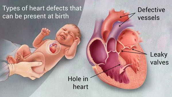
Congenital heart diseases, also called congenital heart defects, are structural problems in the heart or major blood vessels that are present from birth. They range from small defects with few symptoms to complex conditions that affect blood flow and oxygen levels. Some are diagnosed in childhood, while milder defects may only be discovered in adulthood.
The term “heart holes” is a useful patient-friendly description for some congenital conditions. Atrial septal defect (ASD) and ventricular septal defect (VSD) are true openings in the walls between heart chambers. Patent ductus arteriosus (PDA) is different: it is a blood vessel connection between the aorta and pulmonary artery that normally closes after birth but remains open. Cyanotic congenital heart disease is a broader group of defects in which oxygen levels can become low.
What Is an Atrial Septal Defect (ASD)?
An atrial septal defect is an opening in the wall between the two upper heart chambers, called the atria. In many ASDs, oxygen-rich blood passes from the left atrium into the right atrium, sending extra blood through the right side of the heart and lungs.
A small ASD may cause no symptoms and may not need closure. A larger defect can gradually enlarge the right side of the heart and may cause breathlessness, tiredness, palpitations or reduced exercise capacity. Some patients can later develop atrial arrhythmias or pulmonary hypertension.
ASD closure may be considered when the defect causes a significant shunt and right-heart enlargement and when pulmonary vascular conditions are suitable. Many secundum ASDs can be closed with a catheter-delivered device, while other types may require surgery.
What Is a Ventricular Septal Defect (VSD)?
A ventricular septal defect is a hole in the wall between the two lower heart chambers, called the ventricles. Because pressure is normally higher in the left ventricle, blood usually flows from the left ventricle through the VSD into the right ventricle and toward the lungs.
Small VSDs may cause only a heart murmur and can sometimes close naturally during childhood. Large defects can send excessive blood to the lungs, making the heart work harder and increasing pressure in the pulmonary arteries. Infants with a significant VSD may breathe rapidly, feed poorly or have difficulty gaining weight.
Long-standing large shunts can cause irreversible pulmonary vascular disease in some patients. In advanced cases, the direction of blood flow may reverse, causing Eisenmenger syndrome and low oxygen levels. Closure in this setting requires careful specialist assessment. VSD treatment depends on size, location, symptoms, heart enlargement and pulmonary pressure. Some defects need surgical patch closure, while selected VSDs can be closed with a catheter-based device.
What Is Patent Ductus Arteriosus (PDA)?
The ductus arteriosus is a normal blood vessel before birth that connects the pulmonary artery and aorta and allows fetal blood to bypass the lungs. After birth, it normally closes. If it remains open, the condition is called patent ductus arteriosus.
PDA is therefore not a septal hole between heart chambers, although it is commonly grouped with heart-hole conditions. When it stays open, blood can flow from the aorta into the pulmonary artery, sending extra blood to the lungs.
A small PDA may cause no symptoms. A larger PDA can increase the workload on the heart and may lead to chamber enlargement or pulmonary hypertension. Depending on age, anatomy and size, treatment may involve catheter-device closure or surgery.
What Is Cyanotic Congenital Heart Disease?
Cyanotic congenital heart disease is not one single defect. It is a group of congenital heart conditions in which oxygen-poor blood reaches the body, leading to reduced oxygen saturation and sometimes bluish discoloration of the lips, tongue or skin, called cyanosis.
Examples include Tetralogy of Fallot, d-transposition of the great arteries, pulmonary atresia and tricuspid atresia. The circulation problem differs between conditions. Some reduce blood flow to the lungs, while others create abnormal connections or mixing of oxygen-rich and oxygen-poor blood.
Tetralogy of Fallot includes four structural abnormalities, one of which is a VSD. d-Transposition of the great arteries involves the two main arteries being connected to the wrong ventricles. Severe cyanotic defects usually require specialized treatment, and adults treated in childhood still need long-term follow-up.
What Symptoms Can Congenital Heart Disease Cause?
Symptoms depend on the defect and its severity. Small ASD, VSD or PDA may cause no noticeable symptoms. Larger shunts can produce breathlessness, fatigue, reduced exercise capacity, recurrent respiratory symptoms in children or difficulty gaining weight in infancy.
Complex congenital disease can cause low oxygen saturation, cyanosis, breathlessness, dizziness or fainting. Adults with repaired or unrepaired congenital heart disease may later develop palpitations, rhythm disturbances, valve problems, pulmonary hypertension or heart failure symptoms.
Sudden severe breathlessness, fainting, marked cyanosis or major deterioration requires urgent assessment.
How Are Congenital Heart Diseases Diagnosed?
A 2D Echocardiogram with Doppler is one of the most important tests. It can show the location and size of an ASD or VSD, assess blood flow through a PDA, evaluate chamber enlargement and valve function, and estimate pulmonary pressure.
Transesophageal Echocardiography, or TEE, may provide more detailed anatomy before selected catheter-based procedures. ECG can identify rhythm abnormalities. Pulse oximetry measures blood oxygen saturation and is especially useful when cyanotic disease is suspected.
Cardiac CT and Cardiac MRI can provide detailed information about complex anatomy, major blood vessels and previous repairs. Cardiac catheterization may be needed when direct pressure measurements, pulmonary vascular resistance assessment or an interventional procedure is planned. Modern congenital heart care uses imaging and physiological assessment to determine whether and when intervention is appropriate.
How Are Heart Holes and Other CHDs Treated?
Treatment is individualized. Some small defects can be monitored when they are not causing important symptoms or heart enlargement. Other defects need closure to prevent long-term complications.
Selected ASD, VSD and PDA defects can be treated with catheter-based device closure through the blood vessels, avoiding open-heart surgery. Not every defect is suitable for a device. Size, anatomy, surrounding tissue, valve relationships and pulmonary pressure all affect the decision.
Surgery remains important when catheter closure is unsuitable and for many complex congenital heart conditions. Cyanotic diseases such as Tetralogy of Fallot and transposition of the great arteries generally require specialized congenital heart treatment based on the individual anatomy.
Why Is Long-Term Follow-Up Important?
Congenital heart disease can remain relevant throughout life, even after childhood repair. The 2025 ACC/AHA guideline for adults with congenital heart disease emphasizes ongoing follow-up and appropriate involvement of clinicians with adult congenital heart disease expertise.
Follow-up may include ECG, echocardiography, Holter Monitoring, exercise testing, CT or MRI depending on the original defect and repair. Adults may need assessment for rhythm problems, residual shunts, valve abnormalities, pulmonary hypertension and ventricular function. Pregnancy planning can also require specialist evaluation in women with significant congenital heart disease.
Frequently Asked Questions:-
Are ASD and VSD really holes in the heart?
Yes. ASD is an opening between the two atria, while VSD is an opening between the two ventricles.
Is PDA also a hole inside the heart?
Not exactly. PDA is a persistent blood vessel connection between the aorta and pulmonary artery. It is often grouped with heart-hole conditions because it also creates abnormal blood flow.
Do all heart holes need closure?
No. Small defects that do not cause important heart changes may only need monitoring. Closure depends on the defect type, size, symptoms, chamber effects and pulmonary pressure.
Can adults have congenital heart disease?
Yes. Some mild defects are first found in adulthood, and many people treated during childhood still need follow-up as adults.
Can heart holes cause pulmonary hypertension?
Yes. Significant unrepaired ASD, VSD or PDA can expose the lungs to excessive blood flow and may contribute to pulmonary hypertension over time.
Can congenital heart disease be treated without surgery?
Yes. Selected ASD, VSD and PDA defects can be closed through catheter-based procedures. Other conditions may require surgery or monitoring.
Congenital Heart Diseases Treatment in Hyderabad
Patients with a known heart hole, unexplained heart murmur, breathlessness, low oxygen levels, recurrent palpitations or a previously repaired congenital heart defect may need structured cardiac evaluation.
Evaluation may include ECG, 2D Echocardiography, TEE and advanced cardiac imaging when appropriate. Management depends on the exact defect, shunt size, chamber effects, pulmonary pressure, symptoms and previous treatment. Selected ASD, VSD and PDA defects may be considered for catheter-based closure, while complex or cyanotic congenital heart disease may require coordinated care involving congenital heart specialists and cardiac surgeons.
Patients with severe cyanosis, fainting, major breathlessness or sudden deterioration require urgent hospital assessment rather than a routine consultation.