

Pulmonary Hypertension & Pulmonary Thromboembolism in Hyderabad
Home > Services > Pulmonary Hypertension & Pulmonary Thromboembolism in Hyderabad
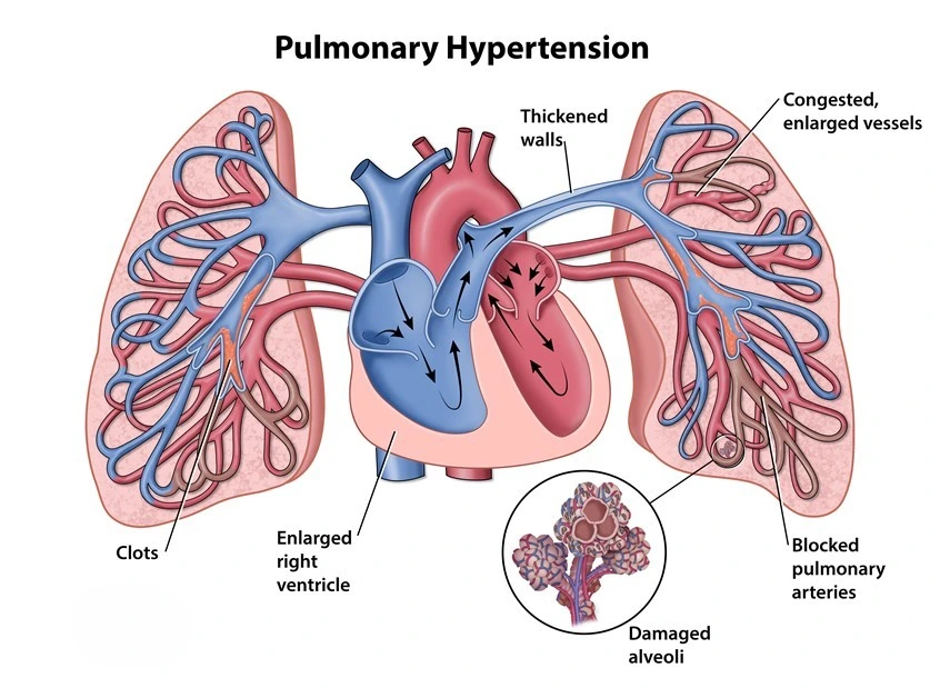
Pulmonary hypertension and pulmonary thromboembolism both affect blood flow between the heart and lungs. Pulmonary hypertension means that pressure within the pulmonary circulation is abnormally elevated. Pulmonary thromboembolism occurs when a blood clot travels through the bloodstream and blocks one or more pulmonary arteries. It is also commonly called pulmonary embolism, or PE.
Although these are different conditions, they can sometimes be related. A previous pulmonary thromboembolism may leave persistent clot material in the pulmonary arteries in a small number of patients, eventually leading to chronic thromboembolic pulmonary hypertension, or CTEPH. Both may cause breathlessness, chest discomfort, reduced exercise capacity, dizziness, or right-heart strain, so accurate diagnosis is important.
What Is Pulmonary Hypertension?
Pulmonary hypertension, often shortened to PH, is a condition in which pressure in the blood vessels carrying blood from the right side of the heart to the lungs becomes elevated. The right ventricle must then work harder to push blood through the pulmonary circulation. If this pressure remains high, the right side of the heart may enlarge and weaken.
Patients may initially notice shortness of breath during walking or climbing stairs. As the condition progresses, fatigue, chest pressure, palpitations, dizziness, fainting, or swelling of the legs or abdomen may occur. These symptoms can occur in other heart and lung conditions, so PH requires structured evaluation.
What Causes Pulmonary Hypertension?
Pulmonary hypertension is not a single disease. Pulmonary arterial hypertension, or PAH, involves changes in the small pulmonary arteries and may occur without an identifiable cause or with connective tissue disease, congenital heart disease, certain medicines, or other conditions.
PH can also result from left-sided heart disease, including heart failure and valve disease, or from chronic lung disease and low oxygen levels. Persistent obstruction after pulmonary thromboembolism can cause CTEPH. Finding the correct cause matters because treatment appropriate for one type may not suit another.
What Is Pulmonary Thromboembolism?
Pulmonary thromboembolism develops when a blood clot, usually from a deep vein in the leg or pelvis, breaks free and travels to the lungs. When it lodges in a pulmonary artery, it blocks blood flow and increases the resistance against which the right ventricle must pump.
Severity depends on the size and location of the clot, the amount of pulmonary circulation affected, and the patient’s existing heart and lung function. A small clot may cause limited symptoms, while a large clot burden can produce severe right-heart strain, low blood pressure, collapse, or cardiac arrest.
Risk factors include recent surgery or hospitalization, prolonged immobility, previous deep vein thrombosis or pulmonary thromboembolism, active cancer, pregnancy and the postpartum period, estrogen-containing medicines, and some clotting disorders.
What Are the Symptoms of Pulmonary Thromboembolism?
Sudden unexplained breathlessness is one of the most important warning symptoms. Chest pain, especially pain that worsens with deep breathing, may occur. Other symptoms can include a rapid heartbeat, dizziness, fainting, coughing, coughing up blood, or unexplained weakness.
Some patients also have symptoms of deep vein thrombosis, such as swelling, pain, warmth, or tenderness in one leg. However, leg symptoms are not always present.
Severe breathlessness, fainting, low blood pressure, bluish discoloration, confusion, or collapse can indicate a high-risk pulmonary thromboembolism and requires emergency hospital care. A suspected acute clot in the lungs should not wait for a routine outpatient consultation.
How Are These Conditions Diagnosed?
In suspected acute pulmonary thromboembolism, doctors consider symptoms, examination findings, oxygen levels, risk factors, and the estimated clinical probability of a clot.
A D-dimer blood test may help exclude pulmonary thromboembolism in selected patients when clinical probability is not high. CT pulmonary angiography, or CTPA, is one of the main imaging tests used to identify clots within the pulmonary arteries. A ventilation-perfusion, or V/Q, scan may be used when appropriate and is particularly important when chronic thromboembolic disease is suspected.
Ultrasound of the leg veins may identify deep vein thrombosis. ECG and cardiac blood tests can provide additional information, while a 2D Echocardiogram can assess right ventricular size and function and look for signs of pressure overload.
For suspected PH, echocardiography is an important initial test because it can estimate the probability of elevated pulmonary pressure and evaluate the right heart, valves, and left ventricular function. Further testing may include chest imaging, pulmonary function tests, blood tests, V/Q scanning, or CT imaging. Right-heart catheterization is used when direct measurement of pulmonary pressures and hemodynamics is required to confirm and classify PH.
How Is Pulmonary Thromboembolism Treated?
Anticoagulation is the main treatment for most patients with acute pulmonary thromboembolism. Anticoagulant medicines reduce additional clot formation and lower the risk of clot extension or recurrent venous thromboembolism while the body gradually clears the existing clot.
The choice and duration depend on clinical condition, kidney function, bleeding risk, pregnancy, cancer history, provoking factors, and whether an intervention is planned. Current guidance recommends direct oral anticoagulants for many suitable patients who require oral anticoagulation, unless contraindicated.
Patients with severe pulmonary thromboembolism and cardiopulmonary instability may require faster restoration of pulmonary blood flow. Depending on severity and bleeding risk, treatment can include systemic thrombolysis, catheter-directed therapy, mechanical thrombectomy, or surgical embolectomy. These options are reserved for selected patients.
How Is Pulmonary Hypertension Treated?
Treatment starts with identifying the underlying PH group. If pulmonary pressure is elevated because of left-heart disease, management focuses mainly on the heart condition. When chronic lung disease or low oxygen levels are contributing, treatment is directed toward the lung disorder and oxygenation when appropriate.
Patients with confirmed PAH may require medicines that target pulmonary vascular pathways. Treatment depends on symptoms, functional capacity, hemodynamic findings, disease severity, and ongoing risk assessment.
Pulmonary hypertension-specific medicines should not be started simply because an echocardiogram shows an elevated pressure estimate. Correct classification is important before disease-specific therapy is selected.
What Is Chronic Thromboembolic Pulmonary Hypertension?
CTEPH occurs when clot material does not fully resolve and becomes organized within the pulmonary arteries. This creates long-standing obstruction, raises pulmonary pressure, and strains the right side of the heart.
Patients may experience persistent breathlessness, fatigue, exercise limitation, chest discomfort, dizziness, or swelling after a previous pulmonary thromboembolism. Some patients do not remember having a clearly diagnosed acute clot.
CTEPH is important to identify because specific treatments are available. Pulmonary endarterectomy can remove organized clot material surgically in suitable patients. Balloon pulmonary angioplasty and targeted medical therapy may be considered for selected patients with inoperable disease or persistent PH after surgery. Management usually requires specialist multidisciplinary assessment.
Follow-Up After Pulmonary Thromboembolism
Recovery does not end when anticoagulation is started. Patients should be reviewed for ongoing breathlessness, reduced exercise capacity, recurrent clot symptoms, bleeding complications, and the appropriate duration of anticoagulation.
Persistent breathlessness or functional limitation deserves further evaluation because it may be related to CTEPH, chronic thromboembolic pulmonary disease, another heart or lung condition, or another cause. Current guidance emphasizes assessment of PE-related symptoms during follow-up.
Frequently Asked Questions:-
Is pulmonary thromboembolism the same as pulmonary embolism?
Yes. Pulmonary thromboembolism is a valid medical term for a thromboembolic clot blocking the pulmonary arteries. Pulmonary embolism, or PE, is the commonly used shorter term.
Is pulmonary hypertension the same as regular high blood pressure?
No. Regular hypertension refers to pressure in the systemic arteries. Pulmonary hypertension refers to elevated pressure in the circulation between the heart and lungs.
Can pulmonary thromboembolism cause pulmonary hypertension?
Yes. Most treated clots do not cause chronic PH, but persistent organized clot obstruction can lead to CTEPH in a small proportion of patients.
Does every pulmonary thromboembolism need thrombolysis?
No. Most patients are treated with anticoagulation. Thrombolysis, thrombectomy, or surgical clot removal is reserved for selected patients based on severity, cardiovascular stability, and bleeding risk.
Can a 2D Echo diagnose pulmonary hypertension?
A 2D Echocardiogram can suggest PH and assess its effect on the right heart. Definitive hemodynamic confirmation and classification may require right-heart catheterization.
Pulmonary Hypertension & Pulmonary Thromboembolism Treatment in Hyderabad
Patients with persistent unexplained breathlessness, reduced exercise tolerance, fainting, leg or abdominal swelling, or evidence of right-heart strain may require assessment for pulmonary hypertension. Sudden breathlessness, chest pain, fainting, coughing up blood, or symptoms of deep vein thrombosis may indicate pulmonary thromboembolism and require urgent hospital evaluation.
Dr. Kiran Lakkireddy’s cardiac practice in Hyderabad provides cardiovascular evaluation for pulmonary hypertension, right-heart strain, and thromboembolic disease using ECG, 2D Echocardiography, cardiac imaging, and additional investigations when appropriate. Treatment is individualized according to whether the problem is acute pulmonary thromboembolism, pulmonary arterial hypertension, PH related to another heart or lung disease, or CTEPH. Suspected acute pulmonary thromboembolism is an emergency and should not be delayed for a routine consultation.