

Cardiac MRI in Hyderabad
Home > Services > Cardiac MRI in Hyderabad
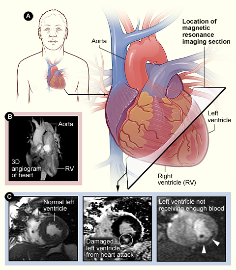
Cardiac Magnetic Resonance Imaging, commonly called Cardiac MRI or CMR, is a non-invasive imaging test that creates detailed pictures of the heart, heart muscle, valves, major blood vessels, and surrounding structures. Instead of X-rays, MRI uses a powerful magnetic field, radiofrequency waves, and computer processing, so it does not expose the patient to ionising radiation.
In Hyderabad, Cardiac MRI may be recommended when a cardiologist needs detailed information about heart structure, pumping function, heart muscle disease, inflammation, scarring, congenital conditions, or the effects of a previous heart attack. It is especially useful when an echocardiogram or another test has not fully answered the clinical question.
What Is Cardiac MRI?
Cardiac MRI is designed specifically to image a moving organ. ECG electrodes placed on the chest allow the scanner to synchronize image acquisition with the heartbeat, and the patient may be asked to hold the breath briefly during some sequences so that movement does not blur the images.
The test can assess heart size, ventricular volumes, pumping function, wall movement, major blood vessels, and tissue characteristics in a single examination. In selected studies, gadolinium-based contrast is injected through a vein. Contrast can help show inflammation, scar tissue, fibrosis, or areas of previous heart muscle injury.
Different MRI sequences provide different information, so the examination can be tailored to the condition being evaluated.
Why Is Cardiac MRI Recommended?
A cardiologist may recommend Cardiac MRI when more precise information about the heart muscle is required. One common indication is cardiomyopathy, a group of conditions in which the heart muscle becomes enlarged, thickened, weakened, or otherwise abnormal. MRI can measure ventricular size and function accurately and may demonstrate patterns of scar or fibrosis that help characterize the condition.
Cardiac MRI is also useful when myocarditis, or inflammation of the heart muscle, is suspected. Patients may develop chest discomfort, breathlessness, palpitations, or reduced heart function. MRI can identify changes within the myocardium that support the diagnosis when interpreted together with symptoms, ECG, blood tests, and other findings.
After a heart attack or in patients with reduced pumping function, MRI may be used to assess myocardial scar and viability. It can also help evaluate selected congenital conditions and the aorta.
What Can Cardiac MRI Show?
Cardiac MRI can provide accurate measurements of the size and pumping function of the right and left ventricles. It can show whether the chambers are enlarged, whether the heart muscle is thickened, and whether particular areas of the heart wall are moving normally.
Another major strength is tissue characterization. Using specific sequences, MRI can assess patterns associated with edema, inflammation, fibrosis, and scar. Late gadolinium enhancement imaging, when contrast is used, can show characteristic patterns of myocardial injury and may help distinguish damage related to coronary artery disease from some non-ischemic heart muscle disorders.
The scan can also evaluate congenital abnormalities, cardiac masses, the pericardium, and parts of the aorta. However, Cardiac MRI is not usually the preferred test for detailed imaging of the smaller coronary arteries. CT Coronary Angiography or invasive coronary angiography may be more appropriate when coronary anatomy is the main concern.
How Should You Prepare?
Preparation varies according to the type of Cardiac MRI. Some centres allow normal food and medicines, while others may provide specific fasting instructions, especially if sedation or a particular protocol is planned. Follow the instructions given by the imaging centre.
MRI safety screening is essential. Tell the team if you have a pacemaker, implantable defibrillator, cochlear implant, aneurysm clip, neurostimulator, metal fragments, previous surgery involving metal implants, or any other implanted medical device. Many modern cardiac devices are MRI-conditional and can be scanned under specific protocols, but the exact device must be identified and checked for compatibility.
You should also inform the team about kidney disease, pregnancy, previous reactions to gadolinium contrast, claustrophobia, or severe difficulty lying flat. Jewelry, watches, phones, bank cards, and other metallic or electronic objects must be removed before entering the MRI room.
What Happens During the Scan?
Before the examination, ECG electrodes are attached to the chest so the scanner can coordinate imaging with the heartbeat. An intravenous cannula is inserted when contrast is required. You then lie on a movable table that slides into the MRI scanner.
The machine makes repeated tapping, knocking, or humming sounds while images are acquired, so ear protection is provided. The technologist remains outside the scan room but can communicate with you through an intercom. You will usually be asked to remain still and perform several short breath holds.
If contrast is part of the study, gadolinium is injected through the IV and additional images are obtained. The complete examination often takes longer than CT because multiple image sequences are required. The duration depends on the clinical question and how easily high-quality images can be obtained.
What Will You Feel During Cardiac MRI?
Cardiac MRI itself should not be painful. The main challenges for some patients are lying still, repeated breath holding, scanner noise, and the enclosed space inside the magnet. People with claustrophobia should tell the team beforehand because reassurance or occasionally medication may make the examination easier.
If contrast is used, you may feel the needle from the IV placement. Significant reactions to gadolinium-based contrast are uncommon. You can communicate with the technologist throughout the test.
Understanding Your Cardiac MRI Results
The report may describe ventricular size, ejection fraction, wall thickness, regional wall movement, congenital anatomy, the pericardium, and major blood vessels. When tissue characterization has been performed, it may also describe edema, scar, fibrosis, or late gadolinium enhancement patterns.
These findings should not be interpreted from one number alone. Ejection fraction is important, but the pattern and location of scar, ventricular size, symptoms, ECG findings, and the underlying diagnosis may be equally important. The cardiologist interprets the MRI together with the patient’s clinical history and previous investigations.
An abnormal Cardiac MRI does not automatically mean that an invasive procedure is required. Some findings are managed with medicines and follow-up, while others may lead to additional testing, rhythm monitoring, coronary evaluation, genetic assessment, or specialist treatment.
Safety and Limitations
Cardiac MRI does not use ionising radiation. The magnetic field is not harmful to the average patient when appropriate safety procedures are followed, but implanted devices and metallic foreign bodies must be screened because some can malfunction, move, heat up, or interfere with image quality.
Gadolinium contrast is generally well tolerated, but kidney function may need to be considered, particularly in people with serious kidney disease. Nephrogenic systemic fibrosis is a rare complication associated with gadolinium in severe renal impairment, and modern practice uses careful screening and appropriate contrast selection.
Cardiac MRI also has limitations. Irregular heart rhythms, inability to hold the breath, movement, severe claustrophobia, or certain implants can reduce image quality or make the study more difficult. MRI may also take longer than CT and is not usually the first choice when detailed coronary artery imaging is the main goal.
Frequently Asked Questions:-
Is Cardiac MRI painful?
No. Cardiac MRI is non-invasive. You may feel the IV needle if contrast is required and may find the scanner noise or enclosed space uncomfortable.
Does Cardiac MRI use radiation?
No. MRI uses a magnetic field and radiofrequency waves rather than X-rays.
Do I always need contrast?
No. Some examinations can provide useful information without contrast. Gadolinium is used when the clinical question requires additional tissue characterization.
Can I have Cardiac MRI with a pacemaker?
Many modern pacemakers and defibrillators are MRI-conditional, but the exact device and leads must be checked before scanning.
How long does Cardiac MRI take?
Many studies take around 30 to 90 minutes depending on the protocol and image quality required.
Can Cardiac MRI detect a previous heart attack?
Yes. Appropriate MRI sequences can identify patterns of myocardial scar associated with previous heart muscle injury.
Can Cardiac MRI show coronary artery blockages?
It is not usually the preferred test for detailed coronary narrowing. CT Coronary Angiography or invasive coronary angiography may be more appropriate.
Cardiac MRI in Hyderabad
If your cardiologist needs detailed assessment of heart muscle structure, function, inflammation, scar, cardiomyopathy, congenital heart disease, or another complex cardiac condition, Cardiac MRI may provide information that complements echocardiography, CT, and other heart tests.
Dr. Kiran Lakkireddy’s cardiac practice in Hyderabad provides evaluation of heart-related symptoms and guidance on appropriate advanced cardiac imaging. Whether Cardiac MRI is suitable should be decided according to your symptoms, previous test results, implanted devices, kidney function, and the specific clinical question that needs to be answered.