Stress-induced ischemia
A defect that appears or becomes more pronounced during stress may suggest reduced blood supply to a region of heart muscle.


A specialised nuclear cardiac scan used to assess blood flow to the heart muscle, identify stress-related ischemia, evaluate previous heart-attack damage and assess viable myocardium when clinically required.
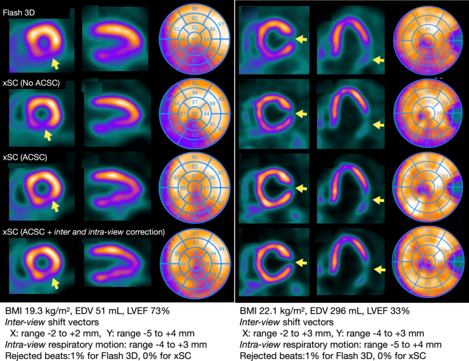
SPECT Myocardial Perfusion Imaging (MPI) is a non-invasive nuclear medicine test that shows how blood reaches different areas of the heart muscle. SPECT stands for Single-Photon Emission Computed Tomography. A small amount of a radiotracer is injected into a vein, and a specialised gamma camera records the tracer distribution around the heart.
The scan is commonly performed at rest and during cardiac stress. Stress may be produced by walking on a treadmill or, when adequate exercise is not possible, by a carefully selected medicine. Comparing the rest and stress images can help identify heart muscle that receives enough blood at rest but not during increased workload.
ECG-gated SPECT may also provide information about left-ventricular pumping function, chamber size and regional wall motion. A viability assessment uses a specifically selected protocol to estimate whether poorly contracting heart muscle may still be living and potentially capable of functional recovery. Not every routine perfusion scan automatically includes a full viability study.
Shows the relative distribution of blood flow through the heart muscle.
Compares heart-muscle blood flow at rest and during increased cardiac demand.
Selected protocols assess whether dysfunctional muscle may still be viable rather than permanently scarred.
The images are interpreted together with the stress ECG, symptoms, blood-pressure response, previous heart history and other cardiac investigations.
A defect that appears or becomes more pronounced during stress may suggest reduced blood supply to a region of heart muscle.
A fixed reduction present on both rest and stress images may represent scar, although attenuation and other technical factors must be considered.
ECG-gated images may estimate ejection fraction, ventricular volumes, wall thickening and regional wall motion.
The amount, location and severity of abnormal perfusion can contribute to clinical risk assessment and treatment planning.
Selected patients may undergo MPI after medicines, angioplasty, stenting or bypass surgery when symptoms or clinical questions remain.
A dedicated protocol may help distinguish potentially viable, underperforming muscle from tissue more likely to be permanently scarred.
The test is ordered only when the expected information is likely to influence diagnosis, risk assessment or treatment. It is not a routine screening test for every person with a cardiac risk factor.
The nuclear medicine team selects the tracer, imaging sequence, timing and stress method according to the clinical question, patient safety and local protocol.
Images obtained after stress are compared with rest images. A change between the two may indicate inducible ischemia, while a persistent defect requires correlation for scar, artefact or another cause.
Selected SPECT protocols, including delayed or redistribution imaging with an appropriate tracer, may be used to estimate whether dysfunctional muscle retains viable tissue.
Synchronising image acquisition with the ECG can provide additional information about ejection fraction, ventricular volumes, contraction and wall thickening.
Preparation depends on whether you will exercise, receive pharmacological stress or undergo a specialised viability protocol. Follow the imaging centre’s written instructions even when they differ from general advice.
You may be asked to fast for several hours, with water permitted. When vasodilator stress is planned, caffeine-containing drinks, coffee, tea, cola, energy drinks, chocolate and some medicines may interfere with the test and often need to be avoided for a specified period. Avoid smoking or nicotine according to the centre’s instructions.
Bring a complete medication list. Beta blockers, nitrates, calcium-channel blockers, theophylline-containing medicines and other drugs can affect stress testing, but do not stop any prescribed medicine unless the cardiologist or testing centre specifically tells you to do so. Report asthma, COPD, seizures, low blood pressure, rhythm problems and previous reactions.
Patients using insulin or glucose-lowering medicines need individual instructions so fasting does not cause low blood sugar. Confirm meal timing and medicine adjustments before the appointment, and carry glucose supplies if advised.
Tell the team before tracer injection if you are pregnant, might be pregnant or are breastfeeding. Nuclear imaging may need to be postponed, replaced or performed with specific precautions. Breastfeeding instructions depend on the exact radiotracer and dose.
The order of rest and stress imaging, tracer type and waiting period vary. Some studies are completed on one day; others may be divided across two visits.
The team confirms symptoms, medical history, medicines, fasting and caffeine preparation, pregnancy status and the planned stress method. Baseline heart rate, blood pressure and ECG are recorded.
A small IV cannula is placed, usually in the arm. The radiotracer is injected at the appropriate point in the protocol. The tracer is not the same as the iodine contrast used for CT angiography.
After a waiting period that allows tracer uptake, you lie on the imaging table with your arms positioned as instructed. The camera rotates or moves around the chest to collect images.
You may walk on a treadmill while ECG, blood pressure and symptoms are monitored. If adequate exercise is unsuitable, a pharmacological stress medicine may be used under supervised monitoring.
The tracer is injected at the target stress point. After the required uptake period, a second set of SPECT images is acquired. Remaining still helps reduce motion artefact.
When viability is the clinical question, delayed, redistribution or other protocol-specific images may be required. The team checks image quality before removing the IV and confirming discharge instructions.
The scan itself is painless, although the IV, exercise or stress medicine may cause temporary sensations. Tell the monitoring team immediately about any symptom.
Walking speed and incline may gradually increase. Breathlessness, tired legs and a faster heartbeat are expected, but chest discomfort, dizziness or unusual symptoms must be reported.
Depending on the medicine, temporary flushing, breathlessness, chest pressure, headache, nausea, palpitations or light-headedness may occur. The team monitors and treats symptoms according to protocol.
Most people feel only the IV needle. Significant allergic reactions to commonly used perfusion radiotracers are uncommon, but pain, swelling, rash or breathing difficulty should be reported immediately.
The test is commonly performed under trained supervision. The clinical benefit should justify the radiotracer exposure and the small but important risks associated with cardiac stress.
SPECT uses ionising radiation from a small amount of radiotracer. The dose depends on tracer, protocol, equipment and patient factors. Modern laboratories aim to use the lowest exposure that can answer the clinical question. Drinking fluids and passing urine regularly may help clear the tracer when permitted.
Exercise or pharmacological stress can rarely trigger a significant rhythm disturbance, marked blood-pressure change, severe bronchospasm, heart attack or another complication. Continuous monitoring, screening and emergency equipment are used to reduce risk and allow prompt treatment.
A result should not be interpreted from one phrase alone. The cardiologist considers image quality, stress response, symptoms, ECG findings and the full pattern of perfusion and function.
Tracer distribution appears broadly similar and adequate through the heart muscle at rest and stress, without a significant inducible perfusion defect.
Reduced uptake on stress images that improves at rest may suggest inducible ischemia in the corresponding heart-muscle territory.
A defect seen at both rest and stress may represent previous infarction or scar, but attenuation, motion and technical artefacts must be excluded.
A partly reversible abnormality may indicate scar with additional ischemia around or within the affected region.
The report may include ejection fraction, end-diastolic and end-systolic volumes, wall motion, wall thickening and transient ventricular dilation.
The report may estimate the location and amount of viable versus non-viable myocardium, while noting protocol limitations and the need for multimodality correlation.
SPECT provides functional information about myocardial blood flow. Other tests may be better suited to coronary anatomy, tissue characterisation, rhythm assessment or precise viability questions.
Body movement, soft-tissue attenuation, bowel or liver tracer activity and incorrect positioning can mimic or hide abnormalities. Repeat or alternative imaging may occasionally be advised.
CT coronary angiography or invasive coronary angiography is used when the clinical question requires direct assessment of coronary narrowing and anatomy.
Cardiac MRI or PET may provide additional information in selected patients when SPECT findings are uncertain or more detailed tissue assessment is required.
Dr. Kiran Lakkireddy provides evidence-based cardiac evaluation with a focus on selecting the right test for the clinical question. SPECT perfusion and viability findings are most useful when correlated with symptoms, ECG, Echo, coronary anatomy, previous heart attacks and prior cardiac procedures.
Clear answers to common questions about SPECT myocardial perfusion imaging and viability assessment.
Book a cardiac consultation to review your symptoms, previous reports, medicine and caffeine instructions, stress suitability and the most appropriate imaging protocol.