Heart chambers
Checks the size, shape and movement of the atria and ventricles, including enlargement or abnormal wall thickness.


A safe, painless and non-invasive heart ultrasound that provides real-time information about the heart’s chambers, valves, pumping function and blood flow.
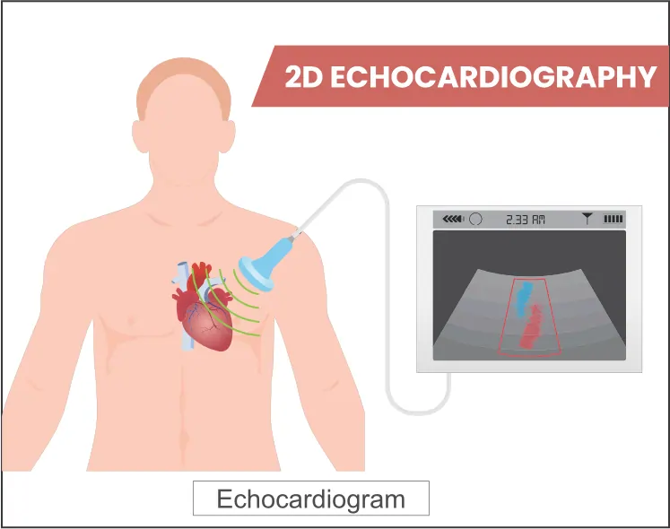
Two-dimensional echocardiography, commonly called a 2D Echo, is a diagnostic ultrasound test that creates moving images of the heart. A small handheld probe called a transducer is placed on the chest to send and receive high-frequency sound waves.
The images help the cardiology team assess the size and movement of the heart chambers, the pumping action of the heart muscle, the opening and closing of the valves, and the direction and speed of blood flow. Colour Doppler, spectral Doppler and M-mode measurements may also be used when clinically required.
Reviews chambers, walls, valves and major connected vessels.
Assesses heart-muscle movement and overall pumping performance.
Doppler imaging helps identify abnormal flow, narrowing or leakage.
The examination provides important information about heart anatomy and function. The exact measurements and views depend on age, symptoms, medical history and the clinical question.
Checks the size, shape and movement of the atria and ventricles, including enlargement or abnormal wall thickness.
Assesses how strongly the heart contracts and may include calculation of left ventricular ejection fraction.
Evaluates valve structure and movement and helps identify narrowing (stenosis) or backward leakage (regurgitation).
Doppler imaging helps assess the direction and velocity of blood through valves, chambers and major vessels.
Helps evaluate structural differences present from birth, including suspected abnormal connections or openings.
Can identify fluid around the heart and may estimate pressures related to pulmonary hypertension.
The basic technology is ultrasound, but the examination is adapted to the patient’s age, body size, heart rate, symptoms and clinical condition.
Used to evaluate symptoms and monitor known heart conditions, including valve disease, heart failure, cardiomyopathy and the effects of high blood pressure.
A specialised examination for infants, children and adolescents. Measurements and interpretation consider the child’s age, height, weight and developing heart.
Used when a newborn requires detailed assessment of heart structure, function, circulation or the cardiovascular changes that occur after birth.
An echocardiogram may be advised because of symptoms, physical examination findings, an abnormal ECG or X-ray, a known cardiac condition, or the need for treatment follow-up.
A standard transthoracic echocardiogram is performed from the outside of the chest. It does not involve surgery or a tube passing into the body.
Your symptoms, medical history and the reason for the test are reviewed.
You lie on an examination bed, usually on your back or left side.
Water-based gel is applied and the ultrasound probe is moved over the chest.
Multiple views and measurements are recorded while you breathe normally or briefly hold your breath.
The findings are interpreted in the context of your symptoms and other clinical information.
An echocardiogram contains many measurements. A single number should not be interpreted alone; the complete report must be matched with symptoms, examination findings and other tests.
An estimate of the percentage of blood pumped out of the left ventricle with each contraction.
Describes whether the atria or ventricles appear normal in size or enlarged.
Shows whether different areas of the heart muscle contract normally.
Narrowing of a heart valve that can restrict forward blood flow.
Backward leakage of blood through a valve that does not close completely.
Fluid collected in the sac surrounding the heart.
How the heart muscle relaxes and fills with blood between beats.
An estimate that may help assess increased pressure in the lung circulation.
Terms used for certain abnormal connections or openings associated with congenital heart disease.
Depending on the clinical findings, a cardiologist may recommend another test to answer a specific question that a routine 2D Echo cannot fully address.
Dr. Kiran Lakkireddy provides evidence-based cardiac evaluation with a focus on clear communication, accurate diagnosis and treatment plans tailored to each patient. His clinical practice includes preventive cardiology, complex coronary interventions, congenital cardiac interventions and advanced structural heart therapies.
Clear answers to common questions about adult, pediatric and neonatal 2D echocardiography.
Book a cardiology consultation and 2D echocardiography assessment based on your symptoms, medical history and clinical requirements.