Aortic aneurysm
CTA can measure an enlarged section of the aorta, show its shape and extent, and assess its relationship to important branch arteries for surveillance or treatment planning.


A detailed CT angiography examination that uses intravenous contrast and rapid CT imaging to evaluate the aorta and the arteries supplying the abdomen, pelvis and limbs.
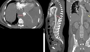
This page describes CT angiography, also called CTA—a non-surgical imaging test that maps blood vessels using a CT scanner and iodine-based contrast injected through a vein.
A CT aortogram focuses on the aorta, the body’s main artery, and may include the thoracic aorta in the chest, the abdominal aorta, or both. A peripheral CT angiogram extends the examination into arteries supplying the pelvis and limbs, most commonly the legs. The exact scan range is selected according to the symptoms and the clinical question.
As the contrast moves through the arteries, the scanner rapidly acquires thin images. Computer processing then creates detailed cross-sectional, multiplanar and three-dimensional views. These images can show the location and length of narrowing, complete blockages, aneurysms, dissection, abnormal vessel anatomy and the quality of blood flow pathways beyond a diseased segment.
This examination is different from catheter angiography. CT angiography generally requires only a small intravenous cannula in the arm. Catheter angiography uses an arterial puncture and a catheter advanced inside the blood vessel; it may be chosen when treatment such as angioplasty or stenting could be performed during the same procedure.
The examination may be requested to diagnose vascular disease, define its severity or plan medical, endovascular or surgical treatment.
CTA can measure an enlarged section of the aorta, show its shape and extent, and assess its relationship to important branch arteries for surveillance or treatment planning.
The scan may identify a tear within the aortic wall, the channels created by the dissection and whether branch vessels or organs are affected.
CTA can map narrowing and blockages in the aortoiliac, femoral, popliteal and below-knee arteries in patients with suspected or known peripheral artery disease.
Detailed vessel measurements can help plan angioplasty, stenting, endovascular aneurysm repair, bypass surgery or hybrid vascular procedures.
CTA may be used to evaluate selected stents, grafts, bypass pathways or repaired aortic segments and to identify complications when clinically suspected.
The examination may help assess arterial injury, congenital vascular differences, compression, inflammatory disease or other selected abnormalities.
The need for CTA depends on symptoms, examination findings, kidney function and results of earlier tests such as Doppler ultrasound.
The exact protocol varies, but a typical examination follows these stages.
The team confirms the scan request, symptoms, allergies, kidney function, pregnancy status and relevant medicines.
A cannula is placed in a vein, usually in the arm, so contrast can be injected at a controlled rate.
You lie on the CT table. The required body area is positioned carefully, and straps or supports may be used to help you remain still.
Iodine-based contrast is injected through the IV. A brief warm sensation, flushing or metallic taste can occur and usually passes quickly.
The table moves through the scanner while images are taken. You may be asked to hold your breath briefly for aortic imaging.
The radiology team checks image quality and creates detailed multiplanar and 3D views for interpretation and treatment planning.
Preparation is important because the examination usually uses iodine-based contrast and ionising radiation.
The report does more than state whether a blockage is present. It describes the anatomy needed for clinical decisions.
The report may describe where a vessel narrows or closes, how long the abnormal segment is and whether several arterial levels are involved.
CTA can show calcified and non-calcified plaque, although very heavy calcification can sometimes make the exact open channel harder to assess.
For leg arteries, the report may comment on vessels beyond the blockage, collateral channels and the number of arteries reaching the lower leg or foot.
Aortic diameter, length, branch-vessel involvement, clot within the aneurysm and surrounding findings may be documented.
When present, the scan may show the intimal flap, true and false channels, entry points and involvement of branch arteries.
Selected scans provide vessel diameters, access-route information and landing-zone measurements used for endovascular or surgical planning.
CTA is widely used because it is fast and detailed, but it involves radiation and contrast. The expected benefit should outweigh the individual risk.
CT uses ionising radiation. The protocol is tailored to the required body area and clinical question, with dose-reduction techniques used when appropriate.
Most patients tolerate iodine-based contrast. Mild reactions can occur, while severe allergic-like reactions are uncommon. Previous reactions must be reported before the scan.
Contrast may pose greater risk in patients with significantly reduced kidney function, acute kidney injury or dehydration. Kidney tests and preventive planning may be needed.
Rarely, contrast can leak from the vein into surrounding tissue, causing pain or swelling. Tell the technologist immediately if the injection site becomes painful.
Because CT uses X-rays, anyone who is pregnant or may be pregnant should inform the team so urgency, alternatives and protective planning can be considered.
Most outpatients can leave after the required observation. Follow hydration advice and seek help for delayed breathing difficulty, facial swelling, widespread rash or severe symptoms.
The most suitable vascular test depends on the artery involved, urgency, kidney function and whether treatment may be required immediately.
The radiology report should be interpreted together with symptoms, pulse examination, blood-pressure findings, Doppler results, kidney function and overall cardiovascular risk.
A report may identify mild plaque that requires risk-factor control, a significant stenosis that needs closer assessment, a complete occlusion with collateral circulation, or an aortic abnormality that requires surveillance or treatment planning. The importance of a finding depends on its location, severity, symptoms and impact on organ or limb blood flow.
Treatment is not decided by the scan image alone. Options may include smoking cessation, diabetes and cholesterol control, antiplatelet or other medicines, supervised exercise, wound care, angioplasty, stenting, endovascular aneurysm repair or open surgery. The treating team will explain which approach is appropriate.
CT angiography is performed and formally interpreted by the appropriate radiology team. Dr. Kiran Lakkireddy reviews the vascular findings in the context of symptoms, cardiovascular risk factors, clinical examination and other investigations to guide further cardiac or vascular management.
Common questions about contrast, preparation, safety and understanding the scan.
Book a consultation to discuss whether CT angiography is appropriate and how the findings may guide further cardiovascular or vascular treatment.