Heart size and pumping function
Measures right- and left-ventricular volumes, ejection fraction, muscle thickness and regional wall motion with high reproducibility.


Detailed magnetic resonance imaging of the heart to assess cardiac structure, pumping function, blood flow, inflammation, scar tissue and complex heart conditions—without ionising radiation.
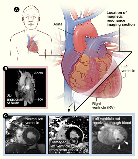
Cardiac Magnetic Resonance Imaging, also called Cardiac MRI or cardiovascular magnetic resonance (CMR), is a non-invasive scan that creates detailed moving and still images of the heart and nearby blood vessels. It uses a strong magnetic field, radiofrequency waves and specialised computer processing rather than X-rays.
The scan can measure the size of the heart chambers, pumping function, wall motion and blood flow. It can also characterise the heart muscle itself—helping doctors look for inflammation, swelling, scar tissue, previous heart-attack damage, infiltration or abnormal thickening that may not be fully explained by an ECG or routine echocardiogram.
Shows the heart chambers, muscle, valves, pericardium and major vessels in multiple planes.
Measures ventricular volumes, ejection fraction, regional wall motion and selected blood-flow patterns.
Selected sequences can identify oedema, fibrosis, scar and other changes within the heart muscle.
Cardiac MRI combines anatomy, motion, blood-flow measurements and tissue characterisation. The findings are interpreted together with symptoms, ECG, Echo, laboratory tests and previous imaging.
Measures right- and left-ventricular volumes, ejection fraction, muscle thickness and regional wall motion with high reproducibility.
Helps assess hypertrophic, dilated, arrhythmogenic, restrictive and other heart-muscle disorders, including patterns of scar or infiltration.
Can identify tissue changes consistent with inflammation, oedema or injury when myocarditis or another inflammatory condition is suspected.
Contrast-enhanced imaging may show the location and extent of myocardial scar and whether damaged muscle may still be viable.
Defines complex anatomy, shunts, repaired congenital defects, right-heart function and major-vessel relationships when Echo is incomplete.
May quantify selected valve leakage, assess pericardial disease and examine the aorta or pulmonary arteries as part of a tailored protocol.
The test is often requested when another investigation has raised a question that needs more detailed structural, functional or tissue assessment.
No. The radiologist and cardiologist select sequences according to the diagnostic question, previous results and patient safety.
Moving cine images can assess chamber size, wall motion, ejection fraction and anatomy without an injection of contrast.
Gadolinium may be used to evaluate perfusion, scar, fibrosis, inflammation or masses when the expected benefit justifies contrast.
Selected patients may receive a medicine that temporarily increases coronary blood flow while MRI looks for areas receiving less blood. This requires specific screening and monitoring.
Preparation varies according to whether contrast, sedation or stress medicine is planned. Follow the imaging centre’s instructions rather than making medication changes yourself.
The exact sequence varies, but most examinations follow the general process below.
The team confirms implants, metal exposure, kidney history, pregnancy status, allergies and whether contrast, sedation or stress medicine is planned.
You change into MRI-safe clothing. ECG stickers are placed on the chest to synchronise images with the heartbeat, and a blood-pressure cuff may be used. An IV line is inserted when contrast or stress medicine is required.
You lie on a padded table with a special receiver coil over the chest. The table moves into the scanner, and ear protection is provided for the loud tapping sounds.
The technologist communicates by intercom and asks you to hold your breath for short periods. Remaining still is important because movement can blur the images.
Gadolinium may be injected to assess tissue or scar. In a stress study, medication is given under monitoring and stopped according to the protocol and your response.
The team reviews the images for completeness before removing the IV. Most patients can leave after the routine scan; additional observation may be needed after stress medication or sedation.
Cardiac MRI is usually painless, although lying still, repeated breath-holds and the enclosed scanner can be uncomfortable for some people.
Rhythmic tapping and thumping are normal. Earplugs or headphones reduce the noise, and you remain in contact with the technologist.
You may be asked to hold your breath repeatedly for several seconds. Tell the team if you cannot follow a breath-hold instruction.
Some people notice temporary warmth, coolness at the IV site or a metallic taste. Report pain, swelling, rash, wheeze or unusual symptoms immediately.
For most appropriately screened patients, Cardiac MRI is a safe test. The main precautions relate to the magnetic field, implanted devices, contrast, kidney function, pregnancy, sedation and stress medicines.
The report is tailored to the clinical question and may include quantitative measurements as well as a description of tissue patterns.
Cardiac MRI answers many complex questions, but image quality and diagnostic value depend on the clinical problem and the patient’s ability to complete the scan.
Severe breathlessness, inability to lie still, frequent irregular beats or difficulty with breath-holds can reduce image quality.
Small coronary arteries are usually assessed more directly with CT coronary angiography or invasive coronary angiography when clinically indicated.
Stress Echo, nuclear perfusion imaging, Holter monitoring or catheter-based tests may be needed depending on symptoms and the MRI findings.
Dr. Kiran Lakkireddy provides evidence-based cardiac evaluation with a focus on accurate diagnosis, clear explanation and individualised treatment planning. Cardiac MRI findings are most useful when they are correlated with the patient’s symptoms, ECG, Echo, laboratory results and previous cardiac procedures.
Clear answers to common questions about Cardiac MRI.
Book a cardiac consultation to review the reason for the scan, previous reports, implant information, kidney function and the most appropriate imaging protocol.