Intra-aortic Balloon Pump

Intra-aortic Balloon Pump (IABP): Temporary Circulatory Support for Heart Conditions

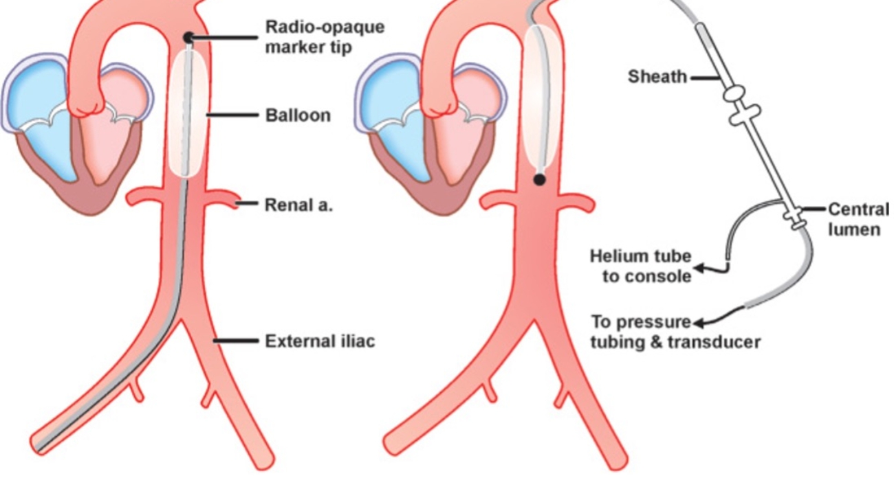
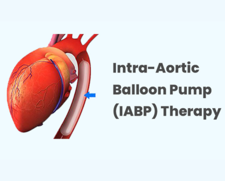
IABP (Intra-aortic Balloon Pump) is a mechanical device used for temporary circulatory support in individuals with certain cardiac conditions. It consists of a long, thin tube called a catheter with an inflatable balloon at its tip.

Here’s how the IABP works:

  1. Placement: The IABP is inserted through the femoral artery, typically in the groin area. The catheter is carefully threaded until it reaches the aorta, the large blood vessel that carries oxygenated blood from the heart to the rest of the body.
  2. Balloon Inflation and Deflation: Once the IABP is positioned in the aorta, the balloon inflates and deflates in sync with the heartbeat. The timing of inflation and deflation is controlled by a console connected to the IABP.
    • Inflation Phase: During the inflation phase, the balloon inflates, temporarily blocking the blood flow in the aorta. This increases pressure in the aorta, which helps improve coronary artery blood flow and reduces the heart’s workload.

    • Deflation Phase: After a brief inflation, the balloon rapidly deflates just before the heart pumps, allowing blood to flow easily from the heart into the aorta and the rest of the body.

The inflation and deflation occur synchronously with the heartbeat, providing mechanical assistance to the heart’s pumping action. This process helps to increase coronary blood flow, reduce the heart’s workload, and improve overall cardiac output.

Clinical Scenarios Where IABP is Used:

  • Acute Myocardial Infarction (heart attack) with cardiogenic shock: The IABP stabilizes the patient’s hemodynamics and improves coronary artery blood flow while awaiting further treatments.
  • Decompensated heart failure: For patients with severe heart failure, the IABP provides temporary support until heart transplantation or ventricular assist device implantation is possible.
  • High-risk cardiac surgeries: The IABP supports the heart during complex cardiac surgeries, ensuring stable blood flow throughout the procedure.

Duration and Monitoring:

The IABP is typically used for a short period, ranging from days to weeks, depending on the patient’s condition. Continuous monitoring and regular assessment of the patient’s clinical status, hemodynamics, and potential complications are essential.

Conclusion:

The IABP is a valuable temporary support device that assists patients with heart failure, acute myocardial infarction, or during high-risk cardiac surgeries. While it provides immediate hemodynamic support and relief, it is not a definitive treatment but a bridge to further interventions, such as heart transplantation or the use of ventricular assist devices.

For patients with severe cardiac conditions, IABP therapy can be life-saving by enhancing coronary blood flow and reducing the heart’s workload. However, its use should always be closely monitored and managed by a specialized healthcare team.

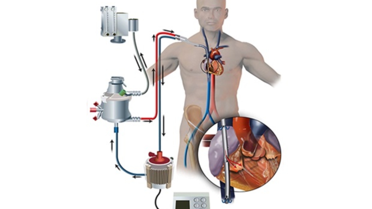
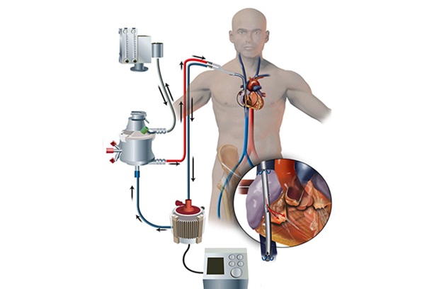
ECMO life support system providing cardiac and respiratory support.

ECMO

ECMO (Extracorporeal Membrane Oxygenation): Life Support for Severe Heart and Lung Failure

ECMO, or Extracorporeal Membrane Oxygenation, is a life support system used to provide temporary cardiac and respiratory support for individuals with severe heart and/or lung failure. This technology temporarily takes over the functions of the heart and lungs, allowing these organs to rest, heal, and recover.

How ECMO Works:

  1. Cannulation: The procedure begins by inserting two tubes called cannulas into large blood vessels, usually through the groin area. One cannula draws blood from the patient, and the other returns oxygenated blood from the ECMO machine to the body.
  2. Blood Flow and Oxygenation: Blood is pumped from the body into the ECMO machine, where it passes through a special membrane that oxygenates the blood and removes carbon dioxide. The oxygenated blood is then returned to the body, ensuring vital organs receive proper oxygenation.
  3. Monitoring and Adjustment: The ECMO machine constantly monitors the blood flow, oxygen levels, and other parameters to ensure that the heart and lungs are receiving optimal support.

Types of ECMO Configurations:

  1. Venoarterial ECMO (VA-ECMO): In this configuration, blood is drawn from a vein and returned to an artery, providing both cardiac and respiratory support. It is used for individuals with severe cardiac failure, respiratory failure, or a combination of both.
  2. Venovenous ECMO (VV-ECMO): This configuration is used when blood is withdrawn from and returned to veins. VV-ECMO primarily provides respiratory support for patients with severe lung failure, giving the lungs time to rest and heal.

Key Benefits of ECMO:

  1. Provides life-saving support when other treatments have failed.
  2. Supports heart and/or lung function, allowing recovery time for the organs.
  3. Short-term therapy, is typically used for a few days to weeks, depending on the patient’s condition.

Risks and Complications:

While ECMO can be life-saving, it carries potential risks, including:

  • Bleeding
  • Infection
  • Clotting
  • Organ injury
  • Cannulation site issues


    Careful monitoring and management by a highly skilled healthcare team, including perfusionists, critical care physicians, cardiothoracic surgeons, and specialized nurses, is essential for patient safety.

Conclusion:

ECMO serves as a bridge to recovery, allowing the heart and lungs to heal, or as a temporary solution until more definitive interventions, such as heart transplantation or lung transplantation, can be performed. The decision to initiate, maintain, or discontinue ECMO support is made based on the patient’s condition, recovery potential, and response to treatment.

Consult Dr. Lakkireddy Kiran Kumar Reddy, an interventional cardiologist at Onus Heart Institute, for expert guidance on ECMO as a treatment option for severe heart and lung failure.

IMPELLA Dr kiran Lakkireddy

IMPELLA

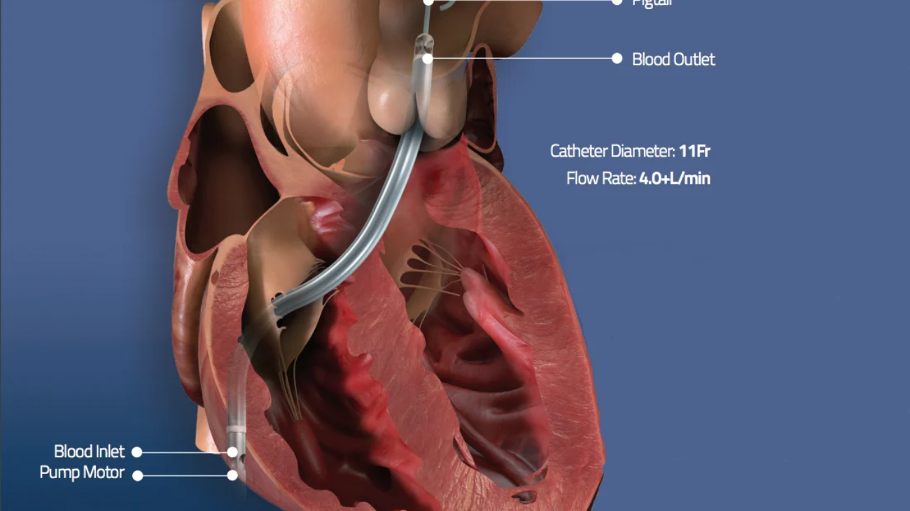
Impella is a type of mechanical circulatory support device used for temporary cardiac support in individuals with severe heart failure or undergoing high-risk cardiac procedures. It is specifically designed to assist the left ventricle in pumping blood, improving cardiac output and perfusion.

The Impella device consists of a catheter with a miniature axial flow pump at its tip. The catheter is inserted into the femoral artery in the groin and advanced into the left ventricle of the heart. The pump is positioned within the left ventricle, drawing blood into the device and then propelling it into the aorta, bypassing the weakened or failing left ventricle.

Key Benefits of Impella:

  1. Continuous blood flow: It helps unload the left ventricle and increases cardiac output, improving organ perfusion.
  2. Temporary cardiac support: It can alleviate symptoms of severe heart failure and aid in recovery.

Clinical Scenarios Where Impella is Used:

  1. High-risk percutaneous coronary interventions (PCI): For patients with complex coronary artery disease or compromised heart function, Impella provides temporary circulatory support during PCI procedures, reducing procedural risks.
  2. Cardiogenic shock: For individuals with severe heart failure or acute myocardial infarction complicated by cardiogenic shock, Impella offers temporary hemodynamic support, buying time for recovery or further interventions.

The Impella device comes in different models, offering varying levels of flow support. The specific model used depends on the patient’s clinical needs and the healthcare provider’s assessment.

Monitoring and Care:

Impella is a temporary support device, usually used for a short duration ranging from hours to days. It requires close management and monitoring by a multidisciplinary team, including interventional cardiologists, cardiac surgeons, and specialized nurses.

Potential Complications:

While Impella can offer valuable support, there are risks involved, including bleeding, vascular injury, infection, device malfunction, and thrombus formation. The decision to use Impella is made on an individualized basis, in collaboration with the healthcare team and the patient, carefully weighing the potential benefits and risks.

Conclusion

Impella offers a powerful solution for patients with severe heart failure or those undergoing high-risk cardiac procedures. It provides vital temporary support, enhancing heart function and improving outcomes in critical situations. However, its use requires careful selection, precise placement, and constant monitoring to ensure patient safety and optimize recovery. At Onus Heart Institute, we work with a team of experts to provide personalized care for patients requiring mechanical circulatory support.

Consult Dr. Lakkireddy Kiran Kumar Reddy at Onus Heart Institute for expert advice on Impella and its role in treating heart failure and related conditions.

Carotid Stenting: Minimally Invasive Treatment for Carotid Artery Stenosis

Carotid stenting, also known as carotid artery stenting, is a minimally invasive procedure used to treat carotid artery stenosis, which is the narrowing of the carotid arteries in the neck. These arteries supply blood to the brain, and when they become significantly narrowed or blocked, it can increase the risk of stroke.

During carotid stenting, a small, flexible tube called a catheter is inserted into an artery, typically in the groin area, and threaded up to the carotid artery that is affected by the stenosis. The procedure is usually performed under local anesthesia.

Here’s a step-by-step overview of the carotid stenting procedure:

Artery access: A small incision is made in the groin, and a catheter is inserted into a large artery, such as the femoral artery. The catheter is then guided through the arteries to the carotid artery.

Balloon angioplasty: A small deflated balloon is advanced to the site of the stenosis in the carotid artery. The balloon is then inflated, compressing the plaque and widening the artery to improve blood flow. This is known as balloon angioplasty.

Stent placement: After balloon angioplasty, a stent is placed in the treated area. The stent is a small, mesh-like tube made of metal or other materials. It acts as a scaffold to keep the artery open and prevent it from re-narrowing. The stent is delivered to the site of the stenosis through the catheter and expanded to fit securely in the artery.

Post-procedure evaluation: Once the stent is in place, the blood flow is assessed to ensure it is restored and the artery is adequately opened. Imaging techniques such as angiography or ultrasound may be used to evaluate the results of the procedure.

Carotid stenting is typically performed in individuals who are considered high risk for carotid endarterectomy, which is a surgical procedure to remove plaque from the carotid artery. High-risk factors may include advanced age, severe medical conditions, previous neck surgery, or previous radiation therapy to the neck.

Following carotid stenting, patients are usually monitored in a hospital setting for a brief period. Medications to prevent blood clotting, such as antiplatelet drugs, are often prescribed to reduce the risk of stent thrombosis.

As with any medical procedure, carotid stenting carries potential risks and complications, including stroke, bleeding, infection, damage to the artery, or allergic reactions to medications or contrast agents used during the procedure. The risks and benefits of carotid stenting should be carefully considered by the healthcare team based on each individual’s specific condition and circumstances.

Regular follow-up visits and monitoring are necessary after carotid stenting to assess the effectiveness of the procedure and manage any ongoing concerns related to the carotid arteries.

Conclusion:
Dr. Kiran Lakkireddy, Carotid stenting is a valuable treatment option for patients with carotid artery stenosis, offering hope for improved quality of life and reduced stroke risk. Our team is dedicated to providing personalized care and expertise to ensure optimal outcomes.

PRIMARY ANGIOPLASTY Drkiranlakkireddy

PRIMARY ANGIOPLASTY

Primary angioplasty, also known as percutaneous coronary intervention (PCI), is a medical procedure used to treat a heart attack (myocardial infarction) caused by a blockage in the coronary arteries. It is considered the gold standard treatment for acute myocardial infarction.

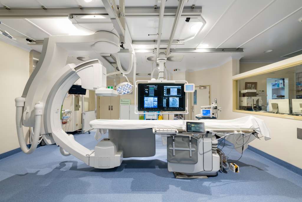
During primary angioplasty, the goal is to quickly restore blood flow to the blocked coronary artery and minimize the extent of heart muscle damage. The procedure is typically performed in a cardiac catheterization laboratory and involves the following steps:

Cath lab image
  1. Access and catheter insertion: A small incision is made, usually in the wrist (radial approach) or groin (femoral approach), to access the artery. A catheter is inserted through the incision and guided to the coronary arteries under X-ray guidance.
  2. Angiography: A contrast dye is injected through the catheter, and X-ray images (angiograms) are taken to visualize the blocked artery and identify the location and severity of the blockage.
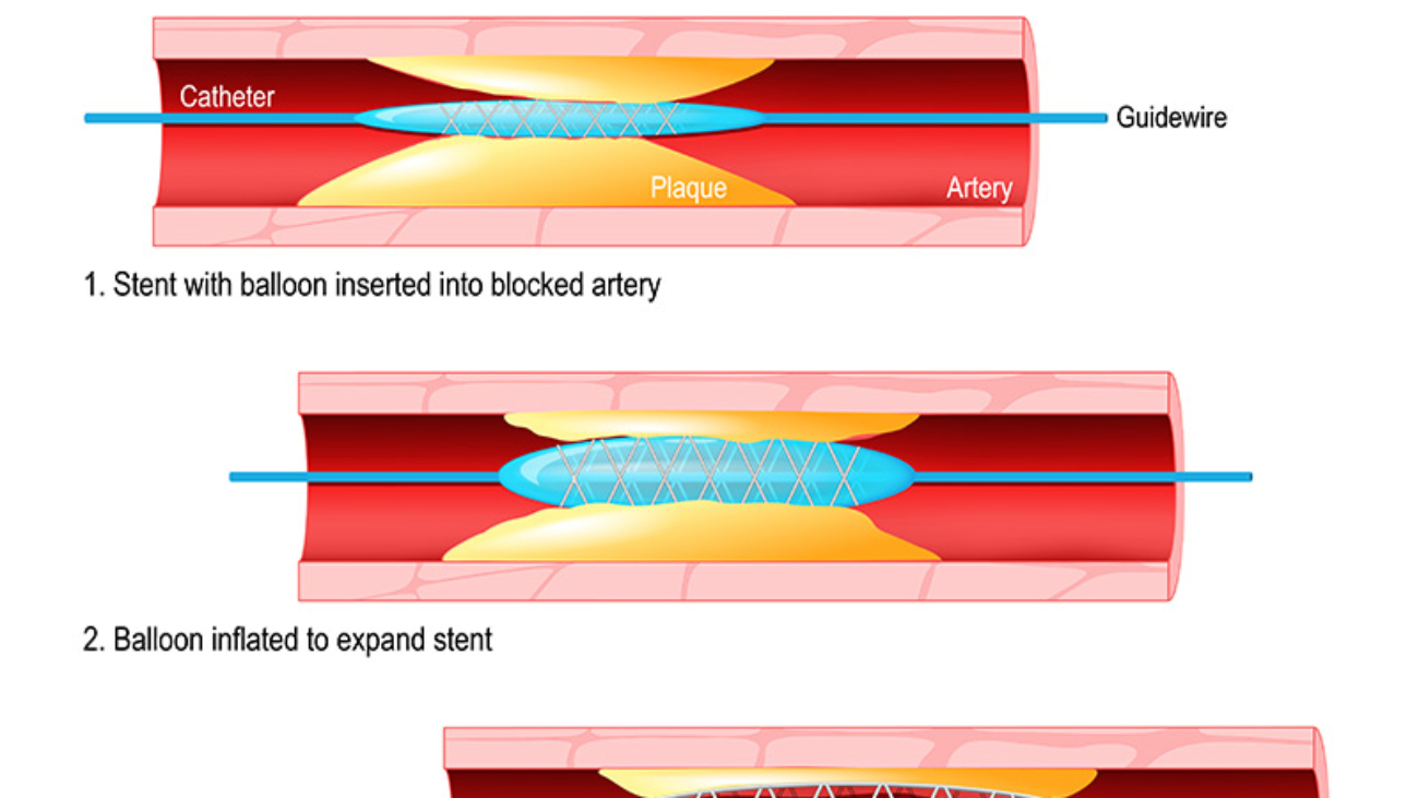
  3. Balloon angioplasty: A thin wire with a deflated balloon attached to it is advanced through the catheter to the site of the blockage. The balloon is then inflated, compressing the plaque against the artery walls and widening the artery to restore blood flow.
  4. Stent placement: In many cases, a stent, which is a small mesh-like tube, is inserted into the treated area. The stent acts as a scaffold to keep the artery open and prevent it from re-narrowing. Some stents are coated with medications (drug-eluting stents) to help prevent re-blockage.
  5. Post-procedure evaluation: Once the blood flow is restored, the success of the procedure is assessed by performing another angiogram to ensure adequate dilation of the artery and good blood flow to the heart muscle.

Primary angioplasty is typically performed as an emergency procedure, and the time from symptom onset to treatment is critical. The goal is to perform the procedure as quickly as possible, preferably within 90 minutes of the patient’s arrival at the hospital (referred to as the “door-to-balloon” time).

Benefits of primary angioplasty include improved blood flow to the heart, reduced heart muscle damage, and improved survival rates compared to other treatments for acute myocardial infarction. However, like any medical procedure, there are risks involved, including bleeding, blood vessel damage, clot formation, and allergic reactions to medications or contrast dye.

After primary angioplasty, patients are closely monitored in the hospital for a period of time. Medications, including antiplatelet agents and anticoagulants, are typically prescribed to prevent clot formation and reduce the risk of future cardiovascular events.

Long-term management after primary angioplasty includes lifestyle modifications, such as a heart-healthy diet, regular exercise, smoking cessation, and adherence to prescribed medications. Regular follow-up visits with the healthcare team are important to monitor the patient’s progress, assess cardiac function, and adjust medications as needed.

Healthy Lifestyle Drkiranlakkireddy

HEALTHY LIFESTYLE

Maintaining a healthy lifestyle is crucial for overall well-being and the prevention of various diseases. Here are some key components of a healthy lifestyle:

  1. Balanced diet: Aim to consume a variety of nutritious foods, including fruits, vegetables, whole grains, lean proteins, and healthy fats. Limit processed foods, sugary drinks, and foods high in saturated and trans fats. Practice portion control and mindful eating.
  2. Regular physical activity: Engage in regular exercise or physical activity for at least 150 minutes per week. Choose activities that you enjoy and that challenge your cardiovascular system, strength, and flexibility. Examples include brisk walking, jogging, swimming, cycling, weightlifting, and yoga.
  3. Adequate sleep: Establish a consistent sleep schedule and aim for 7 to 9 hours of quality sleep each night. Good sleep is essential for physical and mental health, energy levels, and overall functioning.
  4. Stress management: Find healthy ways to manage and cope with stress, such as practicing relaxation techniques (deep breathing, meditation, yoga), engaging in hobbies, spending time with loved ones, or seeking professional support when needed.
  5. Avoid tobacco and limit alcohol consumption: If you smoke, seek resources and support to quit. Limit alcohol consumption or avoid it altogether.
  6. Maintain a healthy weight: Strive to achieve and maintain a healthy body weight through a combination of balanced eating and regular physical activity. Consult with a healthcare professional for personalized guidance.
  7. Regular health check-ups: Schedule regular check-ups with your healthcare provider to monitor your health, screen for potential conditions, and address any concerns or questions you may have.
  8. Hydration: Stay adequately hydrated by drinking plenty of water throughout the day. Limit the intake of sugary drinks and opt for water as the primary beverage.
  9. Social connections: Cultivate positive relationships and social connections with friends, family, and community. Strong social support can contribute to better mental and physical health.
  10. Mental well-being: Prioritize your mental health by engaging in activities that bring you joy, practicing self-care, seeking support when needed, and being mindful of your emotions and mental state.

Remember, adopting a healthy lifestyle is a long-term commitment and involves making sustainable changes. It’s important to listen to your body, make gradual improvements, and seek professional guidance when necessary. Small steps toward a healthier lifestyle can have significant and positive impacts on your overall well-being.

HEALTHY DIET

A healthy diet plays a crucial role in maintaining overall health and preventing chronic diseases. Here are some key principles of a healthy diet:

  1. Eat a variety of nutrient-rich foods: Include a wide range of fruits, vegetables, whole grains, lean proteins (such as poultry, fish, legumes, and tofu), and healthy fats (such as nuts, seeds, avocado, and olive oil) in your diet. These foods provide essential vitamins, minerals, fiber, and other beneficial compounds.
  2. Focus on fruits and vegetables: Aim to fill half your plate with a colorful variety of fruits and vegetables. They are rich in vitamins, minerals, antioxidants, and dietary fiber. Include different types and colors to ensure a wide range of nutrients.
  3. Choose whole grains: Opt for whole grain foods like whole wheat bread, brown rice, quinoa, oats, and whole grain cereals. They contain more fiber, vitamins, and minerals compared to refined grains.
  4. Include lean proteins: Choose lean sources of protein, such as poultry, fish, legumes (beans, lentils, chickpeas), tofu, and low-fat dairy products. These provide important nutrients without excessive saturated fat.
  5. Reduce processed foods: Limit the consumption of processed and packaged foods, which are often high in added sugars, unhealthy fats, and sodium. These include sugary snacks, processed meats, sugary drinks, and packaged snacks.
  6. Limit added sugars and sugary drinks: Minimize the intake of foods and drinks with added sugars, such as soda, candy, pastries, and sweetened cereals. Opt for water, herbal tea, or unsweetened beverages instead.
  7. Control portion sizes: Be mindful of portion sizes to avoid overeating. Use smaller plates and bowls, and listen to your body’s hunger and fullness cues.
  8. Stay hydrated: Drink an adequate amount of water throughout the day. Water helps maintain hydration, aids digestion, and supports overall bodily functions.
  9. Reduce sodium intake: Limit the consumption of high-sodium foods like processed meats, canned soups, and packaged snacks. Opt for herbs, spices, and other seasonings to flavor your meals instead.
  10. Practice mindful eating: Pay attention to your eating habits and enjoy meals without distractions. Eat slowly, savoring the flavors and textures of your food. This can help you recognize feelings of fullness and promote better digestion.

Remember, a healthy diet is about making sustainable choices and finding a balance that works for you. It’s important to consult with a healthcare professional or registered dietitian for personalized guidance based on your specific dietary needs, preferences, and any underlying health conditions.

The Mediterranean diet is a dietary pattern based on the traditional eating habits of people living in countries bordering the Mediterranean Sea, such as Greece, Italy, Spain, and Turkey. It has gained recognition for its potential health benefits and is considered one of the healthiest diets to follow. Here are the key features of the Mediterranean diet:

  1. Abundant plant-based foods: The Mediterranean diet emphasizes the consumption of fruits, vegetables, whole grains, legumes, nuts, and seeds. These foods are rich in fiber, vitamins, minerals, and antioxidants.
  2. Healthy fats: Instead of relying on saturated and trans fats, the Mediterranean diet encourages the consumption of healthy fats, primarily from olive oil, nuts, and seeds. Olive oil, in particular, is a central component and is used as the primary source of fat in cooking and dressing.
  3. Moderate intake of fish and poultry: Fish, especially fatty fish like salmon, mackerel, and sardines, is consumed regularly in the Mediterranean diet due to its omega-3 fatty acid content. Poultry is also included in moderation.
  4. Limited red meat: Red meat consumption is limited in the Mediterranean diet and is often reserved for special occasions. When consumed, it is typically in small portions.
  5. Dairy products in moderation: The intake of dairy products, such as yogurt and cheese, is moderate in the Mediterranean diet. Greek yogurt is a common choice due to its probiotic content.
  6. Fresh fruits as desserts: Instead of sugary desserts, fresh fruits are commonly enjoyed as a healthier alternative. They provide natural sweetness and additional nutrients.
  7. Red wine in moderation: Moderate consumption of red wine, typically with meals, is a characteristic of the Mediterranean diet. This is optional and should be avoided by individuals who cannot tolerate alcohol or have certain health conditions.

The Mediterranean diet has numerous health benefits, including a reduced risk of heart disease, stroke, certain cancers, and chronic conditions like diabetes and obesity. It is believed that the combination of plant-based foods, healthy fats, and moderate consumption of other food groups contributes to these positive health outcomes.

It’s important to note that the Mediterranean diet is not just about the foods consumed but also encompasses a lifestyle approach. Regular physical activity, enjoying meals with family and friends, and adopting mindful eating practices are also emphasized.

While the Mediterranean diet offers many health benefits, it’s important to personalize it based on individual needs, preferences, and any specific dietary considerations. Consulting with a healthcare professional or registered dietitian can provide tailored guidance to help incorporate the Mediterranean diet into your lifestyle.

HEART ATTACK vs HEART BLOCK

A heart attack and heart block are two distinct cardiac conditions with different causes and manifestations. Here’s a brief explanation of each:

  1. Heart attack (Myocardial Infarction): A heart attack, medically known as myocardial infarction (MI), occurs when there is a blockage in one or more of the coronary arteries, which supply oxygenated blood to the heart muscle. This blockage usually results from the buildup of cholesterol plaque that ruptures and forms a blood clot, obstructing the artery. The lack of blood flow and oxygen to a section of the heart muscle can lead to damage or death of the affected tissue. Symptoms of a heart attack often include intense chest pain or discomfort, shortness of breath, pain radiating to the left arm, jaw, or back, nausea, sweating, and lightheadedness. Prompt medical attention is essential for the diagnosis and management of a heart attack, as it is a medical emergency.
  2. Heart block: Heart block, also referred to as atrioventricular block, is a condition that affects the electrical conduction system of the heart. It occurs when the electrical signals that regulate the heartbeat are delayed or blocked as they travel from the atria (upper chambers) to the ventricles (lower chambers). This disruption in the electrical pathway can result in an abnormally slow heart rate, which can cause symptoms such as dizziness, fatigue, fainting, or even cardiac arrest in severe cases. Heart block can have various causes, including age-related degeneration, heart disease, congenital defects, medication side effects, or electrolyte imbalances. Treatment for heart block depends on the severity and symptoms and may involve the use of medications, pacemakers, or other interventions to regulate the heart rate and restore proper conduction.

It’s important to note that while heart attack and heart block are distinct conditions, they can coexist in some cases. For instance, a heart attack may lead to damage in the conduction system, resulting in heart block as a secondary complication. Therefore, it is crucial to seek immediate medical attention if you experience symptoms suggestive of a heart attack or any concerning cardiac symptoms to receive an accurate diagnosis and appropriate treatment.

The management of a heart attack and heart block differs due to their distinct underlying causes and immediate risks. Here’s an overview of the management approaches for each condition:

Management of a Heart Attack (Myocardial Infarction):

  1. Immediate medical attention: A heart attack is a medical emergency, and it is crucial to call for emergency medical assistance (such as an ambulance) as soon as possible.
  2. Emergency medical care: Upon arrival at the hospital, the medical team will quickly assess the situation and provide immediate treatment, which may include:
    • Oxygen therapy: Supplemental oxygen is administered to ensure adequate oxygen supply to the body.
    • Medications: Aspirin or other antiplatelet medications may be given to help prevent further blood clotting. Nitroglycerin and/or pain medications may be provided to relieve chest pain or discomfort.
    • Reperfusion therapy: Time-sensitive procedures such as percutaneous coronary intervention (PCI) or thrombolytic therapy may be performed to restore blood flow to the blocked coronary artery and minimize damage to the heart muscle.
  3. Post-acute care and management: After initial treatment, the patient will typically be admitted to the hospital for further monitoring and management, which may include:
    • Medications: Medications such as antiplatelet agents, beta-blockers, ACE inhibitors, statins, and other appropriate drugs are prescribed to manage risk factors, improve heart function, prevent future complications, and aid in recovery.
    • Cardiac rehabilitation: A structured program that combines exercise, education, counseling, and support to help patients recover, improve cardiovascular fitness, and reduce the risk of future heart problems.
    • Lifestyle modifications: Emphasizing the importance of a heart-healthy diet, regular physical activity, smoking cessation, stress management, and maintaining a healthy weight.
    • Follow-up care: Regular follow-up visits with a healthcare provider to monitor progress, assess the effectiveness of treatment, and adjust medications if needed.

Management of Heart Block (Atrioventricular Block):

  1. Evaluation and diagnosis: A healthcare provider will conduct a thorough evaluation to determine the underlying cause and severity of the heart block. This may involve a medical history review, physical examination, electrocardiogram (ECG), and other tests such as Holter monitoring or electrophysiological studies.
  2. Observation and monitoring: Depending on the severity of the heart block and associated symptoms, the healthcare provider may recommend monitoring the condition to assess any changes or progression.
  3. Medications: In some cases, causative medications such as beta-blockers or calcium channel blockers or other antiarrhythmic drugs should be stopped or medications such as Orciprenaline, Isoprenaline or Atropine my be given to manage symptoms, regulate heart rate, or improve the electrical conduction.
  4. Pacemaker: For more severe cases of heart block, a pacemaker may be implanted. It may be temporary or permanent. Temporary pacemaker is placed from groin to support heart rate temporarily till permanent pacemaker implanted. A pacemaker is a small device that helps regulate the heart’s electrical impulses and ensures a consistent and appropriate heart rate.
  5. Lifestyle modifications: Similar to heart attack management, lifestyle modifications play a crucial role in overall heart health. This includes adopting a heart-healthy diet, engaging in regular physical activity (as advised by healthcare providers), quitting smoking, managing stress, and maintaining a healthy weight.

It’s important to note that the management approach for both conditions may involve personalized and individualized care based on the specific needs, underlying conditions, and severity of the condition. Therefore, it is essential to consult with healthcare professionals for a thorough evaluation, accurate diagnosis, and tailored management plan.